
I. INTRODUCTION
Hughes–Stovin syndrome (HSS) is very rare disorder characterized by the combination of multiple pulmonary artery aneurysms and deep vein thrombosis. Less than 60 cases of HSS have been described in PubMed. The etiology and pathogenesis of Hughes-Stovin syndrome is still uncertain. It usually affects the young adult population and holds a predilection for the male gender. In a critical analysis published in 2021 by the HSS International Study Group, in which they included 57 cases, 43 (75%) were males, with a mean age of 33.8 years and mean disease duration of 54.2 months.
II. CASE PRESENTATION
We present a case of a56-years-old gentleman who suffered formal pha-thalassemia and type 2 diabetes mellitus well controlled on insulin therapy. He was admitted several times in the last two consecutive due to recurrent fever. His first admission was in January 2018. At that time he only presented fever of 38C and pneumonia was found on diagnostic work-up. After antibiotherapy he improved and was discharged. The second episode occurred in February 2019 that was managed in another hospital and no details were given. In March 2020 fever recurred again – maximum 38 – 39C and he was admitted in our Internal Medicine ward. He had ingested raw fish and clonorchissinensis ova were found in his stool culture. He was given praziquantel and discharged upon clinical improvement. After two weeks, he consulted our Emergency Department due to fever again. Blood test showed leukocytosis and C-reactive protein elevation. Blood cultures were sterile. There were fibronodular lesion and small patch lesions in bilateral lung in chest CT scan, considered as previous inflammatory sequelae. He was given broad spectrum antibiotics (meropenem and linezolid) and fever subsided. As he kept a febrile for over one week and as symptomatic, he was discharged.
One month after discharge fever recurred again. On physical examination in our Emergency Department, patient was alert, oriented, cooperative, febrile(39 C), tachycardic, hemodynamically stable, no skin rash or ulcer on the oral and genital mucosa, no palpable peripheral lymph node, cardiopulmonary auscultation was normal, on abdominal palpation there was mild tenderness on right upper quadrant without muscle guarding or rebound tenderness. No joint swelling or edema was noted. Neurological examination was normal. In the initial laboratory investigation, complete blood count showed Hb 10.2g/dL, normochromic and normocytic, leukocytosis, C-reactive protein and procalcitonin were elevated, ESR was 80 mm/h. Diagnostic workup including autoimmune markers, viral serology, LTBI-IGRA, PCR test for COVID-19, bacterial and fungal blood cultures were inconclusive for the fever etiology. Laboratory test results are shown in Table 1. Chest X-ray and ECG were also unremarkable. Several other examinations were performed such as bonemarrow biopsy, colonoscopy, bronchoscopy, echocardiogram and were also inconclusive. Chest CT showed the same fibronodular lesion and small patchy infiltration as previous. PET-CT Scan was performed and showed several patchy opacities with low-grade uptake in the left upper lung lobe which were suggestive of pneumonia. Discussed the case with Pneumologist that suggested to start a 7-day course of amikacin but there

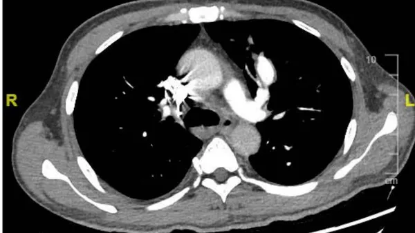
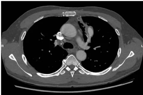
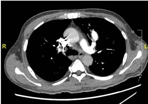
were no clinical improvement and fever persisted. We decided to repeat chest CT and this time there was a new finding of an aneurysm formation and thrombosis in the left upper lobe pulmonary artery segmental branch, thrombosis of the right lower lobe pulmonary artery segmental branches. The venous Doppler ultrasound was negative for lower limbs thrombophlebitis or thrombosis. Pathergy test was performed that was negative. By excluding Behçet disease he was then diagnosed Hughes-Stovin syndrome. He received methylprednisolone pulse followed by oral prednisolone 60 mg/day (1 mg/kg/day), associated with intravenous cyclophosphamide monthly according to 2018 update of the EULAR recommendations for the management of Behçet's disease[7]. Immediately after starting steroids his fever subsided and his clinical condition improved. He was discharged with oral prednisolone and with an indication for hospital admission once monthly for the administration of cyclophosphamide IV which was well tolerated and without any adverse effects. He completed 6 months of the aforementioned plan with gradually tapering down of prednisolone. The control pulmonary angiogram CT showed that the aneurysm in the left upper pulmonary segmental branch remained stable, reduction of thrombosis on the right lower lung segmental branches and the patchy infiltration subsided. At that time, patient refused to continue treatment. One month after stopping the medication he presented with massive hemoptysis and was carried to our ER. He suffered from cardiac arrest and was ressuscitated successfully and transferred to ICU. Chest CT showed left upper lobe pulmonary artery aneurysm rupture with hemorrhage and embolization of left upper pulmonary artery was done by interventional radiologist. However, his clinical condition gradually deteriorated, complicated by severe bilateral pneumonia and was certified dead a few days later.
| Variable | Reference Range, This Hospital | On 3rdAdmission | After amikacin treatment | 6thmonths of diagnosis |
| Hemoglobin (g/dL) | 13.5 - 17.0 | 8.0 | 8.3 | 12.3 |
| HCT (%) | 41 - 53 | 25.5 | 25.8 | 38.0 |
| Mean corpuscular volume (fl) | 80 - 100% | 66.2 | 65.3 | 69.9 |
| White-cell count (x 109/L) | 4.3 - 10 | 15.3 | 26.1 | 8.9 |
| Differential count (x 109/L) | ||||
| Neutrophils (x 109/L) | 1.9 - 7.3 | 12.9 | 22.7 | 6.3 |
| Eosinophils (x 109/L) | 0.0 - 0.7 | 0.0 | 0.0 | 0.1 |
| Basophils (x 109/L) | 0.0 - 2.0 | 0.1 | 0.0 | 0.1 |
| Lymphocytes (x 109/L) | 1.5 - 4.0 | 1.3 | 2.4 | 1.6 |
| Monocytes (x 109/L) | 0.2 - 0.9 | 1.0 | 1.0 | 0.8 |
| Platelets (x 109/L) | 100 - 400 | 273 | 304 | 171 |
| C-Reactive protein (mg/dL) | <0.5 | 14.98 | 16.98 | 0.12 |
| Procalcitonin (ng/mL) | <0.06 | 10.08 | 0.24 | - |
| Erythrocyte sedimentation rate (mm/hr) | 1.0 - 15.0 | 80.0 | - | 7 |
| CEA (ng/mL) | <3.8 | 1.3 | - | - |
| AFP (ng/mL) | <7.9 | 2.27 | - | - |
| CA 125 (U/mL) | <35 | 6.9 | - | - |
| CA 19.9 (U/mL) | <27 | 9.0 | - | - |
| SCC (ng/mL) | < 1.5 | 0.30 | - | - |
| Transferrin (mg/dL) | 174.0 - 364.0 | 117.0 | - | - |
| Variable | Reference Range, This Hospital | On Admission | After amikacin treatment | months of diagnosis |
| Ferritin (ng/mL) | <30-400 | 353.0 | - | - |
| Vitamin B12 (pg/ml) | 197.0 - 771.0 | 373 | - | - |
| Folate (ng/ml) | 3.9 - 26.8 | 7.5 | - | - |
| ANCA | Negative | Negative | - | - |
| Anti-CCP (RU/mL) | <= 5 | 1.52 | - | - |
| Anti-Cardiolipin | Negative | Negative | - | - |
| Immunoglobin A (g/L) | 0.63 - 4.48 | 2.53 | - | - |
| Immunoglobin G (g/L) | 5.40 - 18.22 | 12.94 | - | - |
| Immunoglobin M (g/L) | 0.22 - 2.40 | 0.92 | - | - |
| C3 (g/L) | 0.82 - 1.85 | 1.14 | - | - |
| C4 (g/L) | 0.15 - 0.53 | 0.24 | - | - |
| ANA-Screen | Negative | Negative | - | - |
| COVID-19, PCR assay | Negative | Negative | - | - |
| Cytomegalovirus | ||||
| IgM | Negative | Negative | - | - |
| IgG | Negative | Positive, 134 U/mL | - | - |
| Epstein-Barr virus | ||||
| EA-IgG | Negative | Positive, 1:10 | - | - |
| VCA-IgM | Negative | Negative | - | - |
| VCA-IgG | Negative | Negative | - | - |
| EA+EBNA-IgA | Negative | Negative | - | - |
| Interferon gamma release assay | Negative | Negative | - | - |
| Bacterial blood culture | Negative | Negative | - | - |
| Fungal blood culture | Negative | Negative |
III. Discussion
We presented a challenging case of a 56 years old man with a fever of unknown origin. Initially all the diagnostic work up did not lead to any clue for the etiology of his condition. Even after several courses of antibiotics, some of them of large spectrum, his fever persisted.
Hughes–Stovin syndrome (HSS) is very rare disorder. There is no much publication about this disease. Therefore, there is a lack of diagnostic criteria for Hughes-Stovin syndrome. The symptoms of Hughes-Stovin syndrome included hemoptysis (93.0%), cough (94.7%), dyspnea (86.0%), fever (70.2%), weight loss (47.4%), mouth ulcers (19.3%), genital ulcers (10.5%) and pleuritic chest pain (8.8%). [2]The diagnosis of Hughes-Stovin syndrome is based on the finding of pulmonary aneurysm in CT angiography and the findings of deep vein thrombosis in Doppler ultrasound. [3] In our case, the patient only manifested high-grade fever and did not have systemic involvement. His only manifestation was pulmonary artery vasculitis with aneurysm formation. Therefore, it was an unusual type of Hughes-Stovin syndrome.
Hughes-Stovin syndrome has been variably described as "the cardiovascular manifestation of Behcet's disease" [4], "incomplete Behçet's" [5] and "a rare case of Behçet's disease" [6] in literature. Since Hughes-Stovin syndrome is rare disease, there is not a clinical treatment guideline in current literature. Because of similarities of pulmonary involvement between Behçet's disease and Hughes-Stovin syndrome, the treatment guideline for Behçet's disease can be used to treat Hughes-Stovin syndrome in current medical practice. [7]
The prognosis of Hughes-Stovin syndrome is poor and aneurysm rupture is the leading cause of death, particularly in aneurysms of arterial origin.
IV. CONCLUSION
Hughes–Stovin syndrome (HSS) is very rare disorder characterized by the combination of multiple pulmonary artery aneurysms and deep vein thrombosis. There is no diagnostic criteria and management in current literature due to its rare condition.
We present this case to increase awareness for this condition that unfortunately can lead to death if left undiagnosed and untreated.