Introduction
Non-melanoma skin cancer is a significant public health problem, with an incidence increasing by 4% to 8% annually in developing countries1. Of new cancer cases registered, approximately 20% are located in the skin, making it the most frequent type of cancer in the world2. Primary squamous cell carcinoma (SCC) of the breast is considered a very rare malignant neoplasm, with an estimated prevalence between 0.06% and 0.2% of breast neoplasms2,3. Its occurrence is not well established, but it may be related to squamous metaplasia of the ductal epithelium or even originate from alterations during embryonic development4. Squamous cell carcinoma (SCC) presents rapid progression and a high invasive and metastatic potential when compared to basal cell carcinoma. Because it is an extremely rare neoplasm in the breast region, its clinical diagnosis becomes challenging2.
According to the literature, the prevalence of SCC is associated with white women, ranging from 52 to 64 years of age3. Pure squamous cell carcinoma of the breast can originate from the epidermis, nipple, or even the epithelium of a deep epidermoid cyst or squamous metaplasia5. When it occurs in the breasts, the lesion can mimic breast cancer, with a desquamative and erythematous clinical presentation, of an irregular and verrucous lesion2. It is classified by the World Health Organisation (WHO) as a specific and distinct subtype of metaplastic tumour and presents the following criteria: 90% or more of the lesion composed of keratinising squamous cell carcinoma infiltrating mammary parenchyma; absence of other invasive neoplastic elements; no extensive involvement of the skin overlying the tumour and absence of primary neoplastic skin lesions3.
In addition to its rarity, this carcinoma generally has a poor prognosis due to its aggressive nature and the lack of specific treatments, as there is no established protocol for it3. Core biopsy and immunohistochemistry are used for its diagnosis4. Any lesion that presents a clinical suspicion should be rigorously evaluated, with early biopsy requested to ensure an accurate diagnosis and avoid unnecessary radical therapies2.
Regarding treatment, there is no consensus on the best clinical approach, but chemotherapy remains one of the options used. The chemotherapy regimen with cisplatin and fluorouracil has been described as an excellent option, with reasonable responses reported after its use4. However, surgical removal of the lesion remains the treatment of choice (gold standard), as it allows for complete histopathological examination of the tumour, evaluation of surgical margins, and assessment of prognostic factors such as perineural invasion. In cases of excision of small tumors, where the surgical margins are clear, i.e., 4 mm to 10 mm from the lesion, the cure rate is estimated at almost 99%2.
Therapeutic approaches should be analysed and planned on a case-by-case basis, taking into account the patient's particular factors and preferences. Advanced cases of breast squamous cell carcinoma (SCC) may require mastectomy with axillary lymphadenectomy combined with adjuvant chemotherapy. However, in cases of primary breast SCC, removal with wide surgical excision is recommended, with lymph node dissection only necessary when there is clinical evidence of axillary spread. Histopathological confirmation, along with immunohistochemical evaluation and clinical history, is fundamental for the correct diagnosis of this rare type of tumour2.
Therefore, the rarity of this case, its diagnostic difficulty, and the uncertainties regarding the best treatment to be instituted justify the writing of this article.
Case Report
Female patient, 64 years old, black race, G3P3C0A0, menarche at age 15, menopause at age 53, with no prior use of hormone replacement therapy. She reported being a chronic smoker, with no other reported comorbidities or relevant family history.
The patient was referred to the Mastology Service with a history of a palpable nodule in the left breast, associated with progressive growth. On physical examination, the left breast was larger than the right, due to a large nodule in the left breast, measuring more than 10 cm, associated with an ulcerated lesion, local skin thickening, and the presence of palpable lymph nodes in the ipsilateral axillary region (Figure 1).
Regarding the complementary exams, the patient brought a recent mammogram, classified as BIRADS 3 (Breast Imaging Reporting and Data System), which described a solid, hypoechoic, oval nodule with circumscribed margins, located at the junction of the medial quadrants of the left breast, measuring 4,5 x 4,0 x 4,3 cm.
In light of the clinical findings, a core biopsy of the lesion guided by ultrasound was requested, whose anatomopathological examination revealed a poorly differentiated, solid carcinoma. Immunohistochemistry revealed a triple-negative pattern, with negative estrogen receptor (ER), progesterone receptor (PR), and human epidermal receptor (HER2) and a proliferation antigen (Ki-67) of 70%.
Physical examination showing a large tumor lesion in the left breast, with significant distension and ulceration of the skin, with associated secretion.
The chest CT scan showed a small solid subpleural nodule in the posterior basal segment of the left lower lobe, measuring 0.7 x 0.7 cm, suggestive of secondary pleuropulmonary involvement. Breast ultrasound showed a heterogeneous nodular formation, difficult to measure, occupying the entire left mammary parenchyma, associated with the presence of ipsilateral axillary lymphadenopathy, measuring 2.6 x 1.2 cm, which is atypical. Both bone scintigraphy and abdominal tomography were negative for secondary lesions.
Due to the size of the lesion, the patient was referred to the Oncology Service for neoadjuvant chemotherapy. Treatment with paclitaxel and carboplatin was chosen because this combination is widely recommended and used in advanced cases of breast and gynecological neoplasms and, as neoadjuvant chemotherapy, significantly increases the complete pathological response. However, after four cycles with this weekly protocol, the patient showed a poor response and significant tumour progression and was then submitted to hygienic mastectomy surgery.
Radical mastectomy of the left breast, Halsted type (A, B and C). Intercostal muscle, without the pectoralis major muscle, is already excised (D). Final closure with skin suture and Portovac drain (E). Final view of the surgical specimen (left breast).
The histopathological examination revealed invasive carcinoma with squamous differentiation, ulcerated, grade 3, measuring 17.0 x 12.0 x 10.0 cm, with focal angiolymphatic invasion, infiltration of the pectoral muscle and free margins (pT4b pNx pMx) (Figure 3).
Histopathological examination of the surgical specimen showed invasive carcinoma with Ulcerated Squamous Cell Differentiation.
Only after performing immunohistochemistry on the surgical specimen was the diagnosis of keratinising squamous cell carcinoma (SCC) definitively confirmed (Figure 4).
Immunohistochemistry panel confirming the diagnosis of keratinising squamous cell carcinoma.
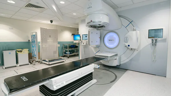
In the immediate postoperative period, the patient developed dehiscence of the surgical wound, in addition to disease progression in the anterior mediastinum and pleura. The case was discussed with the Clinical Oncology and Radiotherapy teams, and it was decided to perform palliative radiotherapy in the anterior hemithorax, followed by capecitabine as a second-line treatment. Upon conducting a new chest CT scan for therapeutic planning, a mass was found in the sternal region, with a significant increase in tumour volume. The adoption of exclusive palliative care was discussed with the patient and her family, and it was agreed that no further interventions would be performed.
Discussion
Primary squamous cell carcinoma of the breast is very rare, with a prevalence of 0.06% to 0.2% in malignant breast neoplasms2,3. It is called primary pure squamous cell carcinoma when the malignant cells are all of the squamous cell type, there is no relation with the skin, and if there is no indication for a primary location somewhere else in the body5. Furthermore, it has a rapid and aggressive course, with a recurrence rate of 25% and metastasis in 50% of cases3,6. SCC typically progresses rapidly and is highly invasive, most frequently found on exposed body surfaces most frequently seen in the sun and is uncommon in the breasts2.
For a more accurate diagnosis, it is essential to differentiate between squamous cell carcinoma (SCC) of the skin and primary squamous cell carcinoma of the breast, which differ in their epidemiology and histopathology. The former is more prevalent in black individuals and in males, and its most common clinical manifestation is an erythematous-squamous or slightly raised plaque. Furthermore, it can present as an ulcerated lesion with irregular borders or as an exophytic nodular lesion, similar to that presented in the case described6. The latter is prevalent in white women and may originate from the epidermis, nipple, or even the epithelium of a deep epidermoid cyst or squamous metaplasia3,5. It is important to distinguish this type from mixed tumours, where some patches of squamous cells can be found in adenocarcinoma of the breast and from metastasis of squamous cell carcinoma that originated elsewhere. The etiology and pathogenesis of squamous cell carcinoma of the breast is still unclear. It has been suggested that it may be a very extreme form of squamous cell metaplasia, developing into an adenocarcinoma7.
The clinical presentation may be painful breast inflammation or a tumor that is always larger than that of breast adenocarcinoma, similar to that found in the case described8.
Regarding imaging studies, there are no typical findings on mammography, and ultrasound may show a complex cyst or an inflammatory process5. In our case, mammography showed only one solid, hypoechoic nodule with circumscribed margins, located at the junction of the medial quadrants of the left breast, measuring 4,5 cm. The patient did not undergo breast ultrasound. It is still debatable whether investigations such as PET scans, in search of distant metastases or a primary squamous tumour site, should be performed9.
For a more precise diagnosis, a core needle biopsy should be performed, as it provides a greater amount of material (tissue) to be analysed6. Regarding the diagnosis, immunohistochemical examination should always be performed, as it allows confirmation of the squamous nature of the tissue and differentiation from other types of breast carcinomas, such as metaplastic carcinoma5. This differentiation is of great importance, as primary squamous cell carcinoma of the breast behaves more aggressively than other breast cancers5.
Squamous cell carcinoma of the breast may require surgery with wide excision to obtain clear margins, as occurred in the reported case2. Regarding lymph node dissection, it should only be indicated when there are signs of axillary invasion, either clinically or by ultrasound2.
Treatment is not specific due to the lack of an established protocol; however, one possible treatment involves adjuvant hormonal therapy with tamoxifen, which is essential when patients are positive for estrogen and progesterone receptors3. Squamous cell carcinomas are generally hormone receptor-negative8,10. For patients with negative expression of these receptors, conventional chemotherapy is necessary, as used in the aforementioned case5.
Postoperative chemotherapy should be recommended and, similar to other cases reported in the literature, the combination of paclitaxel and carboplatin appears to be the best option currently5. This chemotherapy regimen is used in advanced cases of breast, ovarian, and lung cancer. Both cisplatin and carboplatin interact with DNA, inhibiting cell growth (paclitaxel) and damaging tumor DNA (carboplatin), with a high rate of pathological complete response when used in neoadjuvant chemotherapy11. They have a mechanism of action similar to bifunctional alkylating agents, destroying rapidly dividing tumor cells12.
Regarding radiotherapy, it is recommended to administer it as adjuvant therapy, however the radiosensitivity of squamous cell carcinomas is still uncertain. The 5-year survival rate is around 67%13.
Therefore, primary squamous cell carcinoma of the breast is an uncommon finding. Its existence and possible evolution from an apparently benign lesion remain uncertain. For its definitive diagnosis, we emphasize the importance of histopathological examination and immunohistochemistry. Regarding treatment, the role of surgery is well established, but the role of adjuvant therapy is not yet well defined5.
In this particular case, the patient began treatment with neoadjuvant chemotherapy, showing a poor response with disease progression, and subsequently underwent radical breast surgery due to the size of the initial lesion, with clear surgical margins. Despite all the treatment instituted and due to the aggressive nature of the neoplasm, the patient soon experienced significant mediastinal recurrence, considered inoperable, and was referred for palliative and supportive care.