Introduction: Understanding that cancer is a pathology that is challenging to detect, treat, and comprehend is what makes the study of cancer so important. The goal of this case study is to show how a 20-year-old woman was affected by a variety of factors and symptoms that, when combined, raised suspicion of Ewing’s sarcoma, the second most common primary tumor in children and young adults. Ewing’s sarcoma has the characteristics of being an aggressive malignancy originating in the bones.
## I. INTRODUCTION
The main aspect in which a study on cancer is necessary is to understand that lives are transformed by a pathology that is difficult to diagnose, treat, and understand. The objective of this case report is to illustrate how a 20-year-old girl was affected by a whole set of factors and signs that, when compiling the main characteristic points, led to the suspicion of Ewing's Sarcoma, the second most common primary tumor in children and adolescents. Ewing's sarcoma has the characteristics of being an aggressive malignancy originating in the bones.
## II. METHODOLOGY
In this epidemiological study, the utilization of clinical trials to treat and address the epidemiological element is discussed using a case report as a guide. in conjunction with an assessment of the literature, whose sources came from the SciELO and PubMed data sources.The research was made in July 2023, meeting the inclusion criteria, which were articles from the years 2000 to 2023, in Portuguese and English, online texts, and full texts. As strategies for better evaluation of the texts, the following health descriptors (DeCS) were used: "Ewing's sarcoma" and "Chest wall tumor".
### a) Case Report
S.J.J., female, 20 years old, student She arrived at the hospital complaining of chest pain for 2 years—intense pain—and did not present other respiratory symptoms. She denied a fever and weight loss. She reports childhood asthma and denies a cancer history. She denies alcoholism and smoking. She reports that her mother had breast cancer. On physical examination, she was in good general condition, afebrile, anicteric, and eupneic on room air; there were no palpable lymph nodes in the cervical, supraclavicular, and axillary regions; auscultation of the respiratory system showed bilateral physiological vesicular murmurs and no adventitious sounds; there was no palpable tumor in the region of the chest; and she did not present a restriction to the mobilization of the upper limbs. At the time of the first consultation, the saturation was $98\%$ in room air.
A biopsy and several computed tomography images of the chest were done simultaneously. The first separate scout had an expansive lung lesion in the right lower lobe. One characteristic that stands out when all the scouts are analyzed is the nodule development angle, which is larger than 90 degrees and indicates an extrapulmonary lesion. In addition, chest tomography revealed opacity and demarcation in the xiphosternal line. Finally, the immunohistochemical analysis identified a neoplasm as cells with rounded, regular nuclei, coarse chromatin, and scant cytoplasm. The round cells, which in turn present an organoid pattern and fibromyxoid stroma, raise suspicions about the origin of the tumor.
The upper segment of the right lower lobe contained a solid and homogeneous subpleural expansive lesion that measured $6.0 \times 3.4$ cm in the largest axial axes. This lesion was located using a second computed tomography scan of the chest for surgical planning. The anterior cortex of the corresponding fifth and sixth costal arches looked to be in close contact with it, and a minor remodeling of the bones was observed. There is no pleural effusion and normal lung parenchyma.
The patient underwent a treatment that included myocutaneous repair, free margins, and resection from the fourth to the seventh quadrant. Three months after the date of surgery, a computed tomography scan of the chest was performed, showing signs of surgical manipulation on the right chest wall and blurring of the posterior aspect of the fourth to seventh costal arches on that side, with no signs of tumor remaining in the analysis for this one. As a result of the third right costal arch fracture, there was an atelectatic band on the lingula, no pleural effusion, the heart volume was still normal, and the thoracic aorta had a normal course and diameter. The patient was advised to have chest resonance segmentation every six months; however, the patient did not return for follow-up.
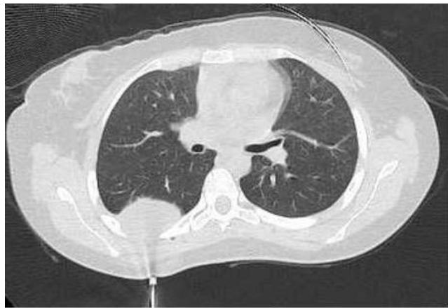 Figure 1: Tomographic scout prior to surgery
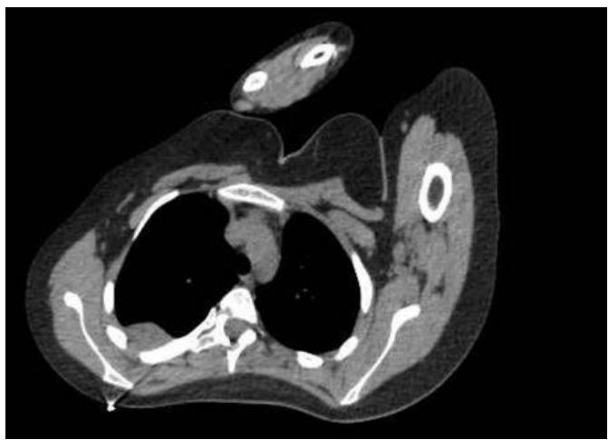 Figure 2: Tomographic scout prior to surgery
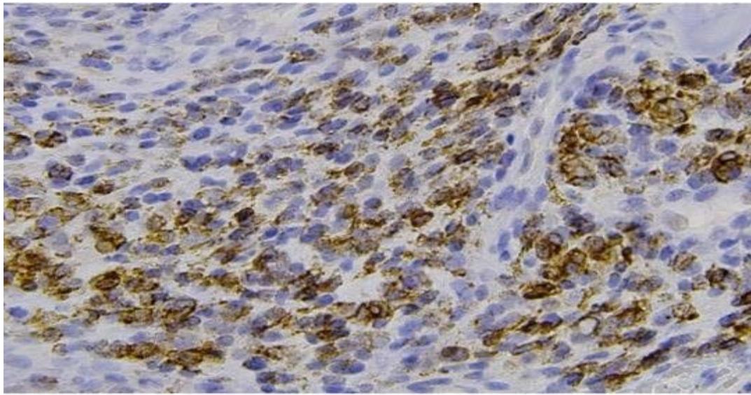 Figure 3: Histological slide
## III. RESULTS & DISCUSSION
In its initial aspect, Ewing's Sarcoma is a malignant bone tumor characterized by round, primitive cells without obvious differentiation. Localized pain, with the potential for a palpable mass depending on the location, characterizes the clinical presentation. Wide surgical resection is the primary form of treatment, possibly with radiotherapy.
Since chest wall tumors are cancerous tumors that can impair lung function, the issue was chosen because of its importance to society. In this regard, primary tumors of the chest wall make up $5\%$ of all tumors and impact bone tissue in roughly $55\%$ of cases. A distinct clinical picture, histological characteristics, specificity, and anatomical location preference. To confirm the diagnostic theory of Ewing's Sarcoma, tomography and biopsies must also be carried out.
Based on the retrospective analysis of prospective studies by the Cooperative Weichteilsarkom Studiengruppe (CWS), carried out with 60 patients from 1997 to 2015, the prevalence of Ewing's sarcoma in men was observed, with a ratio of 4 men to 1 woman. The average age of a person with this sarcoma is 14.5 years old. In addition, the intra-abdominal and retroperitoneal regions are affected in about $93\%$ of cases, with emphasis on the thoracic, paratesticular, and parotid gland regions. In $67\%$ of the cases, they had tumors larger than $10~\mathrm{cm}$, and $88\%$ had locally disseminated tumors; thus, in most cases, sarcoma was characterized by large and disseminated tumors.
$10\%$ of patients had only a single, isolated tumor at the time of clinical presentation; $27\%$ had spread to nearby lymph nodes; and $63\%$ had extraperitoneal metastasis, of which $67\%$ underwent surgical resection. Regarding treatment, of the 60 patients, nine underwent high-dose chemotherapy, six received regional hyperthermia, and 20 received radiotherapy. As a result, 51 patients died, of whom 49 due to the disease and 2 due to abandonment of chemotherapy and peritonitis. Surgery with resection at R0 is the aim; radiotherapy was implemented in patients with extensive or unresectable disease. The VAIA chemotherapy regimen (ifosfamide, vincristine, adriamycin, and actinomycin D) produced the best results overall.
Additionally, $32\%$ of patients had relapsed, $26.7\%$ had disease-free time for three years, and $42\%$ of patients had complete disease remission. The investigation revealed that norisk factor was connected to the progression of the disease, tumor size did not affect mortality, and metastatic sites did not exhibit a statistically significant prevalence. Sarcoma has a proven bad prognosis; often, a patient with a good prognosis has limited extra-abdominal illness, no pleural effusion, has undergone surgery at R0, and has used CT VAIA.
It is possible to observe the proportion of the evolution of cases in several patients, making ageneral analysis and allowing a certain orientation, both for the immediate treatment and for the prognosis of the patients.
In this way, neuroendocrine tumors and Ewing's sarcoma have been combined into a family of sarcomas. Although the latter studies exhibit greater neuroectodermal differentiation than Ewing's sarcoma, this difference is not clinically relevant. Typically, Ewing's sarcoma invades the cortex, periosteum, and soft tissues. They have a sparse cytoplasm that seems cleaner, which is a result of the high glycogen content. They have a sparse cytoplasm that seems cleaner, which is a result of the high glycogen content. It is a sarcoma that primarily affects flat bones like the pelvis, ribs, and vertebrae, as well as the trunk when soft tissues are involved. Immunohistochemical analysis of the samples shows the existence of cells with rounded, regular nuclei, coarse chromatin, and sparse cytoplasm. The round cells themselves have a fibromyxoid stroma and an organoid pattern. As a result, similar characteristics to the reported case are confirmed.
It appears that bone Ewing's Sarcoma is an aggressive tumor, usually affecting individuals in the second decade of life. Note also the appearance of a licit lesion with a periosteal reaction typical of an aggressive lesion on X-RAY. Furthermore, the immunohistochemical study is necessary to differentiate and classify the types of sarcomas in the Ewing family. Thus, the case shows the importance of investigating pathologies that have not yet been well elucidated since rare diseases are not part of the medical routine, so their diagnosis brings the opportunity to obtain more specialized treatments.
## IV. CONCLUSION
In this way, the case highlights the need to research pathologies that have not yet been fully understood because rare diseases are not common and their diagnosis guarantees access to more specialized treatments.
Generating HTML Viewer...
References
7 Cites in Article
Marta Sbaraglia (2020). Ewing sarcoma and Ewinglike tumors.
A Croci,O Camargo,Nrb Oliveira (1996). Tratamento cirúrgico do sarcoma de Ewing: avaliaçãooncológica e funcional.
Giovanna Lopes,Cunha Da,Castro Unknown Title.
Monika Scheer,Christian Vokuhl,Bernd Blank,Erika Hallmen,Thekla Von Kalle,Marc Münter,Rüdiger Wessalowski,Maite Hartwig,Monika Sparber‐sauer,Paul‐gerhardt Schlegel,Christof Kramm,Udo Kontny,Bernd Spriewald,Thomas Kegel,Sebastian Bauer,Bernarda Kazanowska,Felix Niggli,Ruth Ladenstein,Gustaf Ljungman,Kirsi Jahnukainen,Jörg Fuchs,Stefan Bielack,Thomas Klingebiel,Ewa Koscielniak (2019). Desmoplastic small round cell tumors: Multimodality treatment and new risk factors.
John Scott E Kilpatrick,Brian Reith,Rubin (2018). Ewing Sarcoma and the History of Similar ans Possibily RelatedSmall Round Cell Tumors: From Whence Have We Come and Where are We Going?.
Contran,Robbins,Ossos (2016). articulações e tumores de partes moles: Tumores de origem desconhecida.
Davi Bellan,Reynaldo Jesus-Garcia Filho,Jairo Garcia,Marcelo Petrilli,Dan Viola,Murillo Schoedl,Antonio Petrilli (2012). Sarcoma de Ewing: epidemiologia e prognóstico dos pacientes tratados no Instituto de Oncologia Pediátrica, IOP-GRAACC-UNIFESP.
No ethics committee approval was required for this article type.
Data Availability
Not applicable for this article.
How to Cite This Article
Matheus Amorim Grigorio. 2026. \u201cClinical and Epidemiological Profile of Patients with Ewings Sarcoma\u201d. Global Journal of Medical Research - B: Pharma, Drug Discovery, Toxicology & Medicine GJMR-B Volume 23 (GJMR Volume 23 Issue B3).
Explore published articles in an immersive Augmented Reality environment. Our platform converts research papers into interactive 3D books, allowing readers to view and interact with content using AR and VR compatible devices.
Your published article is automatically converted into a realistic 3D book. Flip through pages and read research papers in a more engaging and interactive format.
Subject: Global Journal of Medical Research - B: Pharma, Drug Discovery, Toxicology & Medicine
Authors:
Henrique Hollanda Larangeira, Tábata de Oliveira Silva, Rafaela da Silva Schottz, Ingrid Fabric Gouveia Lima, Lina Borges Cavalcante, Marcus Antonio Studart da Cunha Frota, Janaína Gomes da Rocha, Lorena de Sousa Moura (PhD/Dr. count: 0)
Our website is actively being updated, and changes may occur frequently. Please clear your browser cache if needed. For feedback or error reporting, please email [email protected]
Thank you for connecting with us. We will respond to you shortly.