
"The scientific man does not aim at an immediate result. He does not expect that his advanced ideas will be readily taken up. His work is like that of the planter—for the future. His duty is to lay the foundation for those who are to come and point the way."
I. INTRODUCTION
Surgery, radiation, and chemotherapy can cure about half of cancer patients; nevertheless, in the United States alone, nearly 600,000 people died last year. The US cancer death rate has fallen since 1991, partly due to advances in treatment, early detection, and less smoking [1]. It is better to prevent cancer before than to cure the disease after. A healthy environment and lifestyle can prevent only of cancer-causing mutations [2], while everyone needs cancer prophylactics. Each day apoptosis and the immune system erase billions of mutants and expired cells. The immune system is confused in cancer patients. There is an opportunity to prevent early-stage cancer or metastases by rebooting the immune system with a unique delivery vehicle—alpha-fetoprotein (AFP) and toxins.
From the fertilized egg, trillions of cells grow through duplications. Stem cells are deposited on the way. When stimulated to increase, a stem cell is undergoing an "asymmetric division" [3]. The proliferating daughter cell continues to divide and proceed down the tissue hierarchy, from stem cell to progenitor cell, before becoming a fully differentiated mature tissue cell. Multiple types of stem cells have been identified in a wide range of tissue, sharing multipotency characteristics.
In the bone marrow, hematopoietic stem cells (HSC) exist undifferentiated. They are at the peak of a blood cell differentiation hierarchy. The white blood cell ratio is neutrophils lymphocytes monocytes eosinophils basophils. Myeloid-derived suppressor cells (MDSCs) are a small heterogeneous cell population of immature myeloid progenitors of granulocytes, macrophages, and dendritic cells (DCs) at different stages of differentiation generated from a common HSC [4].
MDSCs can leave the bone marrow and spread throughout the body, becoming immune response calmers during pregnancy, cancer, regeneration, stress, autoimmune and infectious diseases, obesity, age, etc.[5]. Besides the bone marrow, other sites generate MDSCs: the placenta and umbilical cord, the tumor site, and the spleen [6].
MDSCs exist in an undifferentiated state at the peak of the immune cell's hierarchy. They affect innate and adaptive immunity cells directly and indirectly. MDSCs inhibit natural killer (NK) cells, DCs, and T-cells, induce regulatory T cells (T regs) and modulate macrophages, etc. [7-9].
In 1862 Rudolf Virchow was the first to link the origin of cancers from otherwise normal cells correctly: "every cell arises from another cell." Indeed, of tumor-causing mutations are inherited, and tumor cells are activated later in life, while of tumor-causing mutations appear during duplications [2]. According to the hierarchical model, the tumor grows from a single cell. Like in embryogenesis, a core group of stem cells exists at the top of the tumor hierarchy, from which other more differentiated cells are formed. Descending from the undifferentiated cells to the most mature cells that comprise the bulk of the tumor mass. Cancer stem cells (CSCs) are cancer cells with characteristics associated with normal stem cells; specifically, give rise to all cell types found in a particular cancer sample. Approximately of current CSC surface markers appear on embryonic or adult stem cells and are rarely expressed on normal tissue cells. It is believed that the elimination of CSCs could eradicate whole cancer [10].
Pregnancy is a natural phenomenon that ensures the survival of the species. An embryo turns off a critical pathway required for the immune system to attack intruders. The suppression of the immune response in pregnancy is robust since the embryo cells have half the father's proteins that the mother's immune system should recognize as foreign. Moreover, even surrogate motherhood is possible, with no genetic relationship to the embryo. The immunology of pregnancy and cancer is similar [11]. Embryo and cancer cells exploit the mechanisms that allow them to grow despite the host's immune system attacks. During pregnancy, the mother's anti-embryo immune response is neutralized by the oncofetal proteins that re-appear during cancer development.
Some of the oncofetal proteins are AFP, AFP receptor (AFPR), human chorionic gonadotropin (HCG), carcinoembryonic antigen (CAE), and pregnancy-associated protein A. A few molecules regulate the immune tolerance of the mother - AFP, HCG, glycodelin, and pregnancy-specific -glycoprotein [12]. Cancers express oncofetal proteins at "a wrong time in a wrong place" to withstand immune system attacks.
Forty years ago, oncofetal proteins were used for cancer patients' vaccination. The placenta is the first organ that forms after conceiving—before any baby's organs even take shape. Immunotherapy with placental proteins has demonstrated a 5-year survival rate and a 10-year in 35 terminal patients [11]. (The handful of cancer immunotherapy drugs available today have demonstrated robust and durable results only in a minority of patients).
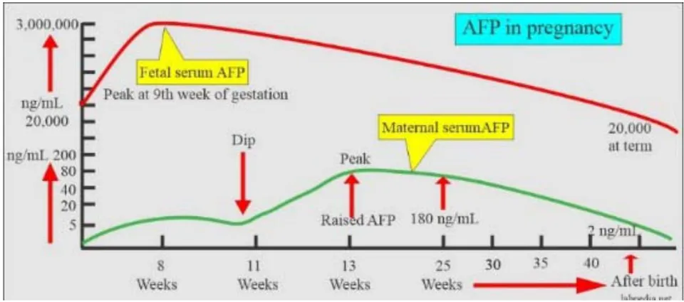
AFP is the major oncofetal protein secreted in early post-implantation embryos of mammals; the ability to synthesize AFP is restricted to the visceral endoderm cells around the embryonic region of the egg cylinder [13]. Later in development, AFP is produced by the yolk sac, liver, and gastrointestinal tract and penetrates the mother's blood (<200 ng/ml) (Fig. 1).
AFP can be re-expressed in adult life in a few cancers: hepatomas, germ cell tumors, yolk sac tumors of the ovary, and gastrointestinal cancers. The AFP structure, functions, and clinical applications are well covered in the literature [14-18].
AFP and its peptides have immunomodulatory properties [19-21]. For example, AFP selectively induced a rapid downregulation of surface MHC class II antigens in their expression on human monocytes, thereby making embryo/tumor cells "invisible" to the immune system. MHC class II antigens on monocytes are the key molecules in antigen presentation. They differentiate between a self-cell and an "alien" embryo or cancer cell. AFP neither alters the expression of MHC I, CD4, CD18, CD45, and Fc receptors for IgG on the surface of monocytes/macrophages nor affects the functional maturation of the macrophages Fc receptors or the ability to express antibody-dependent cell-mediated cytolytic activity. AFP may, by reducing the antigen-presenting capacity of monocytes/ macrophages, function as an essential factor in maintaining a fetal allograft, as well as participate in the downregulation of the entire immune system in cancer [22].
Nevertheless, the primary immune regulatory impact is AFP ligands because AFP delivers dozens of molecules within 3-5 days of its half-life. Moreover, the AFP-binding monocytes play a fundamental role in regulating the immune response.
The AFP's primary function is nutrient delivery in a shuttle manner (like oxygen delivery by hemoglobin). AFP binds different ligands, delivers them to the AFPR-positive cells, and releases them inside the cell compartment with an acidic pH. The 69 kDa AFP can hide 1-2 molecules (<2 kDa) in its hydrophobic cavity [23]. For example, docosahexaenoic acid (DHA) is not synthesized by the mother, who should take essential nutrients with food. AFP grabs DHA from albumin and transports it through the placenta [24]. Polyunsaturated fatty acids are necessary for many purposes, for example, the myelination of nerve fibers in the fast-growing embryo's brain.
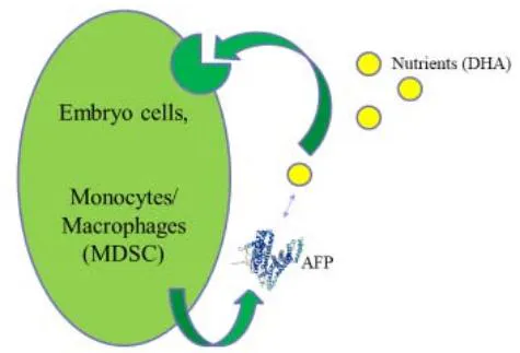
Embryo cells and normal and malignant peripheral monocytes/macrophages use the autocrine AFP/AFPR system for nutrient supply (Fig. 2, A) [25].
Low-differentiated lymphocytes use AFPR during blast transformation [26]. Monocytes/macrophages have specific 62 and 65 kDa AFP-binding receptors, which are involved in the physiological regulation of the immune response [27].
Unlike AFP, many cancers express AFPR [28], which should be considered the tumor marker and oncofetal protein #1. The AFP gene-knockout rodent models have demonstrated that AFP is not obligatory for full-term delivery [29]. The AFPR gene and structure are unknown yet to perform the gene-knockout experiment. In any case, the AFP/AFPR duo is vital for the embryo (and cancer).
The fact that the mother is tolerable to the embryo for nine months demonstrates that from the very beginning, AFP (Fig. 1) delivers nutrients to MDSCs, "corrupting" them to generate a protective shield over an embryo. MDSCs suppress NK cells which are "spontaneous cytotoxic cells" involved in surveillance against tumor cells. NK cells attack "aliens," low differentiated embryos, stem, cancer cells, and CSCs [30, 31]. NK cells can erase cancer at the earlier stages and solve the urgent problem of metastases.
II. INJECTABLE AFP-TOXINS
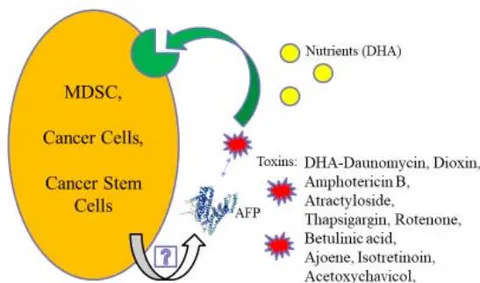
In cancer, MDSCs are "corrupted" also [32]. The effective way to ruin corruption is to eliminate the head. For this purpose, instead of DHA, AFP can deliver toxins to MDSCs (Fig. 2, B). The MDSCs death unleashes NK cells, and the whole immune system, enabling it to recognize and attack cancer the way it does other diseases. In addition, AFP-toxin kills AFPR-positive cancer cells and possibly CSCs (Fig. 2, B).
Like AFP-DHA, AFP complexes with hydrophobic/amphiphilic toxins hidden in the hydrophobic cavity are stable in the bloodstream. They release the toxin only inside the AFPR-positive cells.
After toxin unloading, AFP can work as a shuttle and deliver additional toxins. DHA-daunomycin conjugate binds AFP and inhibits tumor growth in AFP-producing mice [33]. Most cancers do not produce AFP, but are AFPR-positive; hence, exogenous AFP with toxins kills them. Thus, the treatment with AFP and amphotericin B in the excess (1:60-100) for shuttling demonstrated a response in 6 out of 8 cancer patients and increased quality of life [34]. The cytokine storm-like reaction, sometimes observed during the AFP with amphotericin B infusions, preceded cancer cells' death, and it can indicate the consequences of MDSCs death [18]. The potent AFP-binding toxins are expected to provide even better than AFP-amphotericin B response in cancer treatments.
Cancer cells can activate an AFP/AFPR autocrine loop (Fig. 2, B) [35, 36]. MDSCs are progenitor cells with only a few duplication steps from stem and embryo cells, and, as well as CSCs; they should retain an AFP-mediated nutrient delivery system. These need research. At least, AFP is absorbed by MDSCs, stimulating their suppressive activity [37, 38].
AFP attracts MDSCs and T regs through AFP-binding C-C chemokine receptor type 5 (CCR5) [39-41]. These regulatory cells migrate, accumulate, and suppress the immune attack on cancer. Targeting CCR5 reboots immunosuppressive myeloid cells [42, 43].
AFP binds to the neonatal Fc receptor (FcRn) [44, 45]. The FcRn is found in MDSCs in pancreatic cancer monocytes. MDSCs and DCs are elevated in pancreatic cancer patients compared to non-cancer donors [46]. They can be targeted through AFPR and/or FcRn by AFP-toxin drugs.
AFPR, CCR5, and FcRn are valuable MDSCs markers, at least for a transitory period.
Many current drugs do not eliminate CSCs, which may be why many cancers regrow after treatment. Immunotherapy does not act directly on cancer but works on the immune system. Checkpoint inhibitors and CAR T-cells are too unsafe for early-stage cancer; complexity and cost also prevent their application. Dissemination of cancer cells from the primary tumor into distant body tissues and organs is the leading cause of death in cancer patients. While most clinical strategies aim to reduce or impede the growth of the primary tumor, no treatment to eradicate metastatic cancer exists at present [47]. The MDSCs- and CSCs-targeting drugs have a bright future as they are critical in tumor and metastasis prevention [48, 49].
More than 100 years ago, Paul Ehrlich proposed a "magic bullet" that kills cancer cells, sparing the healthy ones. Nevertheless, this approach did not elevate the survival rate of cancer patients. The additional target outside of cancer cells should be hit. This "magic target" is a myeloid suppressor cell. Combining "magic bullets" and the "magic target" approach can cure cancer.
"Magic bullet" can kill "magic target" MDSC. Paclitaxel hits both cancer cells and MDSCs [50], and AFP potentiates its direct cytotoxic and immunotherapy action [51]. Thapsigargin is a more potent toxin than paclitaxel. AFP-thapsigargin complex (ACT-902) depletes MDSCs and tumor-associated macrophages. In mice, chemotherapy using ACT-902 and AFP with paclitaxel demonstrated superior efficacy and safety compared to chemotherapy alone. ACT-902 has led to the complete regression of five out of six highly resistant to chemotherapy POP-92 xenografts by day seven of treatment with no further growth after this period in mice [52]. Or the AFP-maytansine conjugate combines both immunotherapy and targeted chemotherapy with undetectable bone marrow toxicity. It has shown survival with no tumor re-growth after in the mice models [53]. AFP-toxin conjugates might pave a new road to the cancer cure [54-58]. On the other hand, unlike complexes, artificial conjugates have the risk of immune response to themselves.
The neuroblastoma cells may re-express embryonal or fetal antigens, suggesting some reversion towards an earlier stage of differentiation, and they can incorporate AFP [59, 60]. AFP-maytansine conjugate can kill brain tumor cells and CSCs found in human brain tumors [61].
Glioblastoma is the most aggressive, malignant primary brain tumor in adults. Myeloid cells are critical regulators of immune and therapeutic responses to glioblastoma [62]. In the glioblastoma microenvironment, M-MDSCs represent the predominant subset [63]. M-MDSCs can be depleted by AFP-toxin conjugate [54].
It has been found that MDSCs account for approximately of the tumor mass in gliomas. MDSCs are increased following conventional chemotherapy treatments [64]. Targeting MDSCs in combination with other therapies has shown promising therapeutic effects in brain cancer [65], and AFP-toxin drugs can be one of these therapies.
III. ORAL INSTEAD OF INJECTABLE
"Let food be thy medicine, and let medicine be thy food."
Hippocrates
MDSCs can be affected by ingredients from herbs and supplements. For example, withaferin A – a promising anti-cancer constituent of the Ayurvedic medicinal plant Withania somnifera-reduces MDSCs function [66]. Nevertheless, pregnant women should avoid Withania somnifera tonic as it may induce abortion at high doses [67]. In ancient Rome and Greece, women used silphium, an oral herbal contraceptive. This valuable herb is seen on a coin with a crab that once was a cancer disease name (Fig. 3).
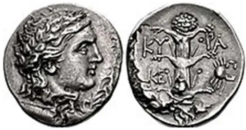
Was silphium used not only for pregnancy prevention but for cancer treatment too? There is no silphium in Nature anymore to check the hypothesis. Still, Artemisia absinthium, also used in Roman times for birth control, contains artemisinin that prevents early embryo implantation in animal models. Supposedly, AFP shuttles silphium ingredients or artemisinin to MDSCs, decreases their immunosuppressive activities, and leads to pregnancy or cancer prevention.
AFP wins the competition with excess albumin for binding embryo toxins such as diethylstilbestrol, dioxin, warfarin, etc., and can lead to pregnancy prevention or loss [18]. It can also be true for orally administrated thalidomide, miltefosine, etc. Toxins directly affect embryo cells, and AFP with toxins activate the mother's immune system like paclitaxel.
Traditional medicines and spices often contain anti-cancer agents, such as withaferin A, ajoene, acetoxychavicol, capsaicin, curcumin, quercetin, all-trans retinoic acid, sinigrin, artemisinin, astaxanthin, scutebarbatine A, etc. Small amounts of AFP naturally existing in the body potentiate their anti-cancer activity by the mechanism discussed earlier. To activate the immune system significantly, AFP-binding anti-cancer agents should be used together with exogenous AFP [68, 69].
Porcine AFP and betulinic acid (1:2) gavage inhibited mouse tumor growth. Tumor inhibition was potentiated by the excess of betulinic acid [70]. The beneficial effect of an extra amount of agent for tumor growth inhibition was supported by the experiment with the porcine AFP-ajeene (1:2) complex and ajoene in excess [18].
In suboptimal doses, the oral porcine AFP- attractyloside (1:2) complex has shown a response in six of twelve metastatic colorectal cancer patients [71]. Additional spices, herbs, or supplements having anticancer properties could potentiate the treatment. In another trial, a woman with stage IV ovarian cancer took elevated doses of the porcine AFP-attractyloside complex and survived over ten years [18].
AFP-toxin complex absorption from the gastrointestinal tract into the lymph nodes needs research. It can be like IgG-antigen complex absorption through the FcRn of the gut enterocytes [18].
AFP with AFP-binding toxins promises helpful in disease prevention, as they require low concentrations of AFP and toxins, like pregnancy prevention. Injectable recombinant AFP is safe in doses higher than in pregnant mothers' blood (0.3 - 0.5 g/mL) [72]. Porcine AFP can be taken orally in high doses safely.
IV. CONCLUSION
AFP-toxin conjugates or AFP with AFP-binding toxins injections deplete MDSCs, reboot the immune system, and can prevent or treat cancer and metastases. As a shuttle delivery vehicle, AFP can potentiate the anti-cancer activity of the AFP-binding toxins or drugs. Oral administration of AFP with herbs or supplements with anti-cancer properties is possible. Like embryo toxins do not hurt the mother but prevent pregnancy, the simultaneous presence of AFP and AFP-binding toxins in the bloodstream can safely prevent early-stage cancers or metastases. Oral preparations do not need high AFP purity, and porcine AFP can be used instead of human protein. Taken once or twice a year course, AFP with AFP-binding toxins can reboot the immune system and prevent cancer and metastasis. No conflict of interests