
Loading menu…

A New Method of Surgical Management for Pseudocyst of Pinna

§ Khulna City Medical College

Send Message

To: Author

A New Method of Surgical Management for Pseudocyst of Pinna

Article Fingerprint

ReserarchID

MR95904

A New Method of Surgical Management for Pseudocyst of Pinna Banner

Key Research Insights

Synthesized scholarly intelligence & interactive research assistant
  • English
  • Afrikaans
  • Albanian
  • Amharic
  • Arabic
  • Armenian
  • Azerbaijani
  • Basque
  • Belarusian
  • Bengali
  • Bosnian
  • Bulgarian
  • Catalan
  • Cebuano
  • Chichewa
  • Chinese (Simplified)
  • Chinese (Traditional)
  • Corsican
  • Croatian
  • Czech
  • Danish
  • Dutch
  • Esperanto
  • Estonian
  • Filipino
  • Finnish
  • French
  • Frisian
  • Galician
  • Georgian
  • German
  • Greek
  • Gujarati
  • Haitian Creole
  • Hausa
  • Hawaiian
  • Hebrew
  • Hindi
  • Hmong
  • Hungarian
  • Icelandic
  • Igbo
  • Indonesian
  • Irish
  • Italian
  • Japanese
  • Javanese
  • Kannada
  • Kazakh
  • Khmer
  • Korean
  • Kurdish (Kurmanji)
  • Kyrgyz
  • Lao
  • Latin
  • Latvian
  • Lithuanian
  • Luxembourgish
  • Macedonian
  • Malagasy
  • Malay
  • Malayalam
  • Maltese
  • Maori
  • Marathi
  • Mongolian
  • Myanmar (Burmese)
  • Nepali
  • Norwegian
  • Pashto
  • Persian
  • Polish
  • Portuguese
  • Punjabi
  • Romanian
  • Russian
  • Samoan
  • Scots Gaelic
  • Serbian
  • Sesotho
  • Shona
  • Sindhi
  • Sinhala
  • Slovak
  • Slovenian
  • Somali
  • Spanish
  • Sundanese
  • Swahili
  • Swedish
  • Tajik
  • Tamil
  • Telugu
  • Thai
  • Turkish
  • Ukrainian
  • Urdu
  • Uzbek
  • Vietnamese
  • Welsh
  • Xhosa
  • Yiddish
  • Yoruba
  • Zulu
Reading Preferences
Font Size
Line Spacing
Background

I. INTRODUCTION

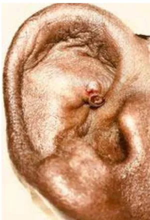
Pseudocyst of pinna is an idiopathic benign painless cystic swelling developed spontaneously on the lateral aspect pinna due to accumulation of fluid. It is an intra cartilaginous cyst without having any epithelial lining. The common sites of origin are cymba concha, scaphoid fossa and triangular fossa of the pinna(1). These lesions are also named as endochondral pseudocyst, intra cartilaginous cyst and benign idiopathic cystic chondromalacia (2).

Histologically these are intra cartilaginous cyst without having any epithelial lining hence it is called pseudocyst. The fluid inside the cyst is yellow or straw colored serous or viscous fluid containing glucose and protein(3). The lesions are mostly unilateral.

These are simple lesions sometimes incidentally found by close contact but difficult to manage either by medical or surgical procedure. Hence there are so many modalities of management described in literature. Whatever modality of treatment is applied the aim of treatment includes restoration and preservation of normal appearance of pinna and prevention of recurrence(3).

We describe our experience of management of these comparatively less common simple lesions of pinna by applying our new and minimal surgical technique.

II. MATERIALS AND METHODS

24 patients were diagnosed clinically as pseudocyst of pinna in my private consultation clinic between 1st January 2015 to 31st December 2018. The procedure to be done was explained to the patients and their written consent taken for enrolment in the study.

Diagnosis was made by clinical examination and confirmed by aseptic aspiration of non-purulent straw or yellow color fluid from the cyst during surgical procedure.

All patients were posted for our minimal invasive new technique of surgical intervention. With all aseptic precaution and under local anesthesia a stab incision was given on selected dependent part of the cyst and through that stab incision instantly made ear grommet type drain tube made up of butterfly needle set inserted. This self-retaining type of drain tubes were kept in situ for two weeks. After two weeks we observed complete resolution of the cyst. Then we removed the drain tube. The patients were followed up once in a month for 3 months to see any recurrence.

III. RESULTS

Twenty-four patients were diagnosed as cases of pseudocyst of pinna from January 2015 to December 2018. The age distribution in our study group ranged from 25 to 45 years. The lesions were seen more in scaphoid fossa followed by triangular fossa and cymba concha. Twenty-two patients had complete resolution within two weeks. Two patients developed secondary infection which was effectively controlled by broad spectrum antibiotics and subsequently cured.

IV. DISCUSSION

The pseudocyst of pinna is fairly an uncommon problem found among adult and predominantly in males (79.16%) in our study group consistent with other reports. The sites of origin were scaphoid fossa, triangular fossa and cymba conchain order of precedence. The predisposing factors and etiology were not known. Its pathogenesis is hypothetical as yet. Hormonal factor may play a part for male predominance (4).

Abnormal release of lysosomal enzymes from chondrocytes give rise to progressive dilatation and formation of intra cartilaginous cavity (5). One hypothesis explained congenital embryonic dysplasia of the auricular cartilage that leads to formation of pseudocyst (6).

There is no single accepted method of treatment for this condition. The various modalities of treatment are close aspiration and pressure bandage, aspiration with buttoning, aspiration and intralesional corticosteroid injection, anterior wall deroofing, incision and curettage of cartilage wall, intralesional sclerosing agents etc. are being practiced (6) (7)(8). Fibrin glue as a sealing material between the two flaps of cartilage has been reported (9). Whatever method is applied to treatment there is every chance of recurrence in significant number of cases (10)(11).

V. CONCLUSION

Almost in all methods of treatment required compression dressing which is difficult to provide and maintain. Our new technique of grommet type plastic tubedrainout insertion through a stab incision does not require any pressure dressing. Moreover, it is simple cost effective and compliant to patient with acceptable outcome.

  • Informed Consent: Duly signed informed consent was taken from all patients.

Financial Disclosure: The authors declare that the study received no fund from any source. There is no conflict of interest.
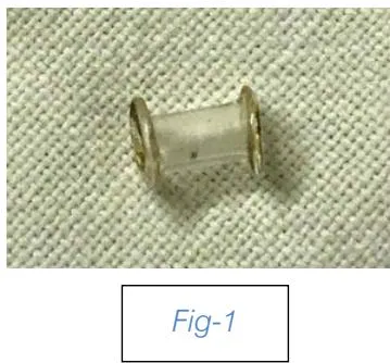

Fig -2:
Fig -2:

References

11 Cites in Article
  1. 1. S Choi,K-H Lam,K-W Chan,F Ghadially,A Ng (1984). Endochondral Pseudocyst of the Auricle in Chinese. Archives of Otolaryngology - Head and Neck Surgery, 110(12), 792-796.
  2. 2. Q Chen,Y Fei,T Zho,D Luo,B Wu,X Yang (2001). Research on the immunological cause of auricular pseudocyst. Lin Chuang Er Bi Yan Hou KeZhi, 15, 304-305.
  3. 3. Klaus Schulte,Norbert Neumann,Thomas Ruzicka (2001). Surgical pearl: The close-fitting ear cover cast? a noninvasive treatment for pseudocyst of the ear. Journal of the American Academy of Dermatology, 44(2), 285-287.
  4. 4. V Bhat,Shipla,Nitha,R Ks (2014). Deroofing of auricular pseudocyst: Our experience. J Clin Diagn Res, 8, 5-7.
  5. 5. Hideaki Miyamoto,Mitsuya Okajima,Ikuyo Takahashi (2001). Lactate dehydrogenase isozymes in and intralesional steroid injection therapy for pseudocyst of the auricle. International Journal of Dermatology, 40(6), 380-384.
  6. 6. D Engel (1966). Pseudocysts of the auricle in Chinese. Arch Otolaryngol, 83, 197-202.
  7. 7. Chao-Hsi Chang,Wen-Rei Kuo,Chih-Hsin Lin,Ling-Feng Wang,Kuen-Yao Ho,Kun-Bow Tsai (2004). Deroofing Surgical Treatment for Pseudocyst of the Auricle. The Journal of Otolaryngology, 33(03), 177.
  8. 8. Serhan Tuncer,Yavuz Basterzi,Reha Yavuzer (2003). Recurrent Auricular Pseudocyst: A New Treatment Recommendation With Curettage and Fibrin Glue. Dermatologic Surgery, 29(10), 1080-1083.
  9. 9. A Rehman,Sangoo,S Hamid,A Wani,N Khan (2013). Recurrent pseudocyst pinna: A rational approach to treatment. Int J Sci Res Publ, 3, 1-4.
  10. 10. T Ramadass,Gopi Ayyaswamy (2006). Pseudocyst of auricle — etiopathogenesis, treatement update and literature review. Indian Journal of Otolaryngology and Head and Neck Surgery, 58(2), 156-159.
  11. 11. C Lim,Y Goh,S Chao,Lim Lynne (2002). Pseudocyst of the Auricle. The Laryngoscope, 112(11), 2033-2036.

Funding

No external funding was declared for this work.

Conflict of Interest

The authors declare no conflict of interest.

Ethical Approval

No ethics committee approval was required for this article type.

Data Availability

Not applicable for this article.

How to Cite This Article

Professor Dr. Sudhangshu Kumar Ballav, Dr. Debnath Talukder, Dr. Shailendranath Biswas, Dr. Susmita Ballav. 2026. "A New Method of Surgical Management for Pseudocyst of Pinna". Global Journal of Medical Research GJMR-F Volume 22 (GJMR Volume 22 Issue F2).

More Citation Formats

Download Citation

Enhances Pseudocystum Management in Pinna.
Journal Specifications

Crossref Journal DOI 10.17406/gjmr

Print ISSN 0975-5888

e-ISSN 2249-4618

Keywords
Classification
GJMR-F Classification NLMC Code: QZ 200
Version of record
v1.2

Issue date
February 28, 2022

Language
English
Order Article Reprint
Experience in AR

Explore published articles in an immersive Augmented Reality environment. Our platform converts research papers into interactive 3D books, allowing readers to view and interact with content using AR and VR compatible devices.

Read in 3D

Your published article is automatically converted into a realistic 3D book. Flip through pages and read research papers in a more engaging and interactive format.

Article Matrices
Total Views: 603
Total Downloads: 54
All Trends

Request Access

Please fill out the form below to request access to this research paper. Your request will be reviewed by the editorial or author team.
X

This is the heading

Lorem ipsum dolor sit amet, consectetur adipiscing elit. Ut elit tellus, luctus nec ullamcorper mattis, pulvinar dapibus leo.

High-quality academic research articles on global topics and journals.

A New Method of Surgical Management for Pseudocyst of Pinna

Professor Ballav
Professor Ballav Khulna City Medical College
Dr. Talukder
Dr. Talukder
Dr. Biswas
Dr. Biswas
Dr. Ballav
Dr. Ballav