
Introduction
Agriculture continues to be the backbone of the Indian economy, providing livelihood to a large portion of the population and serving as the foundation for food security and rural development (FAO, 2023). However, agricultural productivity is frequently affected by various biotic stresses, among which plant diseases are considered the most serious threat (Pantazi et al., 2019). The diseases cause significant yield loss, reducing both the quality and quantity of crops and leading to economic setbacks for farmers. Foliar diseases have a major impact on plant health and crop productivity, widening the gap between food demand and supply (Bhimte & Thool, 2018). Traditional methods of disease detection generally rely on farmers’ observations or expert advice. These visual inspections, although useful, are time-consuming, subjective, and often limited by the availability of experts (Dhaware & Wanjari, 2017). In large-scale farming, manual monitoring becomes difficult, and delays in identifying infections can result in severe crop damage. Hence, there is a growing need for fast, reliable, and automated methods that can assist in the early detection of plant diseases (Rajiv et al., 2020). Recent developments in computational techniques and image analysis have opened new possibilities for agricultural disease diagnosis. Since most plant diseases show visible symptoms on leaves—such as discoloration, spots, and irregular patterns—digital image processing combined with machine learning offers a practical and efficient approach (Arivazhagan et al., 2013). Machine learning models, particularly Support Vector Machines (SVM), have shown strong potential for identifying and classifying plant diseases with high accuracy, even under variable environmental conditions (Sundaram et al., 2023; Jha et al., 2023). These systems allow automatic identification of diseased leaves, reducing human error and improving precision. Pulses are an important component of Indian agriculture due to their nutritional value and soil-enriching properties (Bachhal et al., 2024). Among them, mungbean (Vigna radiata), commonly known as green gram, is a major pulse crop valued for its high protein content and nitrogen-fixing ability. However, mungbean cultivation is often threatened by foliar diseases such as Cercospora Leaf Spot, Powdery Mildew, and Yellow Mosaic Virus, which can severely affect crop productivity (Gavhale & Gawande, 2014). Detecting these diseases accurately in real-world field environments is challenging due to variations in lighting, background, and image quality (Krishnan et al., 2023). The present study proposes a Plant Foliar Disease Identification Model (PFDIM) for mungbean leaves using the Support Vector Machine (SVM) algorithm, focusing specifically on performance under uncontrolled environmental conditions. Leaf images were captured directly from field environments, including natural elements such as soil, stems, and background vegetation. The system involves preprocessing, segmentation, feature extraction, and classification stages. The SVM classifier was selected for its ability to handle high-dimensional feature spaces and its robustness to environmental variations (Youwen et al., 2008; Meunkaewjinda et al., 2008). The findings of this study indicate that SVM can effectively identify mungbean leaf diseases in uncontrolled conditions with accuracy despite variations. The proposed approach offers a practical, scalable, and low-cost method for field-level disease detection. By integrating machine learning into agricultural practices, this research supports early disease diagnosis, reduced pesticide use, and sustainable crop management in India (Kumar & Patel, 2019; Pantazi et al., 2019).
Literature review
Bhimte and Thool (2018) developed a cotton leaf disease detection system using an SVM classifier to identify disorders such as bacterial blight and magnesium deficiency. Hussein and Abbas (2019) proposed a leaf disease detection model using an SVM classifier on a dataset of 799 images, achieving accuracy. Biswas et al. (2016) classified grape leaf diseases such as Anthracnose, Powdery Mildew, and Downy Mildew using image processing and several machine learning algorithms including SVM, achieving accuracy. Arvind et al. (2018) worked on maize leaf disease detection using 2,000 images from the PlantVillage dataset and reported an average accuracy of with SVM. Kumar et al. (2019) implemented a model that predicts the best-suited crop for farmers and detects pest attacks using SVM and Decision Tree algorithms. Meunkaewjinda et al. (2008) employed an unsupervised self-organizing map to cluster leaf colors, followed by SVM to distinguish between healthy and diseased grapevine leaves. Similarly, Youwen et al. (2008) extracted color, shape, and texture features from cucumber leaves and used SVM for disease classification, reporting better accuracy than neural networks. Rajiv et al. (2020) combined Gray-Level Co-occurrence Matrix (GLCM) features with SVM and compared results with KNN, obtaining accuracies between and . Yao et al. (2009) and Jian and Wei (2010) both applied SVM with radial basis function kernels for rice and cucumber disease classification using color- and texture-based features. Arivazhagan et al. (2013) designed a lightweight software solution for leaf disease recognition based on texture and color analysis, while Dhaware et al. (2017) presented a detailed review of image preprocessing, segmentation, and classification algorithms used for leaf disease detection. Pantazi et al. (2019) introduced an automated leaf disease detection system across multiple crops with an accuracy of . Gavhale et al. (2014) used K-means clustering for segmenting leaf lesions and SVM for classification, and Ganatra and Patel (2020) compared various classifiers—Random Forest, SVM, ANN, and KNN—where SVM achieved accuracy. Sundaram et al. (2023) combined CNN and SVM to classify infected plant leaves, showing improved results through hybridization. Krishnan et al. (2023) proposed a computer-aided detection model for plant leaf disease and discussed SVM’s reliability in complex environmental conditions. Bachhal et al. (2024) developed a PRF-SVM hybrid model integrating PSPNet, ResNet50, and fuzzy SVM to identify maize leaf diseases under natural light variations. Jha et al. (2023) presented a Support Vector Machine-based crop disease classification approach using Green Chromatic Coordinate and attention-based features, achieving up to accuracy. These studies collectively demonstrate that SVM remains one of the most effective classifiers for plant disease detection, particularly when applied to images captured under uncontrolled, real-world field conditions where environmental noise and lighting variations present major challenges.
Methods and Procedures
The proposed model PLANT Leaf DISEASES IDENTIFICATION MODEL - (PFDIM) aims to enhance accuracy and robustness in disease classification by utilizing field-captured images that include natural variations in lighting, background, and leaf orientation, ensuring reliable performance under practical agricultural conditions. The Plant Foliar Disease Identification Model (PFDIM) is a proposed system developed to automate the process of leaf disease detection. The model begins by collecting image data from field sources. Once the images are obtained, they are divided into appropriate sets for further training and feature extraction. After preprocessing, the model performs classification to determine whether the leaf is healthy. The system presents the produced results by displaying the result image along with the identified disease status. Figure 1 shows the proposed model for Leaf diseases identification system. The Plant Foliar Disease Identification Model (PFDIM) consists of four main stages designed to achieve accurate identification of mungbean leaf diseases. These stages include data collection, image dataset preparation, the leaf disease recognition system engine, and result generation. The leaf disease recognition system engine forms the central part of PFDIM and is responsible for performing the key operations such as isolating leaf regions. A custom dataset was developed since there is no standard dataset available for mungbean leaves. Although several plant image datasets exist, such as PlantVillage, Flavia, Swedish, New Plant Disease, and Coffee datasets, none of them include samples for mungbean plants. Therefore, images were collected manually from actual field conditions.
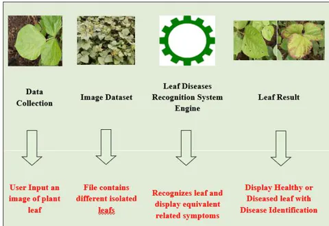
Plant Leaf Diseases Identification Model (PFDIM)
The dataset includes images of both healthy and diseased mungbean leaves belonging to three major diseases—Cercospora Leaf Spot, Powdery Mildew, and Yellow Mosaic Virus. The images were captured from experimental farms at Navsari Agricultural University, Gujarat, and nearby village fields. The process of dataset creation involved selecting healthy and infected plants, capturing high-quality images, and organizing them systematically for analysis. The image dataset serves as the foundation for model development and contains samples captured under natural field environments with variations in lighting, background, and leaf orientation. Each image represents real-world agricultural conditions, which enhances the robustness and practicality of the proposed system. All images were captured using Oppo A5 (13MP) and Mi Note 8 Pro (64MP) smartphone cameras. In total, the dataset comprises 1,307 images, including 367 healthy and 940 diseased samples. This diverse field-based dataset provides realistic input for the PFDIM model, ensuring reliable performance in uncontrolled agricultural environments. Table 1 represents the dataset information.
| Environment | Cercospora | Healthy | Powdery Mildew | Yellow Mosaic |
|---|---|---|---|---|
| Controlled | 224 | 211 | 225 | 223 |
| Uncontrolled | 102 | 156 | 41 | 125 |
Dataset
Figure 2 shows the leaf disease recognition system engine (LDRSE). The Leaf Disease Recognition System Engine (LDRSE) consists of three main stages. The first stage is preprocessing, where the images are enhanced, resized, and noise is removed to improve quality. The second stage is feature extraction, which identifies important visual characteristics of the leaf such as color, shape, and texture. The final stage is identification and classification where the extracted features are analyzed using the SVM classifier to determine the disease status and prepare it for further analysis. This stage involves several key operations such as RGB image acquisition and color transformation, normalization, resizing, masking of green pixels, segmentation, morphological adjustments, and extraction of the region of interest (ROI). Through RGB and color transformation, RGB images are converted into a standardized color space representation to simplify further processing. The images are then resized to maintain uniform dimensions across the dataset. To improve model robustness, data augmentation techniques such as rotation, flipping, scaling, and shifting are applied to artificially increase the dataset size and variability. The segmentation step divides each image into distinct regions to isolate the leaf area and separate it from the background, improving the precision of subsequent analysis. Finally, the ROI extraction isolates the diseased portion of the leaf, which becomes the focus for analysis. In the feature extraction phase, the inherent visual properties of the leaf are identified and measured. This step reduces data complexity while retaining key discriminative characteristics to differentiate between healthy and infected leaves. The classification phase involves training and testing the model using the extracted features. The Support Vector Machine (SVM) classifier is employed to assign specific categories based on identified characteristics. This step determines whether the plant is healthy or diseased, and the system output displays the analysis image along with the classification results. The system accuracy indicates whether the leaf is healthy or affected by a specific disease, completing the automated detection process. Preprocessing plays a crucial role in preparing images for analysis by improving their quality, consistency, and clarity. This stage involves key operations such as image resizing, augmentation, and edge detection. All images are standardized to pixels using the resize() function, ensuring uniform dimensions and eliminating redundant details. Image augmentation expands the dataset and increases its diversity through transformations like rotation, translation, scaling, shearing, and flipping, which enhances model generalization and minimizes overfitting. Edge detection, implemented using the Canny algorithm, identifies boundaries by detecting intensity patterns in the image. Segmentation and feature extraction are essential steps for isolating leaf regions and identifying significant visual features. In this study, HSV color mapping and the GrabCut algorithm were applied to segment leaf images captured under natural conditions. The HSV technique converts RGB images into the HSV colorspace to highlight dominant leaf colors such as green, yellow, and brown, improving region separation. The GrabCut algorithm further refines object boundaries by iteratively distinguishing the leaf from the background. After segmentation, the Histogram of Oriented Gradients (HOG) method is used to extract shape-based features by analyzing gradient directions. Images are then converted to grayscale and stored as NumPy arrays for efficient processing. Grayscale conversion reduces data complexity while preserving brightness information, resulting in faster and more reliable classification performance.

Leaf Diseases Recognition System Engine
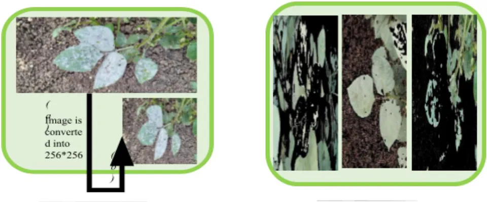
Resizing of Image (Left) and HSV Application (Right)
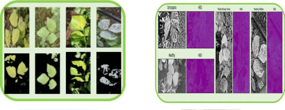
Performing GrabCut (Left) and Applying HOG (Right)
For training the SVM model under an uncontrolled environment, the preprocessing operations and parameter settings were kept consistent with those used in the controlled setup. The training procedure involved several steps, beginning with the import of essential libraries, followed by feature extraction using the Histogram of Oriented Gradients (HOG) method. The dataset was then loaded and divided into training and testing subsets. After this, the Support Vector Machine (SVM) classifier was initialized, trained on the dataset, and later evaluated for performance. The key hyperparameters considered for tuning included the kernel type, regularization factor (C), and gamma value.
Result and discussion
Initially, the model was trained using a polynomial kernel with a regularization parameter (C) of 0.02 and implemented using a one-vs-one classification approach. This configuration achieved training accuracy and testing accuracy, suggesting that the model suffered from overfitting. To address this issue, Grid Search with 5-fold cross-validation was utilized to identify the optimal combination of hyperparameters. The search was conducted over a range of values for C, gamma, kernel type, polynomial degree, and classification strategy. The best-performing setup was found to be , gamma , kernel polynomial, degree , and strategy , which resulted in training accuracy and testing accuracy. This fine-tuned configuration improved the model’s generalization capability while minimizing overfitting. Figure 7 represents the Confusion Matrix for the uncontrolled environment, summarizing the performance metrics such as precision, recall, F1-score, and overall accuracy achieved during evaluation.
In an uncontrolled environment, each image contains the mungbean leaf along with additional background elements such as soil, stems, and surrounding leaves. Table 2 presents the accuracies obtained by different classifiers when evaluated under these natural field conditions. The Support Vector Machine (SVM) classifier achieved a training accuracy of and a testing accuracy of in the uncontrolled environment. This indicates that while the model performed exceptionally well during training, it also maintained strong generalization capability when tested on unseen field data, demonstrating its effectiveness in real-world conditions. A user-friendly application named "Leaf Disease Classification" was developed for the quick and accurate identification of mungbean leaf status. The interface allows users to upload a leaf image, which the system automatically analyzes to determine whether the leaf is healthy or affected by a specific disease. Designed for simplicity, the tool can process images through a quick upload. Users can select an image from their device, view it on the interface, and then click "DETECT" to obtain instant results showing the health status and identifying the disease if present. This application provides farmers and researchers with a tool to determine mungbean disease status quickly and accurately.
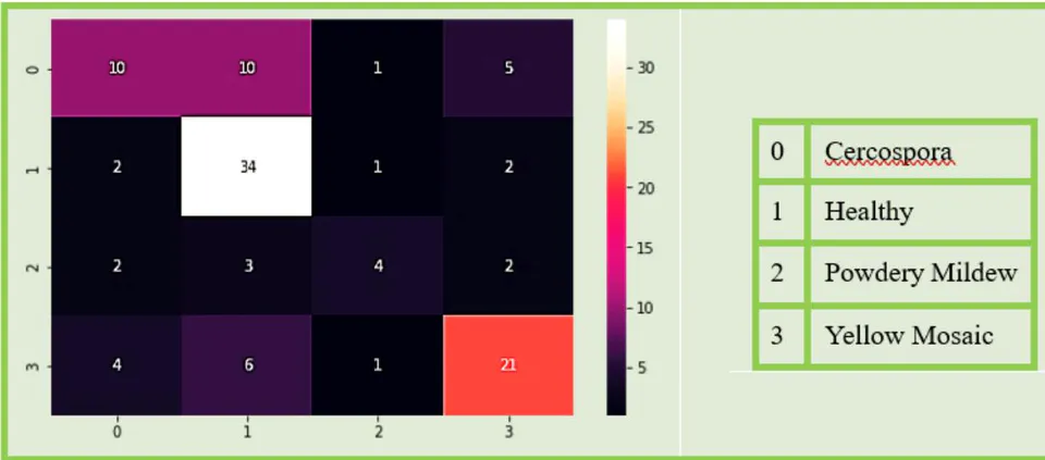
Confusion Matrix for Uncontrolled Environment
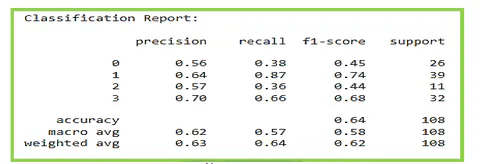
Confusion Matrix for Uncontrolled Environment
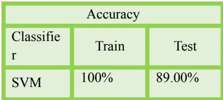
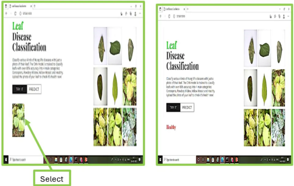
Confusion Matrix for Uncontrolled Environment
Conclusion
In this study, a Plant Foliar Disease Identification Model (PFDIM) was developed to detect mungbean leaf diseases under uncontrolled field conditions. A custom dataset of 1,307 images—including healthy and diseased samples of Cercospora Leaf Spot, Powdery Mildew, and Yellow Mosaic Virus—was created using field-captured images with natural background variations. The model utilized a Support Vector Machine (SVM) classifier with preprocessing techniques such as resizing, augmentation, HSV-based segmentation, and HOG feature extraction to enhance accuracy. After hyperparameter tuning through Grid Search with 5-fold cross-validation, the model achieved training accuracy and testing accuracy, demonstrating strong performance and reliability. The results indicate that SVM effectively identifies leaf diseases in real-world agricultural environments, providing a practical and efficient solution for early disease detection and crop management.