Loading menu…

Anterior Bowing of Tibia in an Adult: A Case Report

Β§ University College of Medical Sciences

Send Message

To: Author

Anterior Bowing of Tibia in an Adult: A Case Report

Article Fingerprint

ReserarchID

X2633

Anterior Bowing of Tibia in an Adult: A Case Report Banner

Key Research Insights

Synthesized scholarly intelligence & interactive research assistant
  • English
  • Afrikaans
  • Albanian
  • Amharic
  • Arabic
  • Armenian
  • Azerbaijani
  • Basque
  • Belarusian
  • Bengali
  • Bosnian
  • Bulgarian
  • Catalan
  • Cebuano
  • Chichewa
  • Chinese (Simplified)
  • Chinese (Traditional)
  • Corsican
  • Croatian
  • Czech
  • Danish
  • Dutch
  • Esperanto
  • Estonian
  • Filipino
  • Finnish
  • French
  • Frisian
  • Galician
  • Georgian
  • German
  • Greek
  • Gujarati
  • Haitian Creole
  • Hausa
  • Hawaiian
  • Hebrew
  • Hindi
  • Hmong
  • Hungarian
  • Icelandic
  • Igbo
  • Indonesian
  • Irish
  • Italian
  • Japanese
  • Javanese
  • Kannada
  • Kazakh
  • Khmer
  • Korean
  • Kurdish (Kurmanji)
  • Kyrgyz
  • Lao
  • Latin
  • Latvian
  • Lithuanian
  • Luxembourgish
  • Macedonian
  • Malagasy
  • Malay
  • Malayalam
  • Maltese
  • Maori
  • Marathi
  • Mongolian
  • Myanmar (Burmese)
  • Nepali
  • Norwegian
  • Pashto
  • Persian
  • Polish
  • Portuguese
  • Punjabi
  • Romanian
  • Russian
  • Samoan
  • Scots Gaelic
  • Serbian
  • Sesotho
  • Shona
  • Sindhi
  • Sinhala
  • Slovak
  • Slovenian
  • Somali
  • Spanish
  • Sundanese
  • Swahili
  • Swedish
  • Tajik
  • Tamil
  • Telugu
  • Thai
  • Turkish
  • Ukrainian
  • Urdu
  • Uzbek
  • Vietnamese
  • Welsh
  • Xhosa
  • Yiddish
  • Yoruba
  • Zulu
Reading Preferences
Font Size
Line Spacing
Background
This converted HTML version may contain rendering inconsistencies. Please refer to the PDF for the authoritative version, or click here to provide feedback.

Abstract

A 35-year-old female presented to us with anterior bowing of her right tibia. The deformity developed in her adolescence and subsequently had not progressed for nearly two decades. The patient had no functional limitation, her only concern being cosmesis. Radiological investigations suggested either fibrous dysplasia or adamantinoma. Biopsy showed fibrous stroma consisting of myxofibrous tissue and woven bone which was confirmatory of fibrous dysplasia. Keeping in mind that it was a dormant benign lesion not hindering with functionality of the limb, it was decided to keep the patient under observation with regular follow-up.

I. INTRODUCTION

Bowing of tibia can be anterior, anterolateral, anteromedial and posteromedial. Anterior tibial bowing, although rare, can be seen in fibrous dysplasia, osteofibrous dysplasia, adamantinoma, congenital pseudoarthrosis, vitamin D deficiency, syphilis, yaws, Paget's disease of the bone, fluorosis and Weismann-Netter-Stuhl syndrome.

We report a case of anterior tibial bowing in a middle-aged female and aim to highlight the importance of differentiation between benign fibrous dysplasia, potentially pre-malignant osteofibrous dysplasia and malignant adamantinoma in such a case.

II. CASE PRESENTATION

A 35-year-old female presented to us with an anteriorly bowed tibia of the right side. She had started noticing it at the age of 8 years, after which the deformity had progressed for a period of 10 years. It has now remained quiescent over the last 17 years. She complains of occasional pain in the right leg, but has no functional limitation. The overlying skin was normal with no dilated veins. She has normal local temperature, mild tenderness over the apex of the deformity, a shortening of 1.5cm on the affected side and the range of motion of the knee and ankle joints on the affected side are comparable to the opposite side. Both feet are comparable in size. There is no distal neurovascular deficit. She doesn't have any other deformity or any hyperpigmentation.
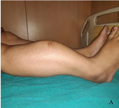
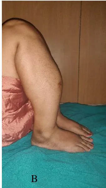
Figure 1: A and B showing anteriorly bowed right leg, fairly good motion at knee and ankle joints with minimal shortening

III. INVESTIGATIONS

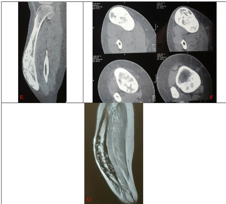
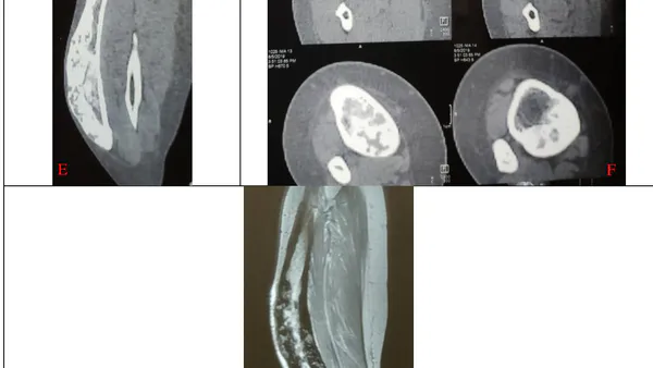
X-ray, CT scan and MRI are shown in the figures. Blood investigations were found to be normal.
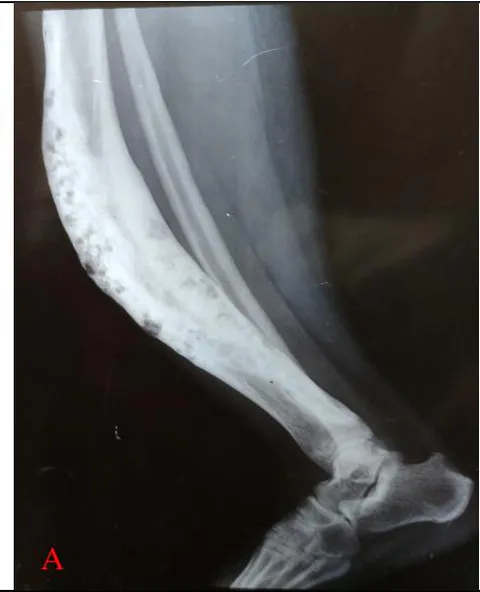
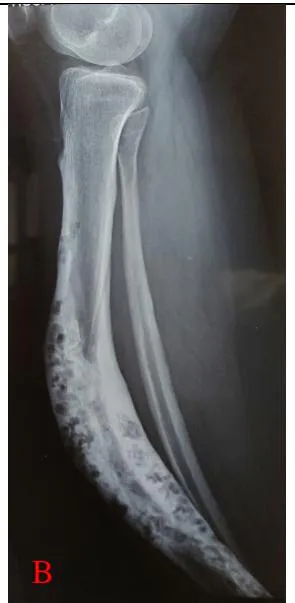
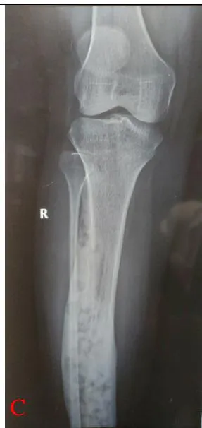
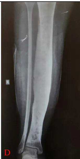
Figure 2: A-E showing anterior bowing of tibia, diffuse expansion of tibial diaphysis, loss of cortico-medullary junction, homogenous ground glass opacities and areas of cystic licences, narrow zone of transition with no cortical breach or soft tissue component

IV. DIFFERENTIAL DIAGNOSIS

Based on the radiological investigations, fibrous dysplasia and adamantinoma were kept as the two possibilities. Moth-eaten type of bone destruction and cortical thickening favour adamantinoma. Non-involvement of fibula and no osseous breach favour fibrous dysplasia.

V. HISTOPATHOLOGY

Characteristic findings indicative of fibrous dysplasia include fibrous stroma consisting of myxofibrous tissue and woven bone. 1 The histologic diagnosis of adamantinoma is made when epithelial-like cells arranged in pallisading nests and strands of cells are identified. Fibrous tissue is abundant in both, so anadamantinoma can remain masked if biopsy is taken from a single lesion site. 2 Precaution must be taken to take samples from lytic as well as dense regions.

Following these principles, we took biopsy with a core biopsy needle. Histopathology was suggestive of fibrous dysplasia.
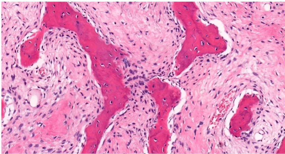
Figure 3: Showing trabeculae of woven bone with fibroblastic stroma and no osteoblastic rimming

VI. MANAGEMENT

Considering the facts that it was a benign lesion, the deformity had not progressed over the last 20 years, patient had no functional limitations or any significant limb length discrepancy, has had no pathological fracture and the lesion was involving almost the whole of tibia making a reconstruction difficult, we decided to keep the patient under observation. We would look for any change in her symptoms or an increase in the deformity and also get yearly X-rays and MRI to look for any enhancement of the lesion. Meanwhile, we started the patient on bisphosphonates and analgesics.

VII. Discussion

Differentiation between fibrous dysplasia, osteofibrous dysplasia and adamantinoma is vital in such a case.

Fibrous dysplasia (FD) is a benign intramedullary fibro-osseous lesion where normal bone is replaced by fibrous tissue. It can involve a single bone (monostotic), a single limb (monomelic) or multiple bones (polyostotic). It is generally an incidental finding. 3 Radiologically, the lesion is intramedullary, expansile and well-defined with an intact cortex. Although typically having a ground-glass appearance, it can also be completely lytic or sclerotic. 4 Characteristic histologic findings indicative of fibrous dysplasia include fibrous stroma consisting of myxofibrous tissue and woven bone.

Osteofibrous dysplasia (OFD) is a bone-forming lesion in the ventral, intracortical area of the tibial shaft with histology different from fibrous dysplasia. Contrary to fibrous dysplasia, the formed, woven trabeculae in osteofibrous dysplasia are rimmed by cuboidal osteoblasts. 5 There is a separate entity called OFD-like adamantinoma, which some believe to be a regressive form of adamantinoma and others believe to be a precursor of adamantinoma. 6

Adamantinoma is a primary low-grade malignant bone tumor most commonly seen involving the tibia. Histologically, adamantinoma shows a biphasic pattern of intermingled epithelial and osteofibrous components. Immunohistochemistry should be done to confirm the diagnosis. 7

It is important to differentiate between the three entities to decide the further line of management. Fibrous dysplasia is a benign lesion, with rare incidences of malignant transformation, and the patient can be kept under observation. 8 Osteofibrous dysplasia has been shown to carry a small but significant risk of containing co-existing adamantinoma or developing into adamantinoma, hence a wide resection of the lesion has been advocated by some. 6 Adamantinoma is a slow-growing, malignant bone tumour and necessitates a wide extraperiosteal resection to prevent recurrence. 9

References

9 Cites in Article
  1. 1. Harsh Mohan,Mittal,&. Preeti,Irneet Mundi,Kumar,&. Sudhir,Harsh Mohan (2011). Fibrous dysplasia of bone: a clinicopathologic review. Pathology and Laboratory Medicine International, 3, 31.
  2. 2. Ramanoudjame M,Guinebretière JM,Mascard E,Seringe R,Dimeglio A,Wicart P (2011). Is there a link between osteofibrous dysplasia and adamantinoma? Orthop Traumatol Surg Res. 2011 Dec, 97(8), 877-880.
  3. 3. Anitha N,Sankari SL,Malathi L,Karthick R (2015). Fibrous dysplasia-recent concepts. J Pharm Bioallied Sci, 7(Suppl 1), S171-S172.
  4. 4. Kushchayeva YS,Kushchayev SV,Glushko TY (2018). Fibrous dysplasia for radiologists: beyond ground glass bone matrix Insights Imaging. Insights into Imaging, 9(6), 1035-1056.
  5. 5. Sakamoto A,Oda Y,Iwamoto Y,Tsuneyoshi M (2000). A comparative study of fibrous dysplasia and osteofibrous dysplasia with regard to Gsalpha mutation at the Arg201 codon: polymerase chain reaction-restriction fragment length polymorphism analysis of paraffin-embedded tissues. J Mol Diagn, 2(2), 67-72.
  6. 6. R. Lee,S. Weitzel,D. Eastwood,F. Monsell,J. Pringle,S. Cannon,T. Briggs (2006). S., Weitzel S., Eastwood D M., Monsell F., Pringle J., Cannon S R., Briggs T W R.Osteofibrous dysplasia of the tibia The Journal of Bone and Joint Surgery. The Journal of Bone and Joint Surgery. British volume, 88-B, 658-664.
  7. 7. Jain D,Jain VK,Vasishta RK,Ranjan P,Kumar Y (2008). Adamantinoma: a clinicopathological review and update. Diagn Pathol, 3, 8.
  8. 8. Qu N,Yao W,Cui X,Zhang H (2015). Malignant transformation in monostoticfibrous dysplasia: clinical features, imaging features, outcomes in 10 patients, and review. Medicine (Baltimore, 94(3), e369.
  9. 9. S. Puchner,R. Varga,G. Hobusch,M. Kasparek,J. Panotopoulos,S. Lang,R. Windhager,P. Funovics (2016). Long-term outcome following treatment of Adamantinoma and Osteofibrous dysplasia of long bones Orthop Traumatol Surg Res. 2016 Nov, 102(7), 925-932.

Funding

No external funding was declared for this work.

Conflict of Interest

The authors declare no conflict of interest.

Ethical Approval

No ethics committee approval was required for this article type.

Data Availability

Not applicable for this article.

How to Cite This Article

Pratyush Shahi, Apoorv Sehgal, aarushi.sudan. 2020. "Anterior Bowing of Tibia in an Adult: A Case Report". Global Journal of Medical Research - H: Orthopedic & Musculoskeletal System GJMR-H Volume 20 (GJMR Volume 20 Issue 3 (H1)).

More Citation Formats

Download Citation

Issue Cover
GJMR Volume 20 Issue 3 (H1)
Pg. 5-9
Journal Specifications

Crossref Journal DOI 10.17406/gjmra

Print ISSN 0975-5888

e-ISSN 2249-4618

Keywords
Classification
GJMR-H Classification NLMC Code: WE 168
Version of record
v1.2

Issue date
January 23, 2020

Language
English
Order Article Reprint
Experience in AR

Explore published articles in an immersive Augmented Reality environment. Our platform converts research papers into interactive 3D books, allowing readers to view and interact with content using AR and VR compatible devices.

Read in 3D

Your published article is automatically converted into a realistic 3D book. Flip through pages and read research papers in a more engaging and interactive format.

Article Matrices
Total Views: 797
Total Downloads: 54
All Trends

This is the heading

Lorem ipsum dolor sit amet, consectetur adipiscing elit. Ut elit tellus, luctus nec ullamcorper mattis, pulvinar dapibus leo.

High-quality academic research articles on global topics and journals.

Anterior Bowing of Tibia in an Adult: A Case Report

Pratyush Shahi
Pratyush Shahi University College of Medical Sciences
<p>University College of Medical Sciences</p>

Request Access

Please fill out the form below to request access to this research paper. Your request will be reviewed by the editorial or author team.
X