

I. INTRODUCTION
Recently published studies have demonstrated the antibacterial properties of monovalent copper ions, suggesting their robust activity in orders of magnitudes compared to silver ion (Ag ) [1]. Divalent copper (Cu ) and metallic copper show no activity in controlled conditions [2]. The Cu ion antibacterial activity is intensified by high temperature, low molecular oxygen concentration, low pH, and poor carbon source [2]. On a minute time scale, ion disinfected bacterial contamination [2]. Recently, patent applications demonstrate semi-hydrophobic ointments generating monovalent copper ions in an aerobic atmosphere in sufficient concentration to disinfect contaminated surfaces [3]. According to a recent study, ions inhibit essential enzymes like DNA/RNA polymerase; it seems that the antibacterial mechanism is via enzymatic inhibition [4]. Copper's antimicrobial activities have been well recognized and exploited since ancient times for medicinal purposes [5]. The interest in antimicrobial applications of copper only increases with time. Currently, copper is widely used as a water purifier, fungicide, and bactericide. Ideas of introducing copper into cotton fibers, polymeric materials, and clothing to provide them biocidal properties were suggested more than a decade ago [6,7,8], and miscellaneous products are on the market already. Copper applications in healthcare might aid in successfully fighting bacterial contamination on solid surfaces and avoiding the spread of multidrug-resistant bacteria in hospitals [9]. All this makes understanding the processes resulting in the potent antibacterial effect of copper highly relevant.
Copper is an essential intracellular element in trace concentrations, while its excess causes toxicity . Due to the ability of copper to exist in metallic and ionic forms, alternating between cuprous and cupric oxidation states, its action on a variety of microorganisms, from fungi to bacteria, is a subject of ongoing research; most of the mechanisms and intracellular targets of this action are not yet elucidated . For instance, in the case of yeasts, metallic copper surfaces mediated toxicity targets membranes, causing extensive membrane and envelope damage while not affecting DNA; this mechanism is known as a contact-mediated killing . In the case of gram-positive bacteria, understanding molecular mechanisms leading to cell death caused by contact with both moist and dry copper surfaces is a controversial topic. It is reported that exposure of Staphylococcus Aureus to copper causes cell death through DNA damage . In addition, cellular respiration is compromised, with little effect on cell membrane integrity .
In contrast, other studies exploring the toxic effect of copper surface contact with Staphylococcus haemolyticus cells suspension point at depolarization of the cytoplasmic membrane as the primary target and suggest that DNA degradation occurs only after cell death . Regarding gram-negative bacteria, it is also shown that E.coli is rapidly killed on copper alloys surfaces . The current model of a contact killing on dry surfaces characterizes this process as a cascade of events, such as successive cell membrane rupture and loss of cell content, copper ions influx into the cells leading to oxidative damage and DNA degradation, while the sequence of these events may differ . In vivo, however, according to the literature, copper ions do not catalyze the formation of oxidative DNA damage . Copper ions use as a weapon in the antimicrobial arsenal of grazing protozoa and phagocytic cells of the immune and affect central carbon metabolism in Staphylococcus aureus , which implies intracellular activity. At the same time, in our view, the role of dissolved mono copper ions that penetrate the cell through cation channels and paralyzes essential enzymes in the killing process should not be underestimated .
In aqueous solution, the common oxidation state of copper ions is bivalent ( , cupric). Copper in the monovalent state ( , cuprous) remains in a low concentration due to a disproportion reaction (self-oxidation of monovalent copper to divalent copper and metallic copper), and due torapidlyoxidized by molecular oxygento divalent copper.
Nevertheless, it is possible to elevate ions concentration; adding reagents that form a more stable complex with ions than with ions may achieve a high concentration of ions in a deaerated aqueous environment. Acetonitrile [23, 24], benzoic acid [25] and ATP [26, 27] are good examples for stabilizing reagents that shift the existing equilibrium between oxidation states to the formation of two ions from one ion and metallic copper.
Our previous research succeeded in exploiting this technique of ions production, thus opening a series of studies devoted to an investigation of the antimicrobial effect of monovalent copper. We have clearly shown the superior efficacy of ions over ions in killing E. coli and Staphylococcus aureus bacteria. Moreover, our studies have revealed that ions had substantially higher efficacy than ions, which are currently widely used as an antibacterial agent . On the whole, our findings suggest that should be considered as a potent antimicrobial agent.
II. MATERIALS AND METHODS
a) Culture Media [28]
E. coli (NCIMB, str. K-12 substrate. MG1655) was stored in vials with 50% glycerol at -80oC until use. The strain was grown either in Luria broth medium (LB broth and agar, Difco). E.coli was grown in LB broth, typically containing 0.5% yeast extract, 1% bactotryptone and 1% NaCl. S.A. Staphylococcus aureus, B.T. Bacillus thuringiensis, E.A. Enterobacter aerogenes, M.L. Micrococcus luteus, S.E. Staphylococcus epidermidis, S.F. Streptococcus faecalis, P.A. Pseudomonas aeruginosa, Delf Delftia tsuruhatensis, S.C. Staphylococcus cohnii, B.B. Brevibacillus brevis, and beer yeast was grown and treated according to the appropriate procedure that appears in the literature.
b) Preparation of Starter and Growth Methods [28]
i. Preparation of starter and bacterial growth in LB broth medium
LB broth was inoculated by an bacteria colony grown on LB agar. The starter was grown overnight in a rotary shaking incubator (37 °C, 170 rpm). The next day, the starter was seeded into fresh LB media at 1:100 dilution and grown to OD600 0.3-0.4 for 2-3 hours to bring the bacteria to the exponential growth phase. The resulting bacterial suspension was finally inoculated into fresh LB at 1:100 dilution.
ii. Preparation of ointments
To make the ointment, heat Vaseline on a water bath until melting, to which add while stirring all the ingredients except ascorbic acid or its Palmitate derivative. The mixture cooled while stirring, and only then, the ascorbic acid was added while stirring, obtaining a homogeneous ointment.
c) Antimicrobial disk-diffusion susceptibility test [29]
With a piece of a wadded disk having 3mm diameter, an amount of 0.1 gr ointment was taken, ensuring ~1 mm thickness layer, and was put in the center of inoculated LB agar in a Petri dish. The test ointment samples and control (inoculated the same way but having no ointment) in Petri dishes were put into the incubator for 18 hours at 37 °C. Each ointment composition was done in a triplicate for standard deviation calculation. Measurements of a zone without bacterial growth, e.g., distance between the edges of ointment and bacterial growth areas, were performed using a ruler.
i. Counting with colony-forming units
For estimates, the number of bacteria or fungal cells in a sample, we used the colony-forming units (CFU) counts. Bacteria were counted using a routine CFU technique, i.e., plating bacteria from serial dilutions onto LB agar and incubating overnight at .
d) Stability test of formulations [30]
To check the stability of the formulations, for hot storage conditions, was incubator was used, and for room storage conditions, samples were stored in the lab. The storage conditions in a closed container were tested and exposed to air conditioning Petri dishes. Before and after storage, each ointment sample was tested for antimicrobial activity by measuring bacterial growth inhibition on triplicated Petri dishes on LB agar.
i. Cooper ions Diffusion test
Eight Petri dishes were prepared for the experiment: four were sterile, and the others were seeded with bacterial inoculation. The bacteria-containing plates were used to determine the bacterial influence on copper diffusion. The ointment was prepared and placed on the center of each dish, and the Petri dishes were placed into the incubator at for the defined time intervals: 1, 2, 3, or 4 hours. After that, each set, consisting of bacteria containing and sterile dish withdrawn from the incubator and four small rings of agar cut off. Each agar sample dissolved with hot nitric acid, and the ICP-OES technique used to measure the copper concentration. The map of a Petri dish used for cutting off agar samples is displayed in Fig. 10, with inner-outer radiuses measured starting from the edge of the ointment sample.
III. RESULTS AND DISCUSSION
a) Antibacterial activity of ointments
Several antibacterial compositions developed, creating a durable and effective concentration of monovalent copper ions.
| Amount (%w/w) | ||||
| Material\ Composition name | A Emulsifying base | B Absorption base | C Absorption base+ S.A | D Encourages diffusion |
| Copper(II) gluconate | 10.0 | 10.0 | 10.0 | 0 |
| Copper sulfate | 0 | 0 | 0 | 3.2 |
| Ascorbyl Palmitate | 6.5 | 6.5 | 7.0 | 0 |
| Ascorbic Acid | 0 | 0 | 0 | 4.0 |
| Vaseline | 20.0 | 27.0 | 42.0 | 27.0 |
| Glycerol | 10.7 | 22.5 | 14.0 | 27.0 |
| Water | 30.0 | 5.0 | 2.0 | 1.8 |
| Stearyl alcohol | 20.0 | 0 | 0 | 0 |
| Lanoline | 0 | 27.0 | 0 | 0 |
| Isopropyl Myristate | 0 | 0 | 8.0 | 27.0 |
| Salicylic acid | 0 | 0 | 17.0 | 0 |
| Sodium lauryl sulfate | 0.8 | 0 | 0 | 11.0 |
| Copper dust | 2.00 | 2.00 | 0 | 0 |
| Acetonitrile | 0 | 0 | 0 | 0 |
Figure 6 displays the inhibition radius of bacterial growth caused by different copper (II) gluconate concentrations for emulsion (A) and absorption (B) bases ointments, respectively.
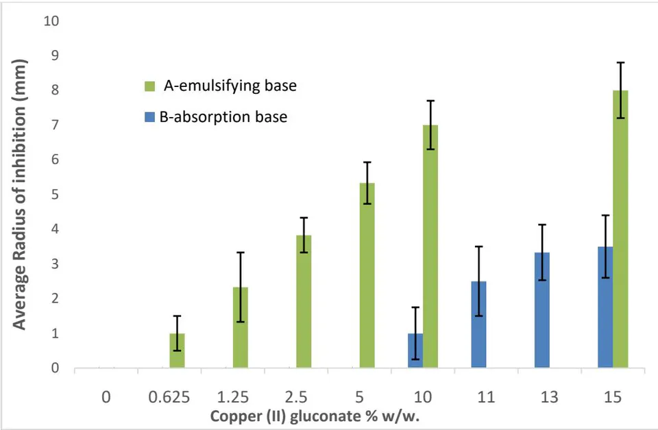
Figure 6 points to the solid correlation of copper (II) gluconate concentration in the ointments (A and B) and its antibacterial effect. That meets our expectations since the ions generation mechanism strongly depends on reactants' concentrations: the more ions are involved in reactions, the more ions are generated and could be diffused throughout agar and create the larger bacterial-free region.
Figure 7 presents the results of E. Coli bacterial growth inhibition radius for ointments A ( ), ointments A with copper (II) gluconate but without the reduction elements (Ascorbyl Palmitate and Copper dust) ( ) that show limited activity, and ( ) resulting from replacing the copper ions in the same molar concentration with silver ions ( ). The last show limited activity as well.
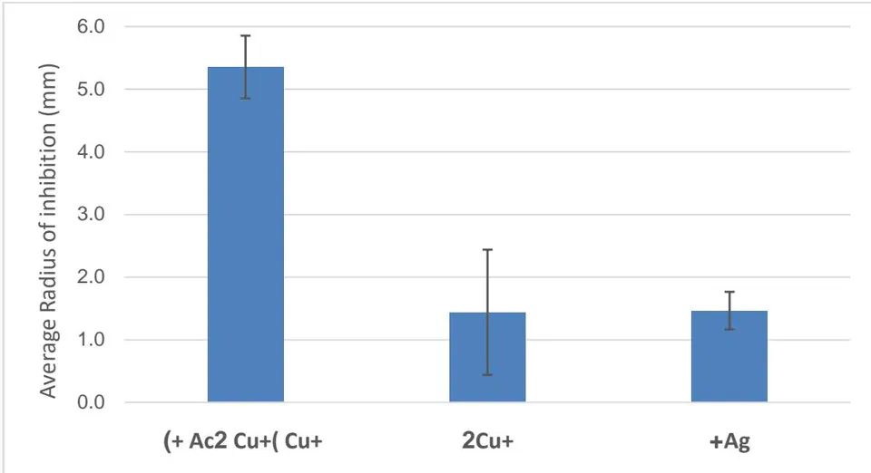
Figure 7 shows that the effect of monovalent copper ions is much more significant than monovalent silver ions used commercially to control bacteria growth. Divalent copper ions have a particular effect attributing to a small concentration of monovalent copper ions obtained from divalent copper ions depending on the nature of the redox potential of the environment.
Comparison of the effect of type A ointment on E. Coli, Bacillus thuringiensis (B.T) bacteria, and beer
Yeast on the growth inhibition radius presented in figure 8.
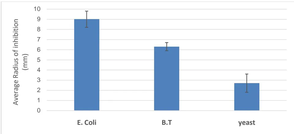
The results show that gram-negative bacteria (represented by Figure 8 by E. Coli) are more sensitive than gram-positive bacteria (represented by Figure 8 by Bacillus thuringiensis (B.T)) and fungi (represented by Figure 8 by beer Yeast) to monovalent copper ions. The ointments (A, B, C) were successfully tested on about 12 types of bacteria: E.C. Escherichia coli, S.A. Staphylococcus aureus, B.T. Bacillus thuringiensis, E.A. Enterobacter aerogenes, M.L. Micrococcus luteus, S.E. Staphylococcus epidermidis, S.F. Streptococcus faecalis, P.A. Pseudomonas aeruginosa, Delf Delftia tsuruhatensis. S.C. Staphylococcus cohnii, B.B. Brevibacillus brevis.
In an attempt to increase the inhibitory capacity of the formula without metallic copper, salicylic acid is added to the formula (ointments C). The choice in Salicylic acid is due to studies indicating that aromatic compounds stabilize monovalent copper and because salicylic acid is FDA approved and is widely used in the cosmetics industry. Figure 9 displays the inhibition radius of bacterial growth caused by different concentrations of salicylic acid in ointments C composition.
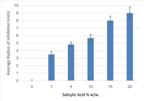
Salicylic acid forms a stabilizing complex with monovalent copper, the stability constant ( ) published in the literature [25]. The results shown in Figure 9 correspond to the above stabilization. Adding a stabilizing agent to monovalent copper ions compensates for the lack of metallic copper that serves as a reducing reservoir.
To test the stability of the ointments (A and B), they were kept for six months at room temperature, and C. Figure 10 presents the results.
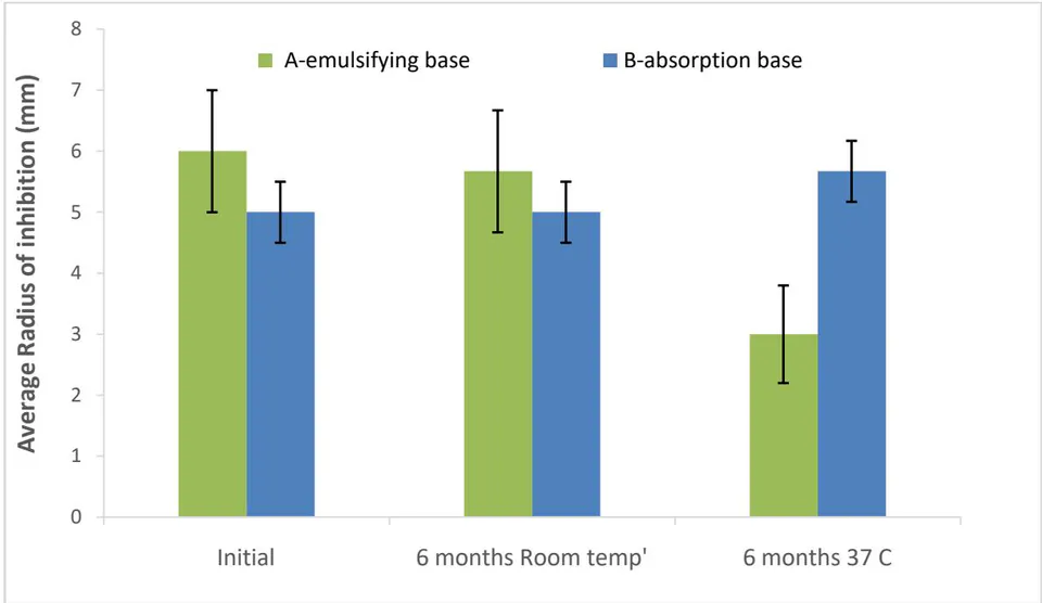
Figure 10 shows that while the effectiveness of type A ointment is deteriorating over time, the effectiveness of type B ointment is maintained and increases over time.
b) Diffusion of Cu(I) ions through agar
Figure 10 displays the concentration of Cu on the agar taken from the LB agar Petri dish. According to the diagram in Figure 10 left, the agar cut into rings, the ring dissolved in nitric acid, and the copper ions were determined using ICP technology. The experiment was performed parallel on a sterile agar and a bacterium-seeded agar.
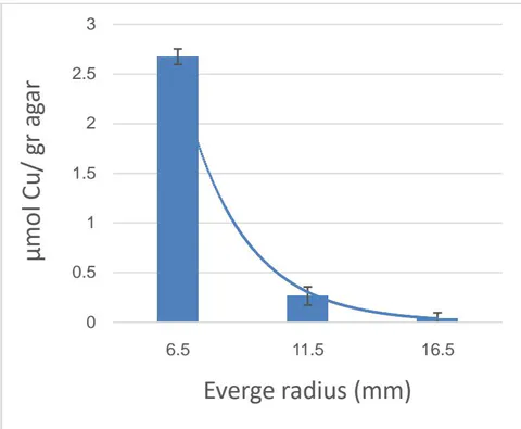

Figure 10 (left) illustrates the diffusion of copper ions into the agar. As expected, there is an exponential dependence on the radius. A good match was obtained between the diffusion of the copper ions, and the radius of bacterial inhibition, the bacterial inhibition radius in the same condition was 9mm. It is possible to conclude that the agar concentration is lethal to E. coli bacteria.
c) Water disinfection based on formula D absorbed on sponge
Figure 11 shows the ability to disinfect water from E.Coli bacteria with the help of a type D ointment. The ointment was absorbed into a medical sponge in which it was in contact with the contaminated water.
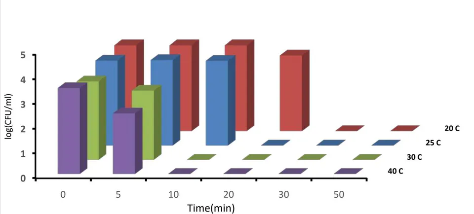
The composition of type D ointment allows the diffusion of monovalent copper ions into the water. The concentrations are sufficient for disinfection (0.2ppm) but below the permitted concentration of copper ions in drinking water (1.3ppm). The temperature has a dramatic effect. Below C, the efficiency drops, and it takes much longer for complete disinfection of the solution. These results are consistent with previous research showing a dramatic effect of temperature on the antibacterial activity of monovalent copper ions [2].
Table 2 indicates the accepted values for water testing and the values measured to the treated water display in fig 11.
| Temp (°C) | Cu ions (ppm) | TN (ppm) | TOC (ppm) | Conductivity (μs/cm) | pH |
| 20 | 0.20±0.05 | 0.54±0.01 | 6.30±0.19 | 283±3.54 | 7.62±0.01 |
| 25 | 0.43±0.02 | 6.10±0.01 | 290±7.78 | 8.04±0.08 | |
| 30 | 0.29±0.03 | 6.48±1.04 | 253±3.54 | 8.40±0.01 | |
| The standard for drinking water [31,32] | 1.3 (USA) 2 (Europe) | 10 | 25 | 500-1000 | 7.5-8.5 |
The water obtained by this method meets the standard; even the amount of organic carbon (TOC) is below the allowed value.
IV. CONCLUSIONS
This study demonstrates how to utilize the understanding that monovalent copper ions are the active form in the antibacterial capacity of copper and its practical developments in room conditions.