

Loading menu…

Appendiceal Abscess in a Young Adult: A Review of the Ultrasonographic Findings: A Case Report

Appendiceal Abscess in a Young Adult: A Review of the Ultrasonographic Findings: A Case Report

Article Fingerprint

ReserarchID

3A3IY

Appendiceal Abscess in a Young Adult: A Review of the Ultrasonographic Findings: A Case Report Banner

Key Research Insights

Synthesized scholarly intelligence & interactive research assistant
  • English
  • Afrikaans
  • Albanian
  • Amharic
  • Arabic
  • Armenian
  • Azerbaijani
  • Basque
  • Belarusian
  • Bengali
  • Bosnian
  • Bulgarian
  • Catalan
  • Cebuano
  • Chichewa
  • Chinese (Simplified)
  • Chinese (Traditional)
  • Corsican
  • Croatian
  • Czech
  • Danish
  • Dutch
  • Esperanto
  • Estonian
  • Filipino
  • Finnish
  • French
  • Frisian
  • Galician
  • Georgian
  • German
  • Greek
  • Gujarati
  • Haitian Creole
  • Hausa
  • Hawaiian
  • Hebrew
  • Hindi
  • Hmong
  • Hungarian
  • Icelandic
  • Igbo
  • Indonesian
  • Irish
  • Italian
  • Japanese
  • Javanese
  • Kannada
  • Kazakh
  • Khmer
  • Korean
  • Kurdish (Kurmanji)
  • Kyrgyz
  • Lao
  • Latin
  • Latvian
  • Lithuanian
  • Luxembourgish
  • Macedonian
  • Malagasy
  • Malay
  • Malayalam
  • Maltese
  • Maori
  • Marathi
  • Mongolian
  • Myanmar (Burmese)
  • Nepali
  • Norwegian
  • Pashto
  • Persian
  • Polish
  • Portuguese
  • Punjabi
  • Romanian
  • Russian
  • Samoan
  • Scots Gaelic
  • Serbian
  • Sesotho
  • Shona
  • Sindhi
  • Sinhala
  • Slovak
  • Slovenian
  • Somali
  • Spanish
  • Sundanese
  • Swahili
  • Swedish
  • Tajik
  • Tamil
  • Telugu
  • Thai
  • Turkish
  • Ukrainian
  • Urdu
  • Uzbek
  • Vietnamese
  • Welsh
  • Xhosa
  • Yiddish
  • Yoruba
  • Zulu
Reading Preferences
Font Size
Line Spacing
Background

I. INTRODUCTION

Appendicitis is the most common cause of acute abdomen, presenting as a triad of pain in the right iliac fossa, fever, and vomiting that often requires surgical intervention.

An appendiceal abscess is a condition in which an abscess is formed around the appendix as a result of appendiceal perforation or extension of inflammation to the adjacent tissues due to aggravation of appendicitis 2. Inflammation of the vermiform appendix is regarded as the most common surgical emergency worldwide with an estimated lifetime risk of about 6 and 8%.

Appendicitis is often a common differential diagnosis among the large number of cases presenting in the emergency unit on account of acute abdominal pain, and more common in the male gender with more than 70% of cases presenting in individuals less than 30 years of age.

The vermiform appendix anatomically has varying location with respect to the caecum and can originate within a 360∘ circumference around the caecum, this is most likely responsible for the varying form of presentation during appendicitis.

The inflammation in acute appendicitis is often fixed by the patient's self-defense mechanism by either formation of an inflammatory mass (an appendiceal phlegmon) or a circumscribed abscess (an appendiceal abscess). These (appendiceal mass and abscess) present as palpable mass days after onset of symptoms and constitute about 2−7% of all cases of appendicitis.

Acute inflammation of the appendix may also be the initial presentation of primary tumors of the appendix in more than 50% of cases. The patients with acute appendicitis typically present with acute central abdominal pain radiating to the right iliac fossa in vast majority of cases.

Appendiceal abscess are managed traditionally by nonsurgical treatment and interval appendectomy, the nonsurgical treatment comprises of the usage of effective antibiotics with subsequent drainage of the abscess under ultrasound or computed tomographic imaging, the surgical treatment is mainly by interval appendectomy.

II. CASE REPORT

A 24-year-old male patient referred from a peripheral health care centre for an urgent abdominopelvic ultrasound on account of persistent colicky right iliac fossa pain, occasional vomiting, increased urinary frequency and general discomfort with fever.

He has had similar right iliac fossa pain in the past with similar symptoms and has had repeated episodes of intake of antibiotics, analgesics and antipyretics on those occasion.

On physical examination, he is febrile to touch, not pale, anicteric, not dehydrated and not in any form of respiratory distress or in any form of altered conscious state. He had some swelling with rebound tenderness over the right iliac fossa and also warm to touch more in the right iliac fossa.

The patient had result of his packed cell volume (37%), full blood count with differentials showing leukocytosis (13500 white blood cells per microliter) in favor of the lymphocytes (6000 lymphocytes in 1 microliter of blood). The erythrocyte sedimentation rate was also normal (12 millimeters per hour).

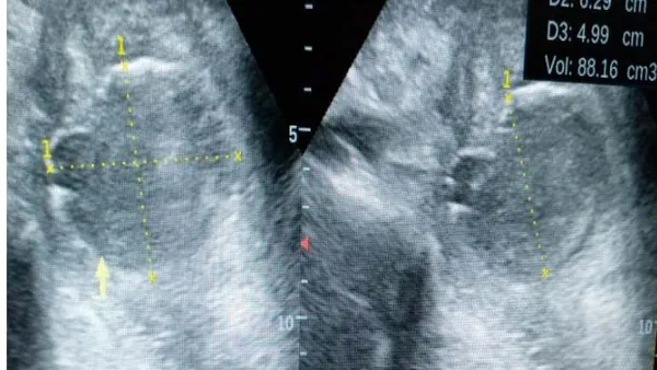
3.5 and 7MHz transducer probes showed a deep seated, oval area of heterogeneous echo-reflectivity with almost an intact wall and with a volume of about 88mls.; see figure 1. There is surrounding prominence of the bowel loops (local ileus), hypoechoic fluid most likely inflammatory in the region of the vermiform appendix. Rebound tenderness was also demonstrated repeatedly with probe pressure. Mobile echoes were also demonstrated within the contents of the urinary bladder.

A diagnosis of a deep seated appendiceal abscess with focal paralyticileus and cystitis was made,

An abdominopelvic ultrasound scan done in both longitudinal and transverse approach using both a the findings of the scan were collated and given to the patient for onward submission to the referring physician.

The patient in the peripheral health care centre had effective course of both intravenous and oral antibiotics with analgesia, intravenous rehydration, adequate monitoring of the vital signs and monitoring of the size of the appendiceal mass per abdominal assessment on hospital admission, and he is been planned for interval appendectomy later following a successful nonsurgical treatment.
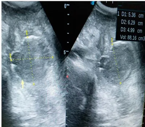
Figure 1: An ultrasonogram of the right iliac fossa region following the use of a 3.5MHz probe transducer showing a near oval area with slight irregularity of the superior walls having a heterogeneous collection with a volume of about 88mls and surrounding hypoechoic fluid; inflammatory and surrounding sentinel loops; focal paralytic ileus. This is the deep seated appendiceal abscess with surrounding focalileus.

III. Discussion

Appendicitis is the most common cause of acute abdomen, presenting as a triad of pain in the right iliac fossa, fever, and vomiting that often requires surgical intervention 1. The index case presented with recurrent right iliac fossa pain, fever, occasional vomiting and dysuria thereby conforming to this literature.

An appendiceal abscess is a condition in which an abscess is formed around the appendix as a result of appendiceal perforation or extension of inflammation to the adjacent tissues due to aggravation of appendicitis 2.

The index case had an abscess cavity in the right iliac fossa in the region of the vermiform appendix thereby conforming to this literature.

Appendicitis is more common in the male gender with more than 70% of cases presenting in individuals less than 30 years of age 3,6, the index case is a young male patient aged 24 years conforming to these literatures.

Imaging which primarily include ultrasonography and computed tomography play vital role in the diagnosis of appendiceal abscess 3, the index case had abdominopelvic ultrasonography following which the abscess was demonstrated, thereby conforming to this literature.

Appendiceal abscess are managed traditionally by nonsurgical treatment and interval appendectomy 3,14, the case under presentation was not an exception, he initially had a course of antibiotics and analgesia with adequate monitoring of the vital signs to ensure a successful nonsurgical treatment which will be followed by interval appendectomy to prevent recurrence.

IV. CONCLUSION

Appendiceal abscess is often as a result of acute appendicitis and common in individuals within the second decade of life, this condition when suspected can be diagnosed ultrasonography and management immediately instituted to prevent further complications (perforation) and save the lives of these individuals.


References

14 Cites in Article
  1. 1. S Daga,S Kachewar,D Lakhkar,K Jethlia,A Itai (2017). Sonographic evaluation of acute appendicitis and its complications. West Afr J Radiol, 24, 152-156.
  2. 2. Shinjiro Kobayashi,Ryoji Makizumi,Kazunari Nakahara,Satoshi Tsukikawa,Nobuyoshi Miyajima,Takehito Otsubo (2014). Appendiceal Abscesses Reduced in Size by Drainage of Pus from the Appendiceal Orifice during Colonoscopy: A Report of Three Cases. Case Reports in Gastroenterology, 8(3), 364-370.
  3. 3. G Sharp,N Railton,S Kadirkamanathan (2014). A mystifying mass. Journal of Surgical Case Reports, 2014(1), rjt123-rjt123.
  4. 4. Andy Petroianu (2012). Diagnosis of acute appendicitis. International Journal of Surgery, 10(3), 115-119.
  5. 5. C Wray,L Kao,S Millas,K Tao,T Ko (2013). Acute appendicitis: controversies in diagnosis and management. CurrProbl Surg, 50, 54-86.
  6. 6. Alexis Schizas,Andrew Williams (2010). Management of complex appendicitis. Surgery (Oxford), 28(11), 544-548.
  7. 7. Baris Yildiz,Osman Abbasoglu (2006). Two unusual presentations of appendiceal masses. International Journal of Surgery, 6(6), e9-e11.
  8. 8. Zaza Demetrashvili,Giorgi Kenchadze,Irakli Pipia,Eka Ekaladze,George Kamkamidze (2015). Management of Appendiceal Mass and Abscess. An 11-Year Experience. International Surgery, 100(6), 1021-1025.
  9. 9. J Tannoury,B Abboud (2013). treatment options of inflammatory appendiceal masses in adults. World J Gastroenterol, 19, 3942-3950.
  10. 10. Jeong-Ki Kim,Seungbum Ryoo,Heung-Kwon Oh,Ji Kim,Rumi Shin,Eun Choe,Seung-Yong Jeong,Kyu Park (2010). Management of Appendicitis Presenting with Abscess or Mass. Journal of the Korean Society of Coloproctology, 26(6), 413.
  11. 11. Frederico Teixeira,Sérgio Couto Netto,Eduardo Akaishi,Edivaldo Utiyama,Carlos Menegozzo,Marcelo Rocha (2017). Acute appendicitis, inflammatory appendiceal mass and the risk of a hidden malignant tumor: a systematic review of the literature. World Journal of Emergency Surgery, 12(1)
  12. 12. S Connor,G Hanna,F Frizelle (1998). Appendiceal tumors. Diseases of the Colon & Rectum, 41(1), 75-80.
  13. 13. Joseph Ting,Rebecca Farley (2008). Subhepatically located appendicitis due to adhesions: a case report. Journal of Medical Case Reports, 2(1), 339.
  14. 14. Roland Andersson,Max Petzold (2007). Nonsurgical Treatment of Appendiceal Abscess or Phlegmon. Annals of Surgery, 246(5), 741-748.

Funding

No external funding was declared for this work.

Conflict of Interest

The authors declare no conflict of interest.

Ethical Approval

No ethics committee approval was required for this article type.

Data Availability

Not applicable for this article.

How to Cite This Article

Sule Muhammad Baba, Sa’idu Sule Ahmed, Ma’aji Sadisu Mohammed, Danfulani Muhammad, Umar Usman Aminu. 2021. "Appendiceal Abscess in a Young Adult: A Review of the Ultrasonographic Findings: A Case Report". Global Journal of Medical Research - D: Radiology, Diagnostic GJMR-D Volume 20 (GJMR Volume 20 Issue D2).

More Citation Formats

Download Citation

Journal Specifications

Crossref Journal DOI 10.17406/gjmra

Print ISSN 0975-5888

e-ISSN 2249-4618

Keywords
Classification
GJMR-D Classification NLMC Code: WN 208
Version of record
v1.2

Issue date
January 16, 2021

Language
English
Order Article Reprint
Experience in AR

Explore published articles in an immersive Augmented Reality environment. Our platform converts research papers into interactive 3D books, allowing readers to view and interact with content using AR and VR compatible devices.

Read in 3D

Your published article is automatically converted into a realistic 3D book. Flip through pages and read research papers in a more engaging and interactive format.

Article Matrices
Total Views: 961
Total Downloads: 49
All Trends

Request Access

Please fill out the form below to request access to this research paper. Your request will be reviewed by the editorial or author team.
X

This is the heading

Lorem ipsum dolor sit amet, consectetur adipiscing elit. Ut elit tellus, luctus nec ullamcorper mattis, pulvinar dapibus leo.

High-quality academic research articles on global topics and journals.

Appendiceal Abscess in a Young Adult: A Review of the Ultrasonographic Findings: A Case Report

Sule Baba
Sule Baba USMANU DANFODIYO UNIVERSITY
Sa’idu Ahmed
Sa’idu Ahmed
Ma’aji Mohammed
Ma’aji Mohammed
Danfulani Muhammad
Danfulani Muhammad
Umar Aminu
Umar Aminu