## I. INTRODUCTION
Patient with systemic lupus erythematosus (SLE) may experience various intestinal disorders such as diarrhea, vomiting, rectal bleeding, tenesmus. However, even if it's not very common, it is important to think of inflammatory bowel diseases especially Crohn's disease as well as Ulcerative colitis. In fact, SLE and Ulcerative colitis (UC) rarely coexist. They may both have intestinal manifestations, laboratory results, and radiographic findings that appear similar, therefore differentiating between intestinal involvement in UC and in SLE may be difficult(1).
Moreover, there are issues establishing true links between UC and SLE because SLE may mimic UC, and some UC treatments may cause drug induced SLE(1).
This makes the diagnosis of both diseases in one patient a real challenge. There are, in fact, few reports suggesting an association between these diseases (1-6).
Current understanding of the pathogenesis of the UC and SLE suggests that immune mechanisms play a prominent role in both diseases. In both diseases, genetic seems also to play an important role. Genetic markers located in the short arm of chromosome 6 have been found associated with both SLE to a lesser degree with UC (7,8).
It is well known that UC is a chronic condition for which therapy is required to induce and maintain remission; therapeutic decisions should be categorized into those for induction and maintenance, with a goal of obtaining and maintaining a steroid-free remission (9).
Ulcerative colitis can be treated by 5-aminosalicylate (5-ASA), steroids or anti tumor necrosis factor alpha (anti-TNF alpha). SLE is known to interfere with some of these treatments, moreover, it can be a complication of these treatments. All this poses a real problematic while managing some UC cases, especially those with resistant to the majority of medical treatments (9).
The aim of our case report is to present one of these problematic cases, to compare it with different experiences in other centers, to review the literature in order to find out therapeutic options for these cases, and why not be a base for ulterior guidelines.
## II. OBSERVATION
At the age of 31, the patient was admitted with a seven month history official and finger erythema and arthralgia. Physical Examination revealed a typical malarrash (Figure 1) and discoid lesions on her fingers (Figure 2). The antinuclear, -antibody (ANA) were positives (1/256), the antibodies to double-stranded (DNA) were positives, the serum complement was low (17.6Vml:normal30-40). The patient was therefore diagnosed with SLE according to American Rheumatism Association criteria for SLE. The patient began treatment with 30 mg/day prednisolone (PSL) and chloroquine 400mg/day, PSL was subsequently reduced to 10 mg/day. The symptoms were controlled in the following 9 years under treatment.
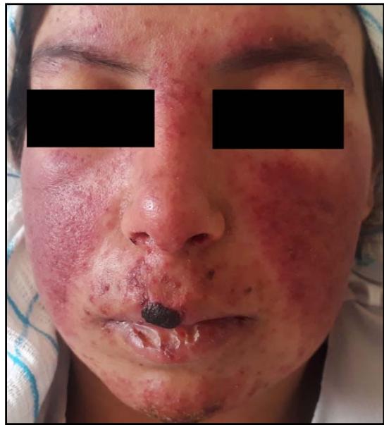 Figure 1: Facial malar rash
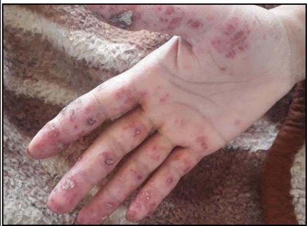 Figure 2: Finger discoid lesions
Bloody diarrhea accompanied with abdominal cramping and rectal bleeding with tenesmus began to occur intermittently when the patient was 40 year old. Intestinal involvement in lupus was first suspected, colonoscopy revealed continuous areas of inflammation and ulcers in the descending colon and in the transverse (figure 3,4,5,6). Stool studies were negative for infection, histological evaluation revealed acute inflammation with crypt architectural distortion diffusely consistent with inflammatory bowel disease, favoring UC.
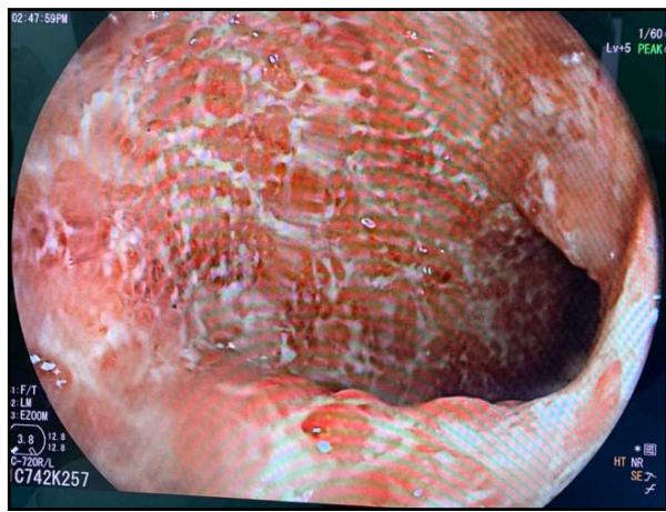 Figure 3
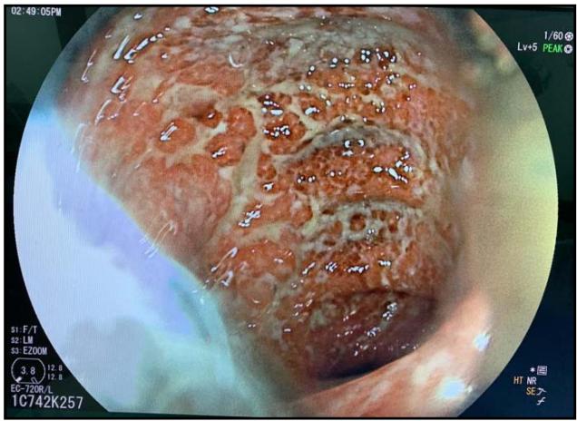 Figure 4
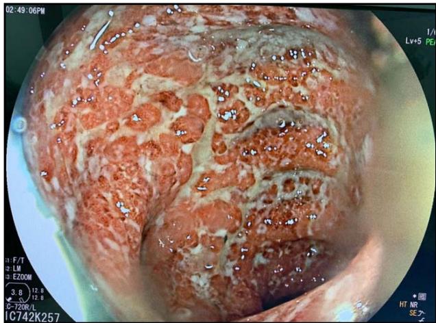 Figure 5
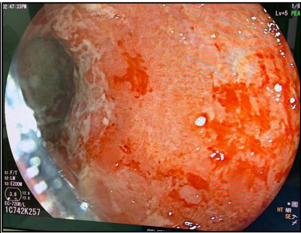 Figure 6
The diagnostic of UC was made and treatment with PSL $50\mathrm{mg}$ daily, azathioprine $125\mathrm{mg/day}$, chloroquine $200\mathrm{mg/day}$ was begun. This was followed by significant improvement. Two months later, remission was maintained with azathioprine, PSL $10\mathrm{mg/day}$ and chloroquine.
The patient was admitted again 6 months later, she developed bloody diarrhea with abdominal pain, and tenesumus, colonoscopy revealed inflamed hyperemic colonic mucosa with multiple active ulcers, Hemoglobin (Hb) was $9.1\mathrm{g / dl}$ (normocytic), white blood cell count was $12.44\times 103 / \mathrm{ml}$ (neutrophilic leukocytes: $90.8\%$, lymphocytes: $7.4\%$ ), and platelets were $3\times 105 / \mu l$ C-reactive protein(CRP) and erythrocyte sedimentation rate were $78~\mathrm{mg / dl}$ and 35 mm/h respectively. The patient was therefore treated with intravenous corticosteroids (1mg/kg/day) for 7 days then with PSL 50 mg/day, and azathioprine was switched to 6 mercaptopurine (6MP) 75mg/day, this was followed by clinical and biological improvement.
The patient developed 3 weeks later, anemia (Hb: 7.5 g/dl) and leucopenia (white blood cell count was 2341/ml), this was related to 6MP hematotoxicity. 6 mercaptopurine (6MP) was stopped and anemia as well as leucopenia improved. The patient was then treated with methotrexate (MTX) to maintain remission. Remission was maintained for the following year under MTX.
A year and two months later, the patient was admitted for an UC attack, she was having Bloody diarrhea as well as severe tenesmus and rectal bleeding, colonoscopy revealed inflamed friable mucosa severely hemorrhagic. Bblood test revealed anemia with Hb: 8.4 g/dl (normocytic), white blood cell count was $15.333 \times 10^{3} / \mathrm{ml}$, CRP was $120 \mathrm{mg} / \mathrm{dl}$, stool studies were negative for infection. Intravenous corticosteroids as well as corticosteroid and 5-ASA enemas were begun and the patient showed clinical and biological improvement (number of bloody diarrhea diminished as well as CRP
(40 vs 120). The colitis was then considered an immunomodulator refractory colitis, and we had to consider another treatment to maintain remission.
As the patient was treated for SLE, and due the risk of aggravating this pathology with anti-tumor-necrosis-factor alpha (Anti TNF alpha), we proposed the patient for curative surgery (Colectomy with ileal pouch-anal anastomosis) as an alternative to biological therapy. After explaining benefits and risks of the procedure, and after getting consent, the patient underwent surgery without major complications. She is now regularly followed up in our hospital (University hospital Mohamed VI of marrakech) for her SLE as well as the UC.
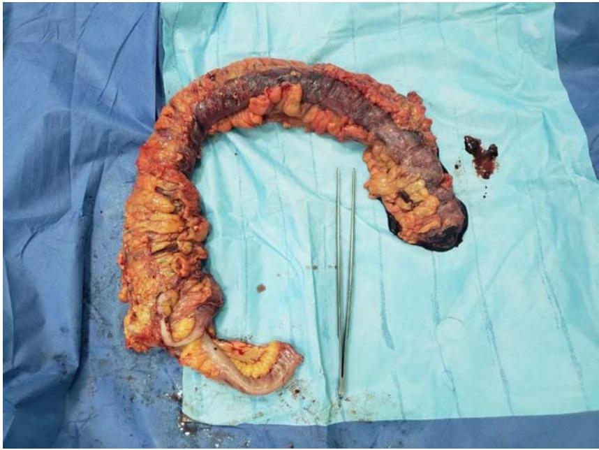 Figure 7: Post-surgery aspect of the colon
## III. DISCUSSION
Autoimmune diseases tend to co-exist; however, systemic lupus erythematosus (SLE) and ulcerative colitis (UC) are rarely described together and a systematic review of the medical literature has seldom been undertaken(10). As reported by several authors, the estimated prevalence of UC in SLE patients is around $0.4\%$ (11).
The precise mechanisms of UC remain undetermined, but autoimmune mechanisms are supposed to be involved in the development of UC as well as InSLE. For example, anti-bodies specific to a Mr 40,000 protein found only incolonic, skin, and biliary epithelia have been demonstrated in patients with UC(2).
Around $40\%$ of SLE patients have gastrointestinal problems, of which gastroduodenal mucosal lesions as adverse effects of nonsteroidal anti-inflammatory agents, corticosteroids or cytotoxic agents are the most common. On the other hand, disease itself causes abdominal pain in only $8\%$ of the SLE patients (12).
Main symptoms include abdominal pain, diarrhea, and bloody stool. The symptomatic feature is indistinguishable from that in inflammatory bowel diseases, especially ulcerative colitis(13).
Coexistence of SLE and UC is difficult to diagnose because both diseases have several similar gastrointestinal symptoms and some drugs used to treat UC may cause drug-induced lupus particularly sulfasalazine, 5-ASA, and infliximab(14). So, it maybe difficult to differentiate whether the symptoms are due to the SLE or other diseases. In Lupuscolitis, ulceration with bleeding or perforation is caused by the inflammatory involvement of small vessels and, sometimes, by necrotizing angitis of small arteries and venules with the deposition of circulating immune complexes (2).
In SLE, Abdominal vascular involvement usually occurs when the disease activity in other organs increases. The evaluation of overall clinical and laboratory findings may help the differential diagnosis (2).
In our case, SLE was considered to be inactive under chloriquine and corticosteroids at the onset of abdominal pain and bloody diarrhea, and typical histopathologic changes of UC such as amicroabscess in the crypt were found in the colo-rectal mucosa.
UC may happen before or after the diagnosis of SLE(14). Our patient was treated for SLE 9 years before developing UC. Many authors described some differences(15)compared to SLE, UC presents more frequently as bloody diarrhea, abdominal pain, and tenesmus.
In all cases, the diagnosis of SLE is made according to the classification criteria for SLE, which necessitate the presence of at least 4 out of 11 clinical and laboratory criteria. The diagnosis of UC is made on the basis of typical clinical, imaging, endoscopic, and histological findings (15).
Our case problematic was related to our therapeutic options, biological therapy was normally indicated for ulcerative colitis, however, the patient was also treated for lupus, and because of the risk of aggravation of SLE after giving anti TNF alpha, we preferred reviewing our options.
To understand the risk of anti TNF alpha induced SLE, we reviewed some cases of lupus like reaction, and DILE (drug induced erythematosus) and its pathogenesis.
Drug-induced lupus erythematosus (DILE) is defined as a lupus-like syndrome temporally related to continuous drug exposure which resolves after discontinuation of the offending drug. There are currently no standard diagnostic criteria for DILE and the pathomechanisms are still unclear. Similarly to idiopathic lupus, DILE can be divided into systemic (SLE), subacute cutaneous (SCLE) and chronic cutaneous lupus (CCLE). Systemic DILE is characterized by typical lupus-like symptoms including skin signs, usually mild systemic involvement and a typical laboratory profile with positive antinuclear and anti-histone antibodies, while anti-double strand (ds) DNA and anti-extractable nuclear antigens antibodies are rare (16).
Sulphasalazine and Anti TNF alpha have been associated with DILE in several cases in the literature (17). Yet this risk of developing Lupus under anti TNF alpha is very low(17), which leads to asking if we should or not use biological therapy in patients with SLE.
Here are some cases of DILE, in patients treated for UC.
Griffiths and al. (18) report a case of a patient who developed a lupus syndrome while receiving sulphasalazine for ulcerative colitis. They first obtained remission, then the patient developed a non-deforming arthritis with active synovitis of shoulders, wrists, metacarpophalangeal joints, proximal interphalangeal joints and digital vasculiticlesions. Anti-nuclear antibodies (ANA) were present in high titres with a homogeneous pattern. DNA antibody concentrations were raised (190 U/ml (normal: \<25 U/ml)).
A drug-induced lupus syndrome was suspected, so sulphasalazine was stopped, cilincal conditions were then slowly resolving. Levels of DNA binding activity and ANA titres remained high for six months after sulphasalazine was stopped, but then fell to normal(18).
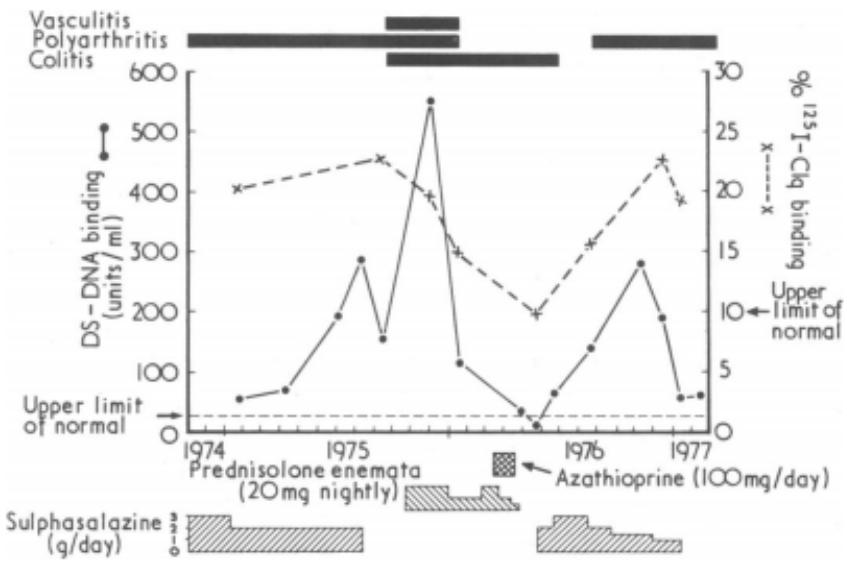 Figure 8: Equential study of clinical features (top), treatment (bottom), and changes in binding activity of double-stranded DNA (DS-DNA) and radioiodinated (1251) complement (Clq)(18).
Another study reviewed 13 cases of DILE due to infliximab in patients with inflammatory bowel diseases IBD(19).
<table><tr><td colspan="2">Patient Characteristics</td></tr><tr><td>Median age (range)</td><td>37 (26–62)</td></tr><tr><td>M:F (numbers)</td><td>2:11</td></tr><tr><td>CD: UC (numbers)</td><td>8:5</td></tr><tr><td>Number of patients on concurrent immunosuppression</td><td>7</td></tr><tr><td>Median (range) duration of infliximab in months prior to DILE</td><td>14 (1–52)</td></tr><tr><td>Median (range) number of infliximab infusions prior to DILE</td><td>10 (2–30)</td></tr><tr><td>Re-treated with second anti-TNF agent</td><td>8/13=61.5%</td></tr><tr><td>Recurrence of lupus like reaction after exposure to second anti-TNF agent</td><td>25%</td></tr><tr><td>Median (range) period of treatment with second anti-TNF agent</td><td>5 months (2–6)</td></tr></table>
Figure 9: Table summarizing patient characteristics (19) In these series of patients, DILE was a female-preponderant disease with a female-to-male ratio of 11:2, and 5 patients were treated for ulcerative colitis (19).
In this same series of patients, Joint manifestations dominated. Symmetric large joint arthralgias were reported by all patients. Fever and malar rash were noted in two and three out of the 13 patients, respectively. The antinuclear antibody titer was elevated in all patients with DILE, The median peak antinuclear antibody (ANA) titer was 1 in 2560, AntidsDNA antibodies were tested in 12 patients(19). AntiTNF therapy was withdrawn in all patients upon diagnosis of DILE, and the resolution of joint symptoms was obtained in a median of 4 weeks (range 3-12 weeks)(19).
In our case, the patient was already treated for SLE before developing UC, and did not receive any the DILE causative drugs.
The following cases report the experience of treating patients with SLE as well as inflammatory bowel disease with biological therapy despite the risk early mentioned.
Yamashita and al.(20) report a case of a 55-year-old Japanese woman with systemic lupus erythematosus (SLE), she developed continuous gastrointestinal bleeding and diarrhea since the patient was aged 30 years that was initially treated as SLE-related colitis. The patient underwent surgery for anal fistulas twice at 50 and 54 years of age and her symptoms were atypical of lupusenteritis. Colonoscopy was performed again when the patient was 55 years of age because we suspected she had some type of inflammatory bowel disease (IBD). Histopathological examination revealed non-caseating granuloma and no evidence of vasculitis, consistent with crohn's disease (CD). Introduction of infliximab dramatically relieved the patient's melena and abdominal symptoms without aggravating SLE symptoms (20).
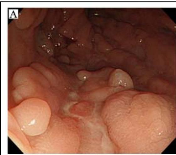 Figure 1-6: endoscopic aspect of ulcerative colitis attack
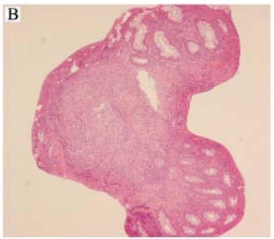 Figure 10: Findings of colonoscopy and colon biopsy specimens. (A) Macroscopic findings of colonoscopy with cobble-stone-like inflammatory polyps and many longitudinal ulcers in the descending colon. (B) Histopathological findings of specimens in A (hematoxylin and eosin, $\times 100$ ) with non-caseating granuloma and no evidence of vasculitis(20)
Principi and al.(21) report a case of CD occurred in a young woman 8 years after a diagnosis of lupus nephritis according to clinical, laboratory and histological criteria. CD was unresponsive to steroids and immunosuppressants and, therefore, the patient was treated with antitumour necrosis factor alpha monoclonal antibody (Infliximab). This therapy led to the remission of both CD (50% of Crohn's Disease Activity Index—CDAI—decrease) and lupus nephritis (disappearance of pyuria in absence of infection, significant increase of serum albumin and improvement of renal function tests)(21).
In case of steroid dependency or steroid refractory TNF-alpha blockers are an effective treatment to induce and maintain remission. The role TNFalpha plays in SLE is controversial and data on the likely effects of blocking TNFalpha on anti-DNA autoantibody production is always of interest. But those antibodies are not generally associated with clinical signs of autoimmunity and there is no indication for monitoring in patients who have no symptoms. There is no clear explanation for this high prevalence of those autoantibodies (22-25).
There are, however, occasional reports describing the efficacy of anti-TNF- $\alpha$ therapy for SLE. It has also been reported that, despite levels of antibodies to ds-DNA and cardiolipin being increased, anti-TNF- $\alpha$ therapy did not exacerbate SLE itself but rather achieved a reduction in disease activity and relief of refractory arthritis, nephritis(26).
TNF- $\alpha$ exerts both deleterious tissue damaging effects mainly through its pro-inflammatory activities and beneficial activities by dampening aggressive autoimmune responses. SLE is a disease with autoimmune disturbance and inflammatory damage, so blocking TNF- $\alpha$ in this autoimmune-prone chronic inflammatory disease may lead to different outcomes, depending on timing and duration of treatment. Thus, infliximab may also be effective for gastrointestinal symptoms associated with SLE(27).
To review our therapeutic options we have to define what an immunomodulator refractory colitis is, and search for possible efficient treatment.
Immunomodulator refractory colitis: Patients who have active disease or relapse in spite of thiopurines at an appropriate dose for at least 3 months (i.e. azathioprine $2 - 2.5\mathrm{mg / kg / day}$ or mercaptopurine $0.75 - 1\mathrm{mg / kg / day}$ in the absence of leukopenia)(28).
Different trials report the benefit of methotrexate and tacrolimus in maintaining remission in UC as second-line immunomodulator Therapies.(28)
The new Zealand society of gastroenterology (28) recommends methotrexate 25 mg weekly for maintaining remission in UC and should be discussed with patients, particularly for those who are steroid dependent.
However, due to the absence of larger, randomised, controlled trials with lengthy follow-up periods tacrolimus cannot yet be considered standard second-line immunosuppression for UC (28).
The American college of gastroenterology recommends using anti-TNF therapy using adalimumab, golimumab, or infliximab to maintain remission in patients with previously moderately to severely active UC (9).
Other therapies have been proposed as an alternative to anti TNF alpha therapy, for patients who are intolerant or who are not responding to it (9).
Vedolizumab is a monoclonal antibody that selectively blocks $\alpha 4\beta 7$ integrin expressed on lymphocytes. A phase III trial investigated the induction and maintenance efficacy of vedolizumab in 895 patients with moderate to severe treatment refractory UC. The study revealed clinical remission rates at week 52 of $44.8\%$ for 4 weekly treatments compared to $15.9\%$ for placebo $\mathsf{p} < 0.0001$.
Vedolizumab has been approved by FDA in 2014, and also in many European countries, for the management of moderate to severe UC(28).
Tofacitinib is an oral inhibitor of Janus Kinases (JAK) 1, 2 and 3, resulting in blocking of interleukin 2, 4, 7, 9, 15 and 21 pathways. Patients were randomised to receive twice daily tofacitinib at doses 0.5, 3, 10 and 15 mg and placebo for 8 weeks. Clinical remission rates at 8 weeks of $48\%$ and $41\%$ of patients were seen at doses of $10\mathrm{mg}$ $(p < 0.001)$ and $15\mathrm{mg}$ $(p < 0.001)$ respectively compared to the placebo rate of $10\%$ (28).
The American college of gastroenterology recommend to facitinib for maintenance of remission in patients with previously moderately to severely active UC now in remission after induction with to facitinib (9).
Thirty percent of patients with ulcerative colitis will eventually come to proctocolectomy and this includes some with troublesome distal colitis. The decision as to whether to proceed to surgery is obviously a big one, with life-long consequences (28).
Removing the colon and rectum in poorly controlled ulcerative colitis restores physical well-being and quality of life (28).
Long-term concerns about neoplasia are put aside. Patients are understandably very concerned about avoiding a "bag". However the starting point of a discussion with the patient about surgery should focus on whether or not the time has come to remove the colon and rectum.
Surgery provides a cure for the colitis, but carries a risk of a variety of short- and long-term complications (28).
There is significant potential for morbidity, but overall greater than $90\%$ of patients are pleased with their resulting health state and bowel function (29).
If optimisation of standard immunosuppression fails in mild to moderate UC, then the main therapeutic options currently available are antiTNF- $\alpha$ therapy and colectomy. While other immunosuppressive strategies exist, they have not been demonstrated to have the same efficacy as antiTNF- $\alpha$ therapy and surgery (28).
To summarize it all, Patients treated for UC as well as SLE can be treated by chloroquine and corticosteroid associated with systemic 5-ASA or immunosuppressive therapy such as azathioprine or 6MP. However, in cases like ours, when the UC is Immunomodulator refractory, treatment to maintain remission can be a real challenge despite different possible options, and differs depending on centers, hospital experience, economic situation as well as patients background.
## IV. CONCLUSION
In conclusion, the diagnostic criteria for UC and SLE overlap, making them difficult to diagnose correctly. Physicians should bear in mind the possibility that a patient may be afflicted with both of these diseases simultaneously. If a patient known to have SLE develops gastrointestinal symptoms such as abdominal pain or diarrhea, it is prudent to rule out UC (30).
Despite some obscure aspects, our case report suggests that the series of UC associated complaints could include a wide range of autoimmune disorders besides those which have been conventionally considered until now. Therefore, a more accurate detection of autoimmune diseases could be suggested in the diagnostic program to be performed in the course of UC (21).
Generating HTML Viewer...
References
30 Cites in Article
H Stevens,L Ostlere,M Rustin (1994). Systemic lupus erythematosus in association with ulcerative colitis: related autoimmune diseases.
I Koutroubakis,H Kritikos,I Mouzas,S Spanoudakis,A Kapsoritakis,E Petinaki (1998). Association between ulcerative colitis and systemic lupus erythematosus: report of two cases.
R Amelia,S Supriono (2014). 17 Year Old Female Patient with Systemic Lupus Erythematosus and Ulcerative Colitis.
I Koutroubakis,A Pena (1997). Genetics of inflammatory bowel disease.
J Batchelor (1993). SYSTEMIC LUPUS ERYTHEMATOSUS AND GENES WITHIN THE HLA REGION.
David Rubin,Ashwin Ananthakrishnan,Corey Siegel,Bryan Sauer,Millie Long (2019). ACG Clinical Guideline: Ulcerative Colitis in Adults.
D Medeiros,D Isenberg (2009). Systemic lupus erythematosus and ulcerative colitis.
D Hallegua,D Wallace (2000). Gastrointestinal manifestations of systemic lupus erythematosus.
D Wallace (1993). Gastrointestinal manifestations and related liver and biliary disorders.
Bruce Hoffman,Warren Katz (1980). The gastrointestinal manifestations of systemic lupus erythematosus: A review of the literature.
X-P Tian,X Zhang (2010). Gastrointestinal involvement in systemic lupus erythematosus: Insight into pathogenesis, diagnosis and treatment.
O Nitzan,M Elias,W Saliba (2006). Systemic lupus erythematosus and inflammatory bowel disease.
Camilla Vedove,Micol Del Giglio,Donatella Schena,Giampiero Girolomoni (2009). Drug-induced lupus erythematosus.
Augusto Vaglio,Peter Grayson,Paride Fenaroli,Davide Gianfreda,Valeria Boccaletti,Gian Ghiggeri,Gabriella Moroni (2018). Drug-induced lupus: Traditional and new concepts.
I Griffiths,S Kane (1977). Sulphasalazine-induced lupus syndrome in ulcerative colitis.
Sreedhar Subramanian,Vijay Yajnik,Bruce Sands,Garret Cullen,Joshua Korzenik (2011). Characterization of patients with infliximab-induced lupus erythematosus and outcomes after retreatment with a second anti-TNF agent.
Hiroyuki Yamashita,Yo Ueda,Hoshimi Kawaguchi,Akitake Suzuki,Yuko Takahashi,Hiroshi Kaneko,Toshikazu Kano,Akio Mimori (2012). Systemic lupus erythematosus complicated by Crohn’s disease: a case report and literature review.
M Principi,A Di Leo,M Ingrosso,A Pisani,S Marangi,A Amoruso,C Panella,A Francavilla,Enzo Ierardi (2004). Lupus Nephritis Improvement After Anti‐tumor Necrosis Factor Alpha Monoclonal Antibody (Infliximab) Treatment for Crohn's Disease: A Case Report.
Paul Rutgeerts,William Sandborn,Brian Feagan,Walter Reinisch,Allan Olson,Jewel Johanns,Suzanne Travers,Daniel Rachmilewitz,Stephen Hanauer,Gary Lichtenstein,Willem De Villiers,Daniel Present,Bruce Sands,Jean Colombel (2005). Infliximab for Induction and Maintenance Therapy for Ulcerative Colitis.
E Garcia-Planella,E Domènech,M Esteve-Comas,I Bernal,E Cabré,J Boix (2003). Development of antinuclear antibodies and its clinical impact in patients with Crohn's disease treated with chimeric monoclonal anti-TNFα antibodies (infliximab).
R Mageed,D Isenberg (2002). Tumour necrosis factor alpha in systemic lupus erythematosus and anti-DNA autoantibody production.
Konstantinos Katsanos,Paraskevi Voulgari,Epameinondas Tsianos (2012). Inflammatory bowel disease and lupus: A systematic review of the literature.
Martin Aringer,Winfried Graninger,Günter Steiner,Josef Smolen (2004). Safety and efficacy of tumor necrosis factor α blockade in systemic lupus erythematosus: An open‐label study.
E Eliadou,A Day,M Thompson-Fawcett,R Gearry,D Rowbotham,R Walmsley (1423). New Zealand Society of Gastroenterology Guidelines for the Management of Refractory Ulcerative Colitis.
V Fazio,R Kiran,F Remzi,J Coffey,H Heneghan,H Kirat (2013). Ileal pouch anal anastomosis: analysis of outcome and quality of life in 3707 patients.
Nacir Oussama,Aboutarik Fatimaezzahra,Aiterrami Adil,Samlani Zouhour,Krati Khadija,Oubaha Sofia (2020). Ulcerative Colitis and Systemic Lupus Erythematosus: An Unusual Association with Diagnostic and Therapeutic Difficulties.
No ethics committee approval was required for this article type.
Data Availability
Not applicable for this article.
How to Cite This Article
Yassine Chait. 2026. \u201cAssociation between Systemic Lupus Erythematosus and Refractory Ulcerative Colitis: What are our Therapeutic Options?\u201d. Global Journal of Medical Research - K: Interdisciplinary GJMR-K Volume 23 (GJMR Volume 23 Issue K3).
Explore published articles in an immersive Augmented Reality environment. Our platform converts research papers into interactive 3D books, allowing readers to view and interact with content using AR and VR compatible devices.
Your published article is automatically converted into a realistic 3D book. Flip through pages and read research papers in a more engaging and interactive format.
Our website is actively being updated, and changes may occur frequently. Please clear your browser cache if needed. For feedback or error reporting, please email [email protected]
Thank you for connecting with us. We will respond to you shortly.