Loading menu…

Chronic Dislocation of the 5th Metatarsophalangeal Joint with Physeal Injury of Metatarsal: A Case Report

§ National Institute of Ayurveda
‡ National Institute of Ayurveda, Madhav Vilas

Send Message

To: Author

Chronic Dislocation of the 5th Metatarsophalangeal Joint with Physeal Injury of Metatarsal: A Case Report

Article Fingerprint

ReserarchID

NQ4DS

Chronic Dislocation of the 5th Metatarsophalangeal Joint with Physeal Injury of Metatarsal: A Case Report Banner

Key Research Insights

Synthesized scholarly intelligence & interactive research assistant
  • English
  • Afrikaans
  • Albanian
  • Amharic
  • Arabic
  • Armenian
  • Azerbaijani
  • Basque
  • Belarusian
  • Bengali
  • Bosnian
  • Bulgarian
  • Catalan
  • Cebuano
  • Chichewa
  • Chinese (Simplified)
  • Chinese (Traditional)
  • Corsican
  • Croatian
  • Czech
  • Danish
  • Dutch
  • Esperanto
  • Estonian
  • Filipino
  • Finnish
  • French
  • Frisian
  • Galician
  • Georgian
  • German
  • Greek
  • Gujarati
  • Haitian Creole
  • Hausa
  • Hawaiian
  • Hebrew
  • Hindi
  • Hmong
  • Hungarian
  • Icelandic
  • Igbo
  • Indonesian
  • Irish
  • Italian
  • Japanese
  • Javanese
  • Kannada
  • Kazakh
  • Khmer
  • Korean
  • Kurdish (Kurmanji)
  • Kyrgyz
  • Lao
  • Latin
  • Latvian
  • Lithuanian
  • Luxembourgish
  • Macedonian
  • Malagasy
  • Malay
  • Malayalam
  • Maltese
  • Maori
  • Marathi
  • Mongolian
  • Myanmar (Burmese)
  • Nepali
  • Norwegian
  • Pashto
  • Persian
  • Polish
  • Portuguese
  • Punjabi
  • Romanian
  • Russian
  • Samoan
  • Scots Gaelic
  • Serbian
  • Sesotho
  • Shona
  • Sindhi
  • Sinhala
  • Slovak
  • Slovenian
  • Somali
  • Spanish
  • Sundanese
  • Swahili
  • Swedish
  • Tajik
  • Tamil
  • Telugu
  • Thai
  • Turkish
  • Ukrainian
  • Urdu
  • Uzbek
  • Vietnamese
  • Welsh
  • Xhosa
  • Yiddish
  • Yoruba
  • Zulu
Reading Preferences
Font Size
Line Spacing
Background
This converted HTML version may contain rendering inconsistencies. Please refer to the PDF for the authoritative version, or click here to provide feedback.

Abstract

Metatarsophalangeal joint dislocations are uncommon injuries. This article describes the surgical management of such injury with six months follow up report. A 13 years old boy presented with the complaints of deformity and shortening of the 5 th toe of the right foot with callosity on plantar aspect since last five years. He sustained this injury by hitting a stone. He was diagnosed to have a compound dislocation of a metatarsophalangeal joint with severely angulated Salter and Harris type II epiphyseal injury of 5 th toe of the left foot. Joint dislocation caused deformed shortened 5 th toe, and epiphyseal malunion resulted in the plantar bony projection, callosity, ulceration, difficulty in walking and wearing the footwear. This case was managed surgically that culminated in an optimum functional and structural outcome. Malunited epiphysis was excised, the metatarsal bone was aligned and fixed with proximal phalanx by Kirschner wire to establish a pseudarthrosis. This method can be useful in such cases; however, needs to be evaluated with future studies.

I. INTRODUCTION

Metatarsophalangeal (MTP) joints of the foot aresmall, very stable; and rarely get dislocated 1,2. Dislocations are usually dorsal in direction, but horizontal and plantar dislocations have also been reported 3. Epiphyseal injuries are the fractures through the growth plates of the bones of the children. Management of such injuries (mal-united fractures and chronic dislocations) is surgical. The present case report describes the management of an ignored dislocation of 5 th metatarso-phalangeal joint with marked ventrally displaced epiphyseal (Salter–Harris type II) injury of head of 5 th metatarsal. The report emphasizes the importance of careful physical examination and assessment of the morbid anatomy of the injury by radiographs; followed by proper management.

II. CASE HISTORY

a) Personal Profile and Present History of the Patient

He was treated in a private clinic (general practitioner) by wound closure that healed in due course of time, but skeletal injury remained ignored. All this resulted in deformity with displaced epiphysis projecting ventrally and base of dislocated proximal phalanx protruding dorsally. Toe as a whole became short, dorsiflexed and made the footwear bearing and walking difficult. Continuous friction over the ventrally projected displaced epiphysis resulted in a painful callosity and ulceration.

III. CLINICAL EXAMINATION

The patient was 13 years old male, student of 6th standard, belonging to middle socio-economic status and Hindu religion. The patient was presented in the hospital with the history of trauma right foot 5 years back having the complaints of deformity and shortening of the 5th toe of right foot with callosity on plantar aspect for last five years. He sustained this injury by accidentally hitting a stone while he was taking a bath outdoors resulting in a wound on the dorsum of foot and injury to joint and bone.

b) Treatment History

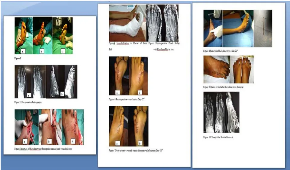
Clinically there was deformity and shortening of 5th toe right foot with scar mark of wound closure dorsally [Figure 1(a)]. On the planter side there was visible protuberance with callosity and ulceration [Figures 1(b) & 1(c)]. On palpation, the plantar protuberance was bony hard. Dorsally the base of the proximal phalanx was palpable. There was minimal tenderness, stiffness, and loss of active and passive movements.

IV. INVESTIGATIONS

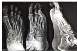
Antero-Posterior and lateral views of X-rays of both the feet were taken and compared. All the required blood investigations along with chest X-ray were done, and were found within normal limits.

V. DIAGNOSIS

Based on the history, clinical findings and radiographic investigation, it was diagnosed as chronic metatarsophalangeal joint dislocation with plantarangulation of malunited Salter and Harris type II epiphyseal injury of head of 5th metatarsal [Figure 2].

VI. MANAGEMENT

a) Planning

Main complaints of the patient were an inability to wear the shoes, difficulty in walking and visible deformity. Conservative reduction was not possible due to fibrosis and malunion of epiphyseal injury owing to a long duration of the injury. So correction by open reduction and internal fixation was planned.

b) Anesthesia and Tourniquet

Procedure was done under the spinal anesthesia. Tourniquet at mid-thigh level was used, and all the precautions were followed.

c) Incision and approach

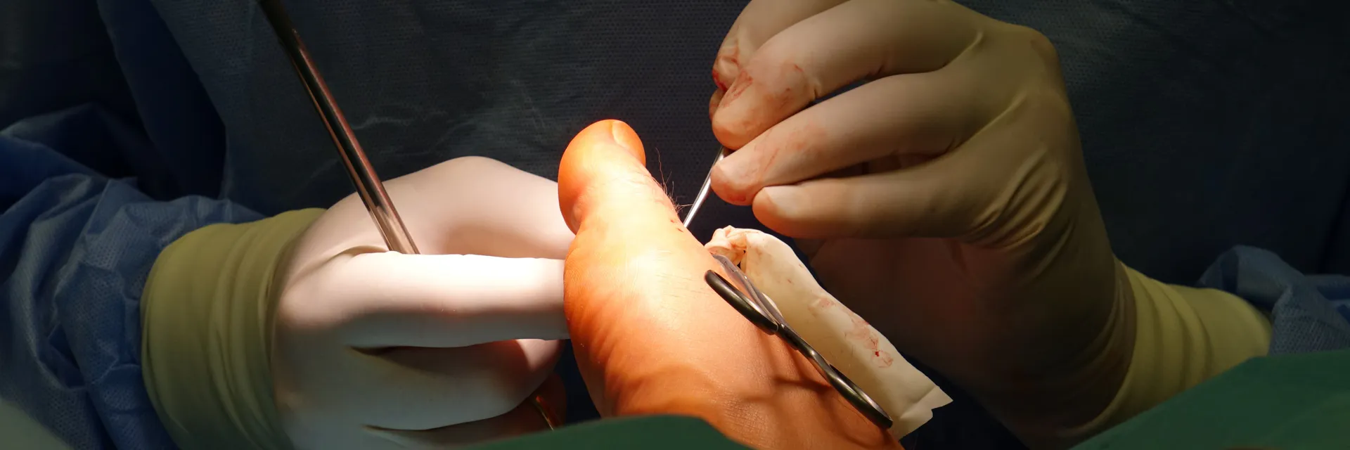
Injury was approached by dorsal and ventral (plantar) two different approaches. Ventrally 2cm straight incision directly over the prominence and on dorsal aspect a zigzag 3cm long incision was made. Angulated mal-united epiphysis was just beneath the skin and could be approached directly. Dorsally joint was approached by the Z-tenotomy of extensor tendon that was short and tense.

d) Procedure

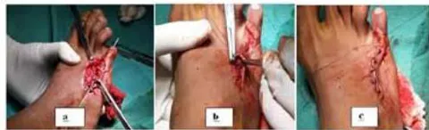
Excision of callosity and displaced distal epiphysis of the head of 5th metatarsal was done. The rough raw area made smooth by bone file. Dorsally after Z-tenotomy of extensor tendon dislocated base of phalanx exposed and mobilized by excising the fibrous tissue. It was aligned with the metatarsal bone and fixed with 1.2mm Kirschner wire [Figure 3 (a)]. After that, lengthening of the extensor tend on was done by performing Z-tenoplasty. Both the wounds were closed [Figures 3 (a), 3 (b) & 3 (c)].

e) Immobilization

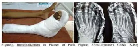
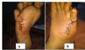
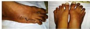
Below knee Plaster of Paris (POP) slab was applied [Figure 4]. The post-operative period was uneventful and Kirschner wire was kept in situ for three weeks. Patient was discharged after suture removal [Figures 7(a) & 7(b)].

f) Follow up

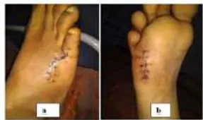
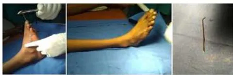
On follow up after three weeks K-wire and below knee Plaster of Paris slab were removed [Figure 8]. There was no deformity except slight shortening of 5th toe.

VII. RESULT

The patient was allowed full weight-bearing at the end of one month. He was able to wear the shoes and walk freely after one month of surgery. After six months follow up, the patient was able to walk comfortably with or without shoes but slight dorsal drifting of the 5th toe with shortening was there. No recurrence of callosity was there and painless movements at metatarsophalangeal pseudarthrosis were present.

VIII. Discussion

Foot injuries if ignored or not properly treated can affect the ability to use the foot and lower extremity and can lead to significant long term problems of stiffness, post-traumatic arthritis, pain, instability, callosities, difficulty in footwear wearing and walking. It is necessary to evaluate these injuries properly and plan treatment accordingly. The present case was of an ignored dislocation of 5th metatarsophalangeal joint with Salter and Harris type II epiphyseal injury of the capital epiphysis of 5th meta-tarsal with marked angulation with planter displacement and mal-union. He was also having painful plantar callosity beneath the projecting displaced epiphysis. Open reduction is best accomplished through a dorsal approach 4−10. Temporary K-wire fixation is only indicated when the reduced joint is very unstable. This case was operated by the authors, five years after sustaining the injury. Capital epiphysis and callosity were excised by direct plantar approach, whereas metacarpal and proximal phalanx were aligned and fixed by K wire for three weeks. A pseudarthrosis developed in between the metaphysis of metacarpal and proximal phalanx with useful movements. Contraction of dorsal surgical wound resulted in slight contracture carrying the toe bit dorsally. As the bony spur was removed, so callosity did not re-appeared and shortening of the toe after surgery was the result of excision of metatarsal head.

IX. CONCLUSION

Traumatic dislocation of the metatarsophalangeal joint and epiphyseal injuries of small joints of foot and toes should be attended, diagnosed, and adequate treatment should be employed early to avoid complications. The case of chronic metatarsophalangeal joint dislocations with or without epiphyseal injuries should be treated surgically by appropriate surgical approaches. This unusual chronic metatarsophalangeal joint dislocation with epiphyseal injury was well managed by surgery with the good functional and structural outcome. This method can be useful in such cases, however, needs to be evaluated with future studies.

Declaration of Patient Consent: The authors certify that they have obtained the consent of the patient and his parents for the clinical history and images to be reported in the journal while maintaining confidentiality.

Financial Support and Sponsorship: Nil

Conflict of Interest

The authors declare that there is no conflict of interest regarding the publication of this paper.

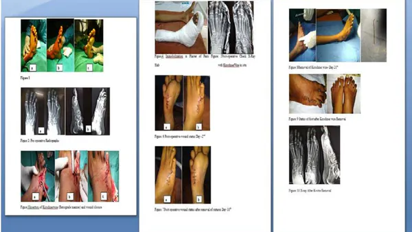
Figure 1

ACKNOWLEDGEMENTS

We are highly thankful to the patient and his attendants for giving us opportunity to manage the case and to consent to the surgical intervention and publication of this case. We are thankful to the Director and the Deputy Medical Superintendent, the anaesthetist and the operation theatre staff of the hospital for their help and co-operation.
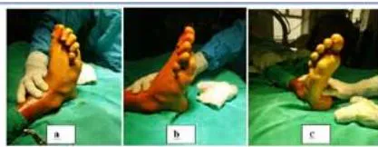
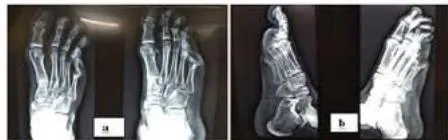
Figure 2: Pre-operative Radiographs

Figure 3 Insertion of Kirschnerwire (Retrograde manner) and wound closure

with KirschnerWire in situ

Figure: 6 Post-operative wound status Day -2 nd

Figure: 7Post-operative wound status after removal of sutures Day-10 th

Figure: 8Removal of Kirschner wire- Day 21 4

Figure: 9 Status of foot after Kirschner wire Removal

Figure: 10 X-ray After K-wire Removal
Figure 1:
Figure 1:
Figure 1: Introduction of Kinchroenter (Retrograde manner) and wound closure

References

10 Cites in Article
  1. 1. H Hey,G Chang,C Hong,W Kuan (2013). Irreducible dislocation of the fourth metatarsophalangeal joint-A Case Report. The American Journal of Emergency Medicine, 31(1), 265-e266.
  2. 2. Devdatta Neogi,Shivanand Bandekar,Vilas Sadekar,Sarthak Patnaik,Trivikram Bhat,Zelio D’mello (2012). Irreducible Dislocation of All the Lesser Metatarsophalangeal Joints of the Foot. Foot & Ankle Specialist, 5(5), 324-326.
  3. 3. Mostapha Boussouga,Jalal Boukhriss,Abdeloihab Jaafar,Khalid Lazrak (2010). Irreducible Dorsal Metatarsophalangeal Joint Dislocation of the Fifth Toe: A Case Report. The Journal of Foot and Ankle Surgery, 49(3), 298.e17-298.e20.
  4. 4. Jacques Brunet,Stanislaw Tubin (1997). Traumatic Dislocations of the Lesser Toes. Foot & Ankle International, 18(7), 406-411.
  5. 5. C Woon (2011). Dislocation of the Interphalangeal joint of the Great Toe: is percutaneous reduction of an incarcerated sesamoid an option? Surgical technique. J Bone Joint Surg Am, 93(Suppl 1), 109-112.
  6. 6. Elliot Sorene,Gilad Regev (2006). Complex Dislocation With Double Sesamoid Entrapment of the Interphalangeal Joint of the Hallux. The Journal of Foot and Ankle Surgery, 45(6), 413-416.
  7. 7. H Leung,B Wong,W (2002). Irreducible dislocation of the hallucal-interphalangeal joint. Hong Kong Med J, 8, 295-299.
  8. 8. J Berger,L Legeyt,M Ghobadi,R (1997). Incarcerated subhallucal-sesamoid of the great toe: irreducible dislocation of the interphalangeal joint of the great toe by an accessory sesamoid bone. Am J Orthop, 26, 226-228.
  9. 9. Isaac Yang,Kelvin Sun,Wai-Leung Sha,Kong-San Yu,Yuk-Yin Chow (2011). Interphalangeal Dislocation of Toes: A Retrospective Case Series and Review of the Literature. The Journal of Foot and Ankle Surgery, 50(5), 580-584.
  10. 10. Antonio Córdoba-Fernández (2012). Management of Nonreducible Lesser Toe Interphalangeal Dislocation. Journal of the American Podiatric Medical Association, 102(3), 252-255.

Funding

No external funding was declared for this work.

Conflict of Interest

The authors declare no conflict of interest.

Ethical Approval

No ethics committee approval was required for this article type.

Data Availability

Not applicable for this article.

How to Cite This Article

Manorma Singh, Sanjeev Sharma, Suman Sharma, Rahul Sharma. 2019. "Chronic Dislocation of the 5th Metatarsophalangeal Joint with Physeal Injury of Metatarsal: A Case Report". Global Journal of Medical Research - H: Orthopedic & Musculoskeletal System GJMR-H Volume 19 (GJMR Volume 19 Issue H3).

More Citation Formats

Download Citation

Journal Specifications

Crossref Journal DOI 10.17406/gjmra

Print ISSN 0975-5888

e-ISSN 2249-4618

Keywords
Classification
GJMR-H Classification NLMC Code: WE 175
Version of record
v1.2

Issue date
October 18, 2019

Language
English
Order Article Reprint
Experience in AR

Explore published articles in an immersive Augmented Reality environment. Our platform converts research papers into interactive 3D books, allowing readers to view and interact with content using AR and VR compatible devices.

Read in 3D

Your published article is automatically converted into a realistic 3D book. Flip through pages and read research papers in a more engaging and interactive format.

Article Matrices
Total Views: 710
Total Downloads: 41
All Trends

Request Access

Please fill out the form below to request access to this research paper. Your request will be reviewed by the editorial or author team.
X

This is the heading

Lorem ipsum dolor sit amet, consectetur adipiscing elit. Ut elit tellus, luctus nec ullamcorper mattis, pulvinar dapibus leo.

High-quality academic research articles on global topics and journals.

Chronic Dislocation of the 5th Metatarsophalangeal Joint with Physeal Injury of Metatarsal: A Case Report

Manorma Singh
Manorma Singh
Sanjeev Sharma
Sanjeev Sharma <p>National Institute of Ayurveda</p>
Suman Sharma
Suman Sharma <p>National Institute of Ayurveda, Madhav Vilas</p>
Rahul Sharma
Rahul Sharma <p>National Institute of Ayurveda</p>