INTRODUCTION
Splenic torsion (ST) is a rare pathology in Canidae species, which is a twisting of the splenic pedicle, leading to vascular obstruction and stasis. Venous blood flow to the affected area is interrupted, leading to infarction. The etiology of ST remains unclear, but in dogs it is thought to be the result of dilatation or volvulus of the stomach, which can stretch and loosen the gastrosplenic ligaments, allowing abnormal splenic mobility. It is also possible that ST may predispose patients to gastric torsion due to gastric motility (Hernandez et al., 2025). ST is most commonly reported in large and giant breeds of dogs (Great Danes and German Shepherds) with deep chests. Acute and chronic forms of splenic torsion have been described. Acute ST presents with signs of severe abdominal pain, weakness, and cardiovascular collapse that occur rapidly, sometimes within a few hours. Chronic ST can be difficult to diagnose, with isolated cases of vomiting, lethargy, weakness, abdominal pain, hematuria, and diarrhea, which are intermittent (Neath et al., 1997). According to Simeonova et al. (2007), the etiology of ST in dogs is poorly understood. The most commonly suggested causes are congenital absence or weakness of the splenic ligaments or acquired laxity of these ligaments as a result of abdominal surgery, trauma, or previous episodes of gastric dilatation. According to other data by Grange et al. (2012), potential causes of ST are splenic inflammation, congenital and acquired laxity of the splenic ligaments, neoplasia, peritonitis, accessory splenic tissue, and trauma. Diagnosis can also be made based on clinical signs, clinical examination results, and abdominal radiography, but abdominal Doppler ultrasonography and computed tomography are usually required for definitive diagnosis (Hughes et al., 2020). Splenectomy is a surgical treatment method in which preoperative stabilization of the patient is necessary, especially in cases of hypovolemic shock and secondary hemoabdomen. Manipulation or dissection of the spleen before its removal is not recommended, as this may release thrombi, sequestered blood, or inflammatory mediators into the central circulation. The prognosis in dogs is generally more favorable in chronic cases due to the lower risk of shock and endotoxemia (Weber, 2000). The human literature refers to a "wandering spleen," a condition in which the suspensory ligaments of the spleen are underdeveloped or lax, characterized by splenic hypermobility and loss of fixation. This is associated with stretching of ligaments and axial rotation and torsion of the spleen. Pregnancy is also thought to be a cause of splenic torsion in women (Virani et al., 2021; Suliman et al., 2024).
Due to the lack of sufficient information in the literature, the aim of this report was to describe in detail the pathoanatomical and histopathological lesions in Doberman splenic torsion with generalized lymphoma. Which will find application and contributions in the scientific and clinical literature and will be of great benefit to practicing veterinarians in small animal clinics in the diagnosis and treatment of spleen diseases and lymphomas.
MATERIALS AND METHODS
Owner consent was obtained for the procedures undertaken and the use of the data for research purposes, while this study was approved by the Ethics Committee at Trakia University (Decision No. 073/2026). This publication presents a clinical case of splenic torsion in a dog with generalized lymphoma. The case is a 7-year-old male Doberman. He was fed pelleted food and meat, was dewormed monthly for internal and external parasites, and was vaccinated annually. The dog was admitted to a private veterinary clinic due to refusal to eat, vomiting for 3 days, and defecating brown-black mushy stools. During the clinical examination, the dog had a subnormal body temperature of , pale mucous membranes, and reduced skin turgor. The abdomen was enlarged and painful on palpation. All cadaveric lymph nodes (mandibular, axillary, prescapular, inguinal, and popliteal) were enlarged. Radiological examination revealed a severely enlarged spleen and liver (Figure 1), and abdominal ultrasound examination showed an enlarged liver with a hyperechoic fine-grained dystrophic structure and an enlarged spleen with multiple subcapsular tumors.
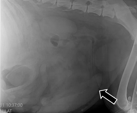
X-ray examination of the abdominal cavity lateral projection enlarged spleen (arrow). Dog with splenic torsion and generalized lymphoma.
Immunochromatographic testing (snap 4dx idexx) was performed on a blood sample, and the result was negative for antibodies to A. phagocytophilum, A. platys, B. burgdorferi, E. canis, E. ewengi, and D. immitis. The changes in the blood count were a strong increase in white blood cells, leukocytes (WBC - ), as well as other blood parameters such as monocytes (MON - ), Monocytes as a percentage of the number of leukocytes (MON% - 16.10%), red cell distribution width (coefficient of variation RDW-CV - 0.21%), and red cell distribution width (standard deviation RDW-SD - 58.30 fL). For humane reasons and after consultation with a veterinarian, the owners requested euthanasia and subsequent necropsy. Euthanasia was performed with 8 mg/kg propofol (Propomitor 10 mg/ml Orion Corporation) and a 0.4 mg/kg combination of embutramide, mebezonium iodide, tetracaine hydrochloride dimethylformamide (T61, MSD Animal Health). After euthanasia, the dog’s corpse was examined pathologically according to the standard autopsy protocol. After analyzing the macroscopic changes in all organs and systems, samples were taken for histopathological examination from different areas of the spleen, liver, stomach, and cadaveric lymph nodes (popliteal and prescapular). The materials for histopathological examination were fixed in 10% neutral formalin solution and processed using the classical histological technique.
RESULTS AND DISCUSSION
External examination:
During the external examination of the corpse, the following changes were found: anemic mucous membranes and conjunctiva. There was plaque on the canines and premolars bilaterally. In the perianal area there were stains with brown-black mushy feces—melena. On examination, the abdomen was greatly enlarged and convex with a doughy consistency. On skin palpation, all cadaveric lymph nodes (mandibular, axillary, prescapular, inguinal, and popliteal) were enlarged bilaterally, and on section, they were juicy with whitish proliferates in the cortex and medulla. During the abdominal section, there were 800 ml of red fluid, free of fibrin and coagulum. The spleen was enlarged with rounded edges, and numerous whitish formations of various-sized metastases protruded from its capsule. In section, the pulp was dark red, juicy, and dotted with whitish proliferates. At one end, we found torsion of the spleen together with part of the omentum (Figure 2).
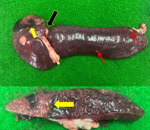
Enlarged spleen with torsion (black arrow) and metastases (red arrows). Hemorrhagic infarction reaching deep into the pulp (yellow arrows). Dog with splenic torsion and generalized lymphoma.
The blood vessels in the hilus were enlarged, and the surrounding tissues were hemorrhagically imbibed and edematous. A large hemorrhagic infarction was observed along one of the edges of the spleen below the torsion zone, reaching deep into the pulp. Authors such as Mergl et al. (2022) have described similar clinical and morphological changes in chronic splenic torsion in a 4-year-old crossbreed dog. They observed a significantly enlarged, swollen, and dark spleen, local peritonitis, and adhesions of the omentum around the splenic pedicle. Hernandez et al. (2025) described changes in the spleen of a wolf with acute torsion: there was a prominent encapsulated lesion on the diaphragmatic surface of the organ and multiple hard, brown intraparenchymal lesions. Histologically confirmed splenic torsion with fibrin deposition, hemorrhages, and mineralization of parenchymal necrosis. No infectious agents or neoplastic processes were present. Researchers such as Angelou et al. (2020) reported splenic torsion in a 5-year-old female Cane Corso dog, with similar clinical data of anorexia, depression, and vomiting for 4 days. Abdominal tenderness, abdominal radiography showed a large mass and splenomegaly. A team led by Melkamu et al. (2025) reported a case of acute abdomen in a 16-year-old girl with gangrene of the spleen due to torsion. The organ was enlarged to about 15 cm in length and had ischemic necrosis along the capsule, twisted about clockwise around the pedicle. With altered topography, hanging on the vascular pedicle. The liver was enlarged with rounded edges and a distended gallbladder. The mesenteric lymph nodes were also enlarged. A report by Seong-won et al. (2024) describes an interesting case of torsion of the caudate process of the caudate lobe of the liver with the spleen in a 3-year-old male German shepherd. On section of the stomach, the mucosa was thickened, in the cardia area there was a flat, concave, ulcerated formation with an oval shape and dimensions of 4 cm in diameter (Figure 4).
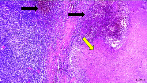
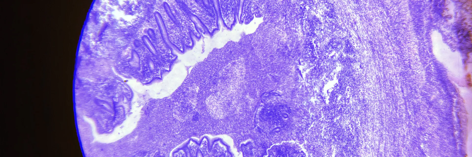
Torsed neoplastic spleen with vascular congestion and hemorrhages (arrows). Ischemic necrosis of the splenic parenchyma - infarction (yellow arrow). dog with splenic torsion and generalized lymphoma. X/E, bar=10 μm.
The blood vessels in the serosa of the stomach, small and large intestines were hyperemic. The kidneys were of a soft, friable consistency and rough in appearance. The lung was compacted on palpation with a violet color, the bronchial lymph nodes were also enlarged. The heart was round, the walls of the left and right ventricles were thinned and relaxed. No other changes were found in the remaining organs and systems during the pathological examination.
Histopathological changes:
The pathohistological changes in the examined organs—spleen, liver, stomach and enlarged cadaveric lymph nodes (popliteal and prescapular) were expressed in hyperemia and neoplastic growths. Structural changes in the spleen were expressed by sharp, distinct boundaries between the torsion zone and the surrounding neoplastic tissue represented by diffuse infiltrative growth of lymphocytes with large nuclei and abundant cytoplasm. Lymphocytes showed signs of anisokaryosis, polychromasia, polymorphism and mitotic figures. Microscopic changes in the infarct area were vascular congestion, fibrinoid thromboses of the vessels and local ischemic necrosis (Figure 3).
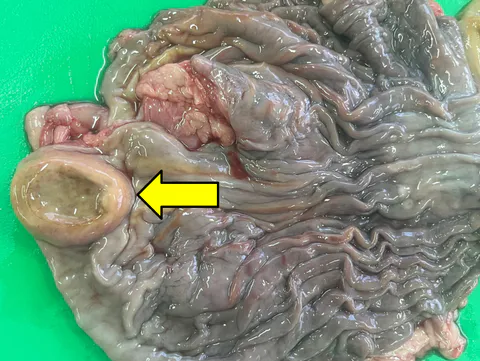
Thickened gastric mucosa with an oval ulcerated formation in the cardia region (arrow). dog with generalized lymphoma.
Activated macrophages were also observed, as well as numerous siderocytes. Ko et al. (2023) described the pathomorphology of 11 cases of splenic lymphoma. According to them, the marginal zone lymphomas had a nodular growth pattern and characteristic architecture, such as a proliferating marginal zone and fading germinal centers of the lymphatic follicles in the white pulp. The neoplastic cells were intermediate-sized lymphocytes, with nuclei 1.5–2 times the diameter of a red blood cell, abundant cytoplasm, and mitotic figures. In 4 cases, a diffgrowth pattern of neoplastic lymphocytes with nuclei twice the diameter of a red blood cell was found. With similar malignant features to those found by us, such as anisokaryosis and up to eight mitotic figures in one field of view. Immunohistochemically, marginal zone lymphomas have been proven to be of B-cell origin, given the strong expression of PAX5 and the lack of CD3 immunoreactivity. Peripheral T-cell lymphomas show a diffuse growth pattern, and the neoplastic cells are diffusely immunoreactive for CD3 but immunonegative for PAX5. According to Valli et al. (2013), there are different subtypes of T- and B-cell lymphomas, of which marginal zone lymphoma is the most common in canine spleens. Accurate diagnosis of lymphoma in the spleen of dogs is of prognostic importance. In the present study, the classification of splenic lymphoma was performed according to this classification, and our findings correlate with the literature that marginal zone lymphoma is the most commonly reported subtype. A group of researchers such as Nazem et al. (2021) and Mergl et al. (2022) have observed identical histopathological changes to ours in splenic torsion in dogs, such as vascular congestion, multiple vascular fibrin thromboses, ischemic necrosis and extensive hemorrhage in the parenchyma. In addition, they also found extensive omental adhesions, with degeneration and necrosis of the omental fat tissue, hemorrhages, chronic inflammation, and formation of reparative granulation tissue with scars. There were no lesions of malignant neoplasia and pathogenic microorganisms in the examined spleen tissue sections. Changes in the liver were severely dilated and blood-filled sinusoidal capillaries, Kupffer cell reaction, siderocytes, and disintegration of hepatocyte beds. The mucosa, submucosa, and partly the muscularis mucosa of the stomach were edematous with diffuse infiltrates of neoplastic lymphocytes and single plasma cells (Figure 5).
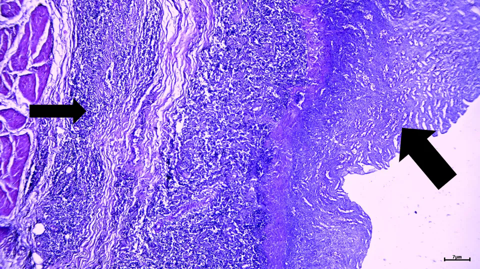
Infiltrates of neoplastic lymphocytes and single plasma cells in the mucosa and submucosa (arrows) of the stomach. Atrophy of the glands and intramural edema of the gastric wall. Dog with generalized lymphoma. X/E, bar=10 μm.
In isolated areas there was fibrosis with glandular atrophy and mononuclear inflammatory infiltration. In the lymph nodes, the changes were expressed in numerous neoplastic lymphocytes of various shapes, sizes and colors, single mitotic figures, medullary edema and vascular hyperemia. Based on the results of the conducted studies, we made a pathohistological diagnosis of lymphoma. Similar to us, Kojima et al. (2021) described generalized alimentary lymphoma in 6 dogs, where neoplastic cells such as small B cells and plasmacytoid cells diffusely infiltrated the submucosa, muscularis layer, and lamina propria of the gastrointestinal tract. The plasmacytoid cells were small with stained nuclei and a moderate amount of stained cytoplasm. Medium to large neoplastic lymphocytes were seen in 2 dogs. Mucosal ulcerations were also present in all 6 cases.
CONCLUSION
In conclusion, we believe that splenic torsion in dogs is a disease with a diverse etiology. The reported clinical, paraclinical, imaging, and pathomorphological changes in the spleen in the presented clinical case. Prove the theory that tumors in the spleen in dogs can be the cause of its torsion with complications, which may be a prerequisite for the rapid death of the animal. These data will contribute to the etiopathogenesis, diagnosis, and treatment of lymphomas and all spleen tumors in the dog. As well as will assist the clinical activity and prognosis of veterinarians in diseases of the spleen and the indications for its removal.