Introduction
The face is highly vulnerable to injury, and zygomaticomaxillary complex (ZMC) fractures are among the most common maxillofacial injuries due to the prominence of the zygomatic region1. The zygoma, derived from the Greek word “Zygon,” plays a critical role in facial structure, aesthetics, and function, with fractures documented as early as 1670 B.C2.
The zygoma articulates with frontal, maxillary, temporal, and sphenoid bones through five key processes, making ZMC fractures more accurately termed “pentapod fractures.” ZMC fracture patterns depend on force direction and magnitude, with disruption occurring at weak articulations due to force transmission through its processes.
ZMC fractures predominantly affect young adult males due to road traffic accidents, assaults, and falls3. CT imaging is the gold standard for diagnosis and treatment planning4.
Management remains controversial, but the goal is precise three-dimensional anatomical restoration5. Open reduction and internal fixation (ORIF) is the gold standard for unstable fractures6,7. It is performed via intraoral access for the zygomaticomaxillary buttress and external incisions for the infraorbital rim and frontozygomatic suture8. Microplates offer advantages such as flexibility, minimal palpability, and reduced soft tissue disruption9.
This study aims to compare two-point versus three-point fixation using microplates to achieve stable fixation with minimal morbidity.
MATERIAL AND METHODOLOGY
Source of data
This study is a randomized controlled trial (RCT) with a parallel-group design, conducted in the Department of Oral & Maxillofacial Surgery at NIMS Dental College & Hospital, Jaipur, for the management of zygomaticomaxillary complex fracture during the period of March 2024 to November 2025.
Sample of data
The clinical study included 56 patients with zygomaticomaxillary complex fractures, selected based on specific inclusion criteria. Informed consent was obtained from all participants prior to enrolment. Ethical approval was granted under protocol no. IEC/P-552/224. on March 20, 2024. Statistical analysis was performed using Student’s paired and unpaired t-tests.
Inclusion criteria
1.Patient between 18 year to 50 years both male and female.
2. Patients having zygomatic complex fracture.
3.ASA I & II
Exclusion criteria
1. Patients who are medically compromised and are not fit to undergo the surgery
2. Patients with old facial fractures, who were referred for reconstructive surgery.
3.Patients not consenting for the study.
4. ZMC Fractures due to pathology.
Sample Size
A total of 56 patients with zygomaticomaxillary complex fractures were included in the study.
Data were collected using a standard proforma, and informed consent was obtained after explaining the study in the patients’ language.
Preoperative, intraoperative, and postoperative photographs were taken, and all necessary haematological investigations were performed.
They were randomly divided into two equal groups of 28 patients each:
Group A (Two-point fixation): fixation at the zygomaticomaxillary buttress and frontozygomatic (FZ) area.
Group B (Three-point fixation): fixation at the zygomaticomaxillary buttress, FZ area, and infraorbital rim.
Surgical technique
After intubation, the eyes were protected with sterile gauze and ointment, and a throat pack was placed. Patients were draped following cleansing with 5% povidone iodine, and the oral cavity was irrigated with normal saline and povidone iodine. Fractures were accessed via intraoral and extraoral incisions, including existing lacerations, and infiltrated with 2% lignocaine with 1:80,000 adrenaline. Subperiosteal dissection exposed the fracture site and infraorbital neurovascular bundle. Fracture reduction was achieved using intermaxillary fixation or manual approximation.
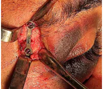
Group A received internal fixation with 0.4–0.6 mm microplates at the zygomaticomaxillary buttress (ZMB) and frontozygomatic (FZ) region, while Group B also included the infraorbital rim. Screw holes were drilled with a guiding sleeve to prevent thermal damage, and monocortical screws were placed. After fixation, the site was irrigated, maxillomandibular fixation released, and the wound closed in two layers with 3-0 Vicryl for deeper tissue and 5-0 Prolene for skin and mucosa.
Intraoperative stability
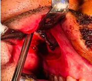
The stability of the repositioned zygomaticomaxillary fracture was assessed intraoperatively by digital manual palpation to decide whether fixation devices were required (Figures 1 and 2).
Figure 1
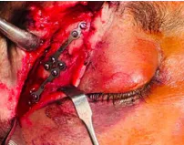
Group A – Intraoperative stability and fixation at fracture site
Figure 2
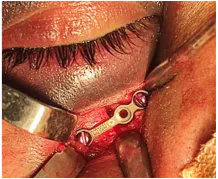
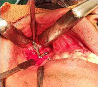
Group B - Intraoperative stability and fixation at fracture site
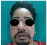
Facial assessment
Assessment of malar asymmetry was performed using frontal and bird’s-eye views.
Assessment of malar asymmetry was carried out in accordance with the Holmes and Mathews classification.
Grade 1: Excellent cosmetic result, no malar asymmetry with any visible scar formation
Grade 2: Good cosmetic result, malar asymmetry on careful inspection with very minimal scar formation
Grade 3: Poor cosmetic result, noticeable malar asymmetry with minimal scar formation
Grade 4: Gross malar asymmetry with noticeable scar formation [Figures https://pmc.ncbi.nlm.nih.gov/articles/PMC8848716/#F3 and https://pmc.ncbi.nlm.nih.gov/articles/PMC8848716/#F4].
Figure 3
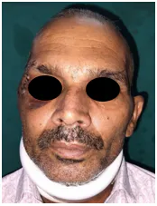

Group A – Facial assessment preoperative and postoperative
Figure 4
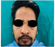
Group B – Facial assessment preoperative and postoperative
Mouth opening
Assessment was carried out using a Vernier calliper preoperatively and at 1-month and 3-months follow-up interval [Figures 5 and 6].
Figure 5
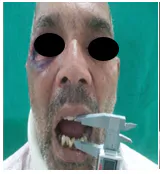


Group A – Interincisal mouth opening preoperative, 1 and 3 months
Figure 6
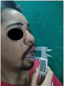
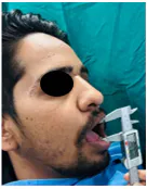
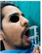
Group B – Interincisal mouth opening preoperative, 1 and 3 months
RESULTS
After completion of the clinical study, measurements and data obtained from all patients were tabulated for statistical analysis, and observational data were derived following comparison of the two groups (Group A and B).
The treatment outcomes of two techniques were evaluated and compared with regard to the following variables:
1. Intra-operative stability
2. Facial assessment
3. Interincisal mouth opening
4. Post-operative complications.
Intra-operative stability
Intra-operative stability was observed in 19 (67.9%) patients in Group A and 23 (82.1%) patients in Group B, with no statistically significant difference between the groups (p = 0.217). Overall, 75% of patients demonstrated intra-operative stability.
Facial assessment
Post-operative facial aesthetic outcomes at 1 week, 4 weeks, and 12 weeks using the Mann–Whitney U test. At the 1-week follow-up, Group A had a higher mean rank (31.52) compared to Group B (25.48). Since a lower mean rank indicates better facial aesthetics, Group B demonstrated superior facial aesthetic outcomes at 1 week, and the difference was statistically significant (p = 0.041).
At the 4-weeks follow-up, the mean ranks of Group A (29.00) and Group B (28.00) were almost identical, indicating that both groups had comparable facial aesthetic outcomes, with no statistically significant difference (p = 0.788).
At the 12-weeks follow-up, Group A again showed a higher mean rank (31.00) compared to Group B (26.00), indicating that Group B demonstrated better facial aesthetic outcomes at 12 weeks, with the difference reaching borderline statistical significance (p = 0.05).
Interincisal mouth opening
Post-operative mouth opening between Group A and Group B at 1 week, 4 weeks, and 12 weeks using the independent t-test. At 1 week, the mean mouth opening was 24.93 ± 3.23 mm in Group A and 26.93 ± 2.49 mm in Group B. The difference was statistically significant (p = 0.012), with Group B demonstrating greater mouth opening.
At 4 weeks, Group A showed a mean mouth opening of 30.96 ± 3.24 mm, while Group B showed 34.93 ± 2.49 mm. This difference was highly statistically significant (p < 0.001), again indicating better mouth opening in Group B.
At 12 weeks, the mean mouth opening increased to 36.11 ± 3.60 mm in Group A and 40.96 ± 2.55 mm in Group B. The inter-group difference remained highly statistically significant (p < 0.001).
Post-operative complications
Post-operative complications at 1 week, 4 weeks, and 12 weeks using the Mann–Whitney U test. At the 1-week follow-up, Group A had a mean rank of 30.84, while Group B had a lower mean rank of 26.16, indicating that Group B experienced fewer and less severe complications, although the difference was not statistically significant (p = 0.237).
At the 4-weeks follow-up, mean ranks were 29.00 for Group A and 28.00 for Group B, showing no meaningful difference in complication severity between the two groups (p = 0.739).
At the 12-weeks follow-up, both groups demonstrated identical mean ranks (28.50), indicating that post-operative complications were comparable between the groups (p = 1.00).
DISCUSSION
The zygomaticomaxillary complex (ZMC) is a critical component of the midfacial skeleton, contributing to facial structure, function, and aesthetic contour¹. ZMC fractures are among the most frequently encountered injuries in maxillofacial trauma, largely due to the prominent and exposed position of the zygomatic region, which increases its susceptibility to facial injury10.
Over the years, multiple management strategies have been described, ranging from closed reduction without fixation to open reduction with internal fixation using different fixation techniques.
In the present study, 56 patients were randomly allocated to two treatment groups: two-point fixation (Group A) and three-point fixation (Group B). Participants were aged 18–50 years.
The mean age was significantly higher in Group A (41.89 ± 11.16 years) compared with Group B (32.25 ± 6.65 years) (p < 0.001). Group A included 8 females and 20 males, whereas Group B comprised 7 females and 21 males. Overall, males accounted for 73.21% of cases, and gender distribution was comparable between the groups (p = 0.763). Similar male predominance has been reported in previous epidemiological studies of facial fractures11 .
Intraoperative stability after reduction was assessed by digital palpation to determine the requirement for fixation. Stability was observed in 19 patients in Group A and 23 patients in Group B, with no significant difference between groups (p = 0.217). Overall, 75% of cases demonstrated intraoperative stability. Improved stability with three-point fixation in complex fractures has been reported by Nasr et al.12.
Facial aesthetic outcomes were evaluated using frontal and bird’s-eye views, and malar asymmetry was graded according to the Holmes and Mathews classification. At 1 week, Group B demonstrated significantly better aesthetic results than Group A (p = 0.041). At 4 weeks, outcomes were comparable between groups (p = 0.788). At 12 weeks, Group B again showed superior results with borderline statistical significance (p = 0.05). Overall, three-point fixation yielded improved early and late aesthetic outcomes.
Maximum mouth opening was measured using a Vernier caliper. Group B demonstrated significantly greater mouth opening at 1 week (p = 0.012), 4 weeks (p < 0.001), and 12 weeks (p < 0.001), indicating superior functional recovery.
Postoperative complications, including pain, swelling, malar flattening, and wound infection, were graded on an ordinal scale. Although Group B showed fewer early complications, the differences were not statistically significant at 1 week (p = 0.237), 4 weeks (p = 0.739), or 12 weeks (p = 1.00). Both groups showed similar complication profiles at later follow-up.
Biomechanical studies suggest that microplates provide stability comparable to miniplates in maintaining fracture reduction and resisting displacement, although their smaller cross-sectional area results in higher stress distribution9. Their use is advantageous in regions with thin soft tissue coverage, such as the infraorbital rim, nasomaxillary region, and frontozygomatic area, where improved palpability and aesthetics are desirable12.
The optimal number of fixation points in ZMC fractures remains debated and depends on fracture severity and displacement. Experimental studies by Davidson et al. demonstrated that three-point fixation at the frontozygomatic suture, infraorbital rim, and zygomaticomaxillary buttress provides maximum resistance to physiological forces. Similar findings were reported in a biophysical study by O’Hara et al.13.
CONCLUSION
The zygoma and maxilla constitute an essential part of the orbital framework and therefore play a significant role in the residual deformities that may persist following fracture management. Three -point microplate fixation demonstrated superior functional and aesthetic outcomes compared with two-point fixation in the management of zygomaticomaxillary complex fractures. Facial aesthetics, evaluated using the Holmes and Mathews grading system, showed improved cosmetic results at early and late follow-up, with better restoration of malar symmetry. Postoperative mouth opening was also significantly greater at all time points, reflecting enhanced functional recovery. Intraoperative stability and complication rates were similar between the groups. These findings are consistent with previous comparative and biomechanical studies indicating greater stability with fixation at three anatomical buttresses. While two-point fixation may be suitable for minimally displaced fractures, three-point fixation is recommended when optimal stability and aesthetic outcomes are desired.