
I. INTRODUCTION
Oral rehabilitation through implants has been growing significantly in recent years and reaching high rates of clinical success. Currently, bone-implant contact is considered predictable, safe, and lasting . The long-term success of dental implants depends on osseointegration . With the advancement of success also comes the problems related to implant dentistry .
Failures can be classified as biological, mechanical or iatrogenic or due to insufficient patient adaptation .
Among these faults, the most dangerous is the biological fault, which can be defined as the impossibility of bone tissue to establish osseointegration . This lack of osseointegration that requires implant removal is considered a biological failure .
Biological faults are classified as primary and secondary. If the failure occurs during the osseointegration process, it is considered a primary failure; if it occurs after charging it is a minor fault . Primary failures result from lack of bone repair, where the connection between the implant surface and the bone does not occur. Instead, fibrous tissue forms between the implant and bone, causing the implant to lose its stability .
Failures can be prevented by appropriate patient selection; proper treatment planning is critical to successful implant dentistry .
Some information has been reported on factors that influence implant osseointegration, such as: biocompatibility, implant design, surface conditions, surgical site, a surgical technique for implant installation, and loads applied to them .
Studies relate early loss in short-length implants in posterior regions where space and volume are insufficient .
To acquire adequate healing conditions, the implant, after insertion, must exhibit good primary stability, which may correspond to the clinical manifestation of osseointegration. Primary stability is primarily determined by factors related to bone biomechanical properties, implant design, and surgical technique; while secondary stability is also determined by bone tissue response to surgical trauma and implant surface . Primary stability is achieved when the implant locks into the apical or marginal portion of the site due a sufficient amount of compact bone or cancellous bone .
The world population has been aging over the years and inevitably, the increase in individuals with low bone density . Several studies have shown a positive correlation between low bone density and implant loss . The primary stability of the implant, as well as its survival, is affected by low bone density . The high rate of implant loss is related to bone type IV when compared to bone type I, II, III .
The aim of this study was to correlate radiomorphometric indices and poor bone quality in osseointegration failure in oral rehabilitation.
II. MATERIAL AND METHOD
The project was submitted to the Research Ethics Committee (CEP) of the Metropolitan University of Santos and approved. It was used the database of the postgraduate course in implantology of the Metropolitan University of Santos, the data of 104 lost implants of various diameters in 74 individuals aged 33 - 84 years of both genders with an average of 59 years, in addition to their images and clinical records. Individuals with a history of hormone replacement therapy or calcium therapy under the age of six months, and those who did not have all the tests necessary for this study were excluded.
The implants lost in the upper and lower arch in the anterior and posterior regions were separated, also grouped by diameter and length.
All digital panoramic radiographs images used in this study are from a partner institute of the Metropolitan University of Santos and performed by the ORTHOPHOS XG 3D PAN/TELE/TOMO device (Sirona Dental Systems GmbH, Bensheim, Germany) following the same protocol of acquisition: The Panoramic Radiography 69 kV, 15 mA, and exposure time 14.1 s.
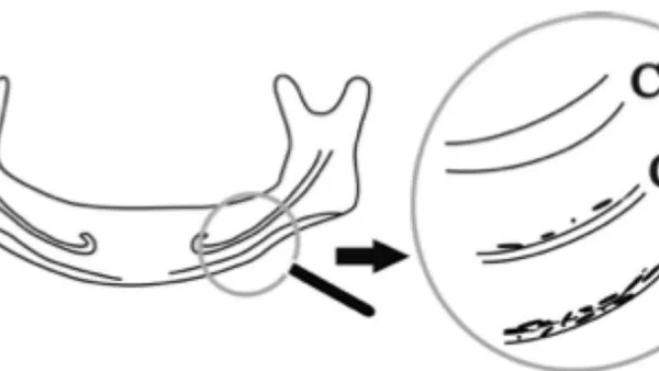
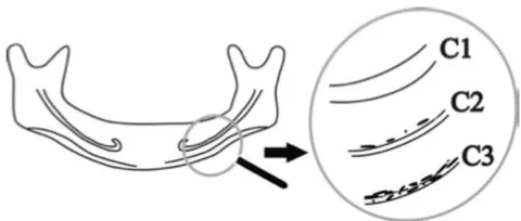
In panoramic radiographs, radiomorphometric indices were evaluated. Among, them the mandibular cortical index (MCI) a bilateral evaluation with results established in: C1 - clear and sharp posterior mandibular cortical, C2 - the endosteal surface presents semilunar defects (lacunar resorption), or the surface presents cortical residues, C3 - a cortical layer is extremely porous (Figure 1).
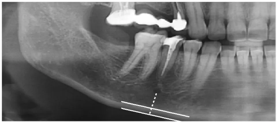
Mental index (MI) bilateral assessment determined by the width of the mandibular cortex, measured on the line perpendicular to the base of the mandible, at the height of the center of the mental foramen (Figure 2).
III. RESULTS
We evaluated data from 104 implants of various diameters in 74 individuals, 48 women and 26 men, aged 33 - 84 years with a mean of 59.4 years. As for the upper and lower arches, 46 implants lost in the lower arch and 58 in the upper arch. We evaluated 76 implants in the posterior region and 28 in the anterior region. Spearman's correlation coefficient test was performed to assess the relationship between the variables of the lost implants (Table 1).
| Age | Sex | MI | MCI | ARCADE | Region | Diameter | Lenght | |
| Age | ||||||||
| Sex | R:0,209*P:0,034 | |||||||
| MI | R:-0,106P:0,282 | R:-0,038P:0,701 | ||||||
| MCI | R:0,198*P:0,044 | R:-0,021P:0,833 | R:-0,721**P:0,000 | |||||
| ARCADE | R:0,214*P:0,029 | R:0,052P:0,597 | R:0,275**P:0,005 | R:-0,249*P:0,011 | ||||
| Region | R:0,116P:0,242 | R:0,158P:0,110 | R:0,054P:0,587 | R:0,035P:0,724 | R:0,235*P:0,016 | |||
| Diameter | R:-0,028P:0,779 | R:-0,128P:0,196 | R:-0,05P:0,616 | R:0,044P:0,655 | R:0,038P:0,700 | R:0,072P:0,467 | ||
| Lenght | R:-0,032P:0,751 | R:-0,040P:0,889 | R:-0,060P:0,547 | R:0,104P:0,292 | R:-,241*P:0,014 | R:-0,339**P:0,000 | R:0,050P:0,615 |
According to the result of the correlation coefficient, it was possible to verify the correlation (-0.721) for p<0.001 inverse relationship between the MI and the MCI. The ratio (0.275) to p<0.001 between MI and the arch where bone quality was evaluated in the upper arch showed worse quality when compared to the lower arch.
It was also correlated (-0.339) to p<0.001 the length of the lost implant and the region, smaller length implants were lost in the posterior region.
Age was correlated (0.198) with the MCI to p<0.05 where the older, the age the worse, the bone quality evaluated.
The MCI correlated with the arch (-0.235) for p<0.05 a worse bone quality when correlated with the implants lost in the upper arch.
IV. DISCUSSION
The use of radiomorphometric indices to assess bone quality is an effective and inexpensive instrument for this detection since the evaluation test is difficult to access and high for the population.
Several studies corroborate our results in the high correlation between radiomorphometric indices evaluated in panoramic radiographs, where these exams may be predictive in helping to assess bone quality .
Some studies contradict these findings showing that there is inconsistency in the data obtained when in smaller groups, although the mandibular cortical index is among the most reproducible radiomorphometric indices .
The influence of age with the loss of bone quality was evidenced in this study. Older individuals had worse bone quality when evaluated by the mandibular cortical index. This result was also found in other studies. Zlataric et al. 2002, verified the values of radiomorphometric indices in elderly individuals and showed that the values of these indices decreased in both sexes up to 78 years . Edgerton et al. 1999 evaluated British women and observed that radiomorphometric indices gradually decreased with increasing age .
When the arches were evaluated, the upper arch showed worse bone quality compared to the lower arch, implants installed in the posterior regions of the upper arch showed a higher loss rate when compared to the other regions.
It was evidenced in this study when the radiomorphometric indices IM and ICM were evaluated and correlated with the length of the implants and the region where the highest concentration of loss occurred in the posterior maxilla and implants of shorter lengths. These data were also evidenced by other studies where the incidence of loss of short posterior maxillary implants was higher when compared, for example, with the mandible .
Contrary to the results obtained in this study, implants installed in the posterior maxilla of short length and diameter presented excellent fixation results .
V. CONCLUSION
According to the results of this study, radiomorphometric indices may be used the preoperative evaluation to assist in the detection of patients with poor bone quality.