## I. INTRODUCTION
Ruminants are suffered from numerous kinds of infections caused by the corynebacteriaceae family. Corynebacteriaceae belong to the Actinomycetales order with many nomenclatural updates in recent years (Markey et al., 2013; Algammal, 2016). This genus belongs to the CMNR group (Corynebacterium, Mycobacterium, Nocardia, and Rhodococcus), a heterogeneous group of pathogenic bacteria (Dworkin et al., 2006) characterized by diverse pyo-or granulomatous clinical infections that affect human and animals (Tauch and Sandbote, 2014; Thiago et al., 2021).
They are intracellular, cocco-bacilli, gram-positive microorganisms, non-capsulated, non-motile, non-sporulating, and facultative anaerobes (Markey et al., 2013; Chandran et al., 2016; Juliana et al., 2019). Under the microscope, they are arranged in the form of palisades or similar to Chinese letters (Markey et al., 2013) or short coryneform rods (Park et al., 2022). Corynebacteria are pyogenic bacteria found externally on the skin, and mucous membranes and hidden internally in the intestinal tract of animals and humans (Bernard, 2012; Feßler and Schwarz, 2017; Park et al., 2022).
Some Corynebacterium species which have been detected in animals, documented to have zoonotic transmission as by the close contact with domestic or wild animals, i.e., during the occupational handling of animals, by animal bites, or by other means. Raw milk or dairy products without pasteurization often used for increase the risk of transmission of zoonotic pathogens as Corynebacteriaceae to humans (Langova et al., 2022) CLA has passive socio-economic effects that are chiefly defined in zoonosis, decrease the hide and market value, reproductive disorders and reduction of wool crop. It leads to dwindling in body weight, in wool and milk production even in the reproductive performance with elevation in the culling rate resulting to death of the infected animals with losses to the farm (Oreiby et al., 2014 and Burmayan and Brundage, 2021).
All ranchers in many countries suffer financial losses due to herd infection with Corynebacteria spp. as they cause numerous forms of pyogenic diseases (Dorella et al., 2006; Abd El Tawab et al., 2019) in addition to emaciation One of them is Corynebacterium (C.) pseudotuberculosis that is the main causative agent for Caseous lymphadenitis in Goat and sheep (Algammal, 2016; Thiago et al., 2021; Sting et al., 2022). 2- Mastitis in Cattle (Markey et al., 2013). 3- Ulcerative lymphangitis and ventral abscess in cattle (Markey et al., 2013; Vikas et al., 2017), Horses, and pigs (Markey et al., 2013). Also Corynebacteria spp. are reclassified into another genus including C. pyogenes as Trueperella (T.) pyogenes (Dworkin et al., 2006; Yassin et al., 2011; Magdalena et al., 2019). Conversely, T. pyogenes could cause a primary pathogen, infection usually follows a physical or microbial trauma that disseminates the organism (Rosenberg et al., 2014). T. pyogenes can cause numerous economically significant suppurative infections involving the skin and visceral organs including mastitis (Quinn et al., 2002; Unnerstad et al.,
2009), pneumonia (Fulton et al., 2009), endometritis (Williams et al., 2005), liver abscess (Dore' et al., 2007), peritonitis and Pleurisy in sheep, goats, and cattle (Rosenberg et al., 2014; Abdallah, 2016). Many authors recorded the affected LNs illustrates marked enlargement with either thick creamy green pus with a central caseated core surrounded by dense fibrous capsule as (Torky et al., 2023).
This study aimed to discuss the conventional and the modern techniques to identify Corynebacteria spp. isolated from ruminants. Then further determination to some virulence factors and sensitivity to some antimicrobial agents would be done.
## II. MATERIAL & AMP; METHODS
### a) Ethical Approval
The research was carried out with the approval and in accordance with the guidelines of the local Ethics Committee at the Faculty of Veterinary Medicine, Benha University, Egypt. All the procedures in the study respect the ethical standards and the protocol was approved by the Ethics Committee with the code BUFVTM 12-10-22.
## i. Samples Collections
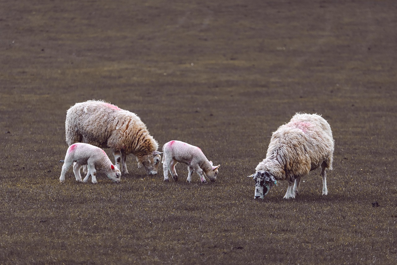
One hundred ninety-seven swabs were taken from diseased farm animals (sheep, goat, cattle) and eighteen lesions from slaughtered one from different farms and slaughtered houses within Gharbiya & Menoufia Governorates from 8, 2021 till 4, 2022 (Table 1) (Fig. 2). Samples were taken as biological swabs from swollen lymph nodes or any superficial abscess and the whole affected lymph nodes from a slaughtered animal. The extraneous surface of the struckd lymph node was cleaned, disinfected by povidone-iodine $7.5\%$ then incised by a sterile blade. After that, the samples were taken from the inner surface and the pyogenic membrane after a gentle squeeze and were taken in a tube with brain heart infusion broth in an ice tank as quickly as possible.
Bacteriological examination according to (Markey et al., 2013, Zasada and Mosiej, 2018).
All collected samples were streaked in triplicate for each sample into plates of brain heart infusion (BHI) agar (Hi-media), $5\%$ fresh heparinized sheep blood agar, and Baird Parker agar. All plates were supplemented by tween 80 by $1\%$ for Lipophilic corynebacteria in addition of utilization of C. pseudotuberculosis (ATCC® $19410^{\text{TM}}$ ) as control positive, then incubated for 24-48 h at $37^{\circ}\text{C}$. Suspected colonies were tested for color, motility, consistency, catalase, oxidase, and stained Gram's stain (EDM) then all colonies with Gram-positive forming cocco-bacilli, negative for oxidase and nitrate, and nonmotile in semi-solid agar were selected, then tested for traditional biochemical tests, and VITEK test.
## ii. Identification of Corynebacterium Species
Identification by vitek 2 compact system (biomerieux, 2006).
A sterile swab was used to transfer many colonies of fresh pure colonies to 3ml sterile saline in a clean plastic test tube. The turbidity should be calibrated by a turbidity meter called VITEK® 2 DensiCHEKTM then insert the ANC card in suspension in less than half an hour. The test tube with the inoculated card was loaded in a vacuum chamber station then the vacuum was applied and the micro-organism suspension was forced into micro-channels. The final data were obtained and automatically printed within 24 hrs.
Identification of some isolates of C. Pseudotuberculosis by PCR acc. to Pacheco et al., 2007).
By using Master Mix and Oligonucleotide primers sequences from Metabion (Germany). forward (ACCGCACTTTAGTGTGTGTG), reverse (TCTCTACG CCGATCTTGTAT) at 816 bp with Cycling conditions: Primary denaturation (94°C 5min) 2nd denaturation (94°C 30sec), Annealing (58°C 40sec), Extension (72°C 50sec), Final extension (72°C 10 min) for 35cycles.
For sequencing reactions, 16Sr RNA PCR product of sample no.1 (540 bp length) was purified using QIAquick Gel Extraction Kit Protocol: (Qiagen Inc. Valencia CA and PCR Clean up sequenced in the forward and reverse directions on an Applied Biosystems 3130 automated DNA Sequencer (ABI, 3130, USA). Using a ready reaction Bigdye Terminator V3.1 cycle sequencing kit. (Perkin-Elmer/ Applied Biosystems, Foster City, CA), with Cat. No.
4336817. according to (Sanger et al. 1977) and the instruction of the manufacturer by Thermo Fisher Scientific, Schwerte, Germany. The sequences were edited and aligned using BioEdit version v7.0.9. Software A BLAST® analysis (Basic Local Alignment Search Tool) (Altschul et al., 1990) was initially performed to establish sequence identity to GenBank accessions.
Detection of some phenotypic virulence factors:
### b) Biofilm Formation
According to (Hassan et al., 2011)
All samples were tested by the tube and the congo red agar method for the detection of biofilm formation. Assessment of biofilm-forming quantitatively by using tube method and according to results lay down by. Bacteria are classified as weak, moderate, and strong biofilm producers. The strong Biofilm formation was detected using the CRA method, color of bacterial colonies was checked as black colonies with dry crystalline consistency indicating a positive result, weak slime producers usually remained pink. A darkening of the colonies with the absence of a dry crystalline colonial morphology indicated an indeterminate result. The test was repeated three times with the reference strain Edwardsiella tarda MW362142 as a positive control for biofilm formation (Abd El-Tawab et al., 2021).
Phenotypic virulence factors of Corynebacteria isolated according to (Yang and Fang, 2003, Markey et al., 2013) The hemolytic; amylase; proteolytic (caseinase); lipolytic and lecithinase activities for Corynebacterium Spp. isolated and CAMP test detection of the synergistic or the reverse hemolytic activity with Staphylococcus aureus ATCC® 6538.
### c) Antimicrobial sensitivity test by Determination of minimum inhibitory
Concentration (MIC) method according to CLSI (2006) and The European Committee on Antimicrobial Susceptibility Testing (2019). ZnO-NPs and Egyptian propolis were obtained from the animal health research institute, El Doki, Egypt, and their concentrations were according to (Hegazi and Abd El Hady, 2002). ZnO-NPs was with a particle size of $371\mathrm{nm}$ with1.137ODwave length in nm and its cytotoxicity was IC50 $100\mu \mathrm{g} / \mathrm{ml}$ on monkey kidney cell line assessed by SRB assay according to (Skehan et al., 1990). The MICs of antibiotics, Egyptian propolis, and ZnO-NPs against Corynebacterium isolates were determined by the broth micro-dilution method. The concentrations used in the MIC test were as follows; ZnO-NPs (100 to 10 ug/ml); Egyptian propolis (10mg/ml to 1,25mg/ml), Oxytetracycline (4 to 16ug/ml), pencilin G (.25 to 8ug), levofloxacin (.25- 8ug/ml).
The MIC of selected Antibiotics was defined as the lowest concentration of antibiotic that inhibits bacterial growth and no visible growth is observed as compared with both a positive control (culture broth containing bacteria only that should appear turbid) and a negative control (culture broth without bacteria that should remain clear). In addition, the half maximal inhibitory concentration (IC50) of ZnO-NPs against Corynebacterium isolates was determined. The Experiment was performed in triplicate and quantitative assessment of biofilm formation on conge red agar.
## III. RESULTS
### a) Detection of Corynebacterium Species
Positive suspected corynebacterium species isolated were 27 from 215 samples with specific greenish colored pus (fig.3) as 19 out of 144 sheep, 5 out of 61 goats, and 3 out of 10 cattle by $12.56\%$ of the total number.
Distribution of Corynebacterium species isolated from abscess locations as table (2).
Out of 19 strucked sheep, the distribution of C. pseudotuberculosis was 9 males (5 of them were old and 4 were from dead animals), 1 parotid lymph node $(11.1\%)$, 1 fatty tail $(11.1\%)$ and 3 pre-scapular lymph node $(33.3\%)$ and 10 females (5 of them were old and 5 were young) as 2 sub-mandibular lymph nodes $(20\%)$, 2 parotid lymph node $(20\%)$ and 2 pre-scapular lymph node (20%),1 retro-pharyngeal lymph node (10%), 2 pre-femoral lymph node (20%) and 1 in the fatty tail (10%). However, the distribution of C. pseudotuberculosis in 3 strucked goats, was 2 males (1 young and the other was old) by 1 in pre-femoral lymph node (50%) and 1 in sub-mandibular lymph nodes (50%) whilst 1 young female by 1 retro-pharyngeal lymph node (100%). We found T. pyogenes in cattle in 3 cows with abdominal abscess (100%) while we found it in two lactating does as non-specific infection in pre-scapular lymph node (100%). These results revealed many points: Sheep was the most affected species mainly after manual shearing by scissors in fixed breeding. The main affected L.N in sheep was in the head region- due to fighting- despite it having fewer affections in goats and being absent in cattle.
### b) Cultural and Biochemical Characteristics of Isolates
All Corynebacterium species isolated were Gram-positive non (sporulated-capsulated-motile) cocco-bacilli to short-chain bacilli which can appear in pairs or single arrangements. The most characteristic club shape was in form of acute angles or Chinese-like appearance as in fig. 4. After $48\mathrm{h}$ of incubation, their colonies appear as minute, white, smooth, dry colonies on BHI agar surrounded by a thin zone of $\beta$ hemolysis on $5\%$ fresh heparinized sheep blood agar. The colonies were waxy and splashing with flame with hard collecting as they were swaying on the agar surface. On Egg-yolk Tellurite or Baird Parker agar, the colonies were small, dark grey, and opaque in appearance after 24-48 h (fig. 5). All tested isolates were non-motile, negative for oxidase, and nitrate reduction tests whilst 22 isolates were positive for Urease and catalase tests and 5 isolates were negative for them.
Identification of Corynebacterium species by vitek 2 compact system. As in fig. 6 & amp; 7 C. pseudotuberculosis was positive for esculin, urease, catalase, Ellman and Beta-D-Fucosidase whilst T. pyogenes was negative for all of them.
## i. The result of 16Sr RNA of C. pseudotuberculosis
Out of identified C. pseudotuberculosis, 8 selected isolates were tested for 16Sr RNA and all were positive at 816 bp as in (fig. 8). The sequence of 16Sr RNA has accession number ON899860 partial sequence and its length 539 BP (780 BP), has similarity with 31 accession number in gene bank with $99.49\%$ as in (fig. 9).
Fig. 8: Agarose gel electrophoresis of PCR for amplification products of The 16S ribosomal RNA gene (16S rRNA) for 8 C. pseudotuberculosis isolates. All Lanes show positive amplification of 16S rRNA gene at 816 bp. Lane L: DNA ladder at 100-1000bp. N.: Negative control (sterile DNase/ RNase free DEPC water). P.: Positive control (C. pseudotuberculosis (ATCC® 19410 TM).
## ii. The Results of Phenotypic Virulence Factors
All isolates had hemolytic, lipolytic, proteolytic (caseinase), and Lecithinase activities. The CAMP test justifies that C. pseudotuberculosis inhibits the staphylococcal beta-hemolysin reaction (reverse CAMP test) whilst T. pyogenes enhances it (pos. CAMP-like reaction). Addition to $90\%$ of isolates produces biofilm as shown in Tables (3, 4) and fig.10 & amp;11.
c) The Result of Antimicrobial Sensitivity Tests
All isolates were resistant to all tested kinds of anti-microbial except levofloxacin at $8\mu \mathrm{g} / \mathrm{ml}$. But nano zinc oxide $100\mu \mathrm{g} / \mathrm{ml}$ and propolis extraction at $10\mathrm{mg} / \mathrm{ml}$ can eradicate the biofilm formation of C. pseudotuberculosis (fig 12.).
## IV. DISCUSSION
The pyogenic bacteria are ferocious and annoying bacteria for animals and ranchers. Not also it affects animal health, but also causes financial losses for the ranchers. One of them is C. pseudotuberculosis which causes Caseous lymphadenitis (CLA) in sheep and goats. CLA is a zoonotic, contagious, and chronic bacterial disease which causes granulomas in lymph nodes (superficial and visceral) extending to several internal organs [Banke et al., 2021; Burmayan and Brundage, 2021; Santos et al., 2021].
Another one is T. pyogenes which causes various suppurative infections in domestic animals as mastitis, abscesses, pneumonia, and lymphadenitis as in (Ribeiro et al., 2015; Rogovskyy et al., 2018). In this research, the prevalence of C. pseudotuberculosis isolated from alive and dead animals was in a congenial ratio with the result reported by (Oreiby et al., 2015; Abd El Tawab et al., 2019, and Torky et al., 2023). Conversely, it is a minor ratio compared with the one reported by (Al-Gaabary et al., 2009). In in-contrast that it is a major ratio reported in (Al-Gaabary et al., 2013;
Al-Gaabary et al., 2015). The prevalence of T. pyogenes was reported in cattle and goats as reported in (Abdallah, 2016; Rogovskyy et al., 2018) with different ratios. Accurate identification of CLA relies on PCR detection methods such as 16S rRNA gene as reported in Public Health England (2014) and Algammal, 2016 that was present in all tested isolates. The molecular genetic sequence analysis of 16S rRNA gene has facilitated a much tighter circumscription of the genus Corynebacterium. The availability of comparative 16Sr RNA gene sequence improved phenotypic data has resulted in much improved and more reliable species identification Public Health England (2014).
The result of MIC test showed the sensitive of C. pseudotuberculosis to levafloxacin and resistance to pencilline which agree with Torky et al., 2023 and disagree with Markey et al., 2013 recorded its sensitivity to penicillin. The propolis and ZnO-NPs exhibited resistance from tested C. pseudotuberculosis but they have a good anti-biofilm activity against it as reported by (Santos et al., 2021 and Abdelghafar et al., 2022). That may be due to their unreasonable use of them during the rearing period. Subsequently, it was a necessity to detection of their ability for biofilm formation as there is a close connection between them. From this study, most infections occur in fixed and mixed breeding, especially after the shearing season as reported by (Oreiby et al., 2014). The sensitivity and specificity for biofilm formation were much more accurate by the tube method than CRA which was confirmed by (Hassan et al., 2011).
## V. CONCLUSION
Corynebacteria cause obstinate infections with unidentified rules for treatment. CLA leads to a dwindling in body weight, wool, and milk production even in the reproductive performance with an elevation in the culling rate resulting in losses to the farm. Further modern researches should be done to know how to handle this microorganism.
### ACKNOWLEDGEMENT
My Sincerest gratitude to Prof. Dr. Magdy Al-Gaabary, Department of Animal Medicine, Faculty of Veterinary Medicine at Kafer El-Shiekh University for all his efforts and his parenteral advice. I appreciate everything you've done.
Conflict of Interest
The authors declared no conflict of interest.
Generating HTML Viewer...
References
48 Cites in Article
Abd El Tawab,A Rizk,A Afifi,S Mohamed,S (2019). Corynebacterium Pseudotube-rculosis infection in small ruminant and molecular study of virulence and resistance genes in Beni-Suef governorate.
Abd El-Tawab,A Rizk,A Selim,A Elwakil,R (2021). Biofilm of Edwardsiella tarda isolated from fresh water fishes and its role in the bacterial Virulence.
S Abdallah (2016). Phenotypic and genotypic characteristics and epidemiological relation of Trueperella pyogenes isolated from various origins.
A Abdelghafar,N Yousef,M Askoura (2022). Zinc oxide nanoparticles reduce bioflm formation, synergize antibiotics action and attenuate Staphylococcus aureus virulence in host; an important message to clinicians.
M Al-Gaabary,Y Hegazy,D Gerges (2015). Epidemiological and preventive studies on caseous lymphadenitis in sheep and goats.
Magdy Al-Gaabary,Salama Osman,Atef Oreiby (2009). Caseous lymphadenitis in sheep and goats: Clinical, epidemiological and preventive studies.
M Al-Gaabary,S Osman,Y Ghanem,A Oreiby (2013). Studies on Corynebacterium infection in Ruminants.
M Algammal (2016). Molecular Characterization and Antibiotic Susceptibility of Corynebacterium pseudotuberculosis Isolated from Sheep and Goats Suffering from Caseous Lymphadenitis.
Stephen Altschul,Warren Gish,Webb Miller,Eugene Myers,David Lipman (1990). Basic local alignment search tool.
I Banke,A Abdul Kadir,F Firdaus,A Jesse,S Ramanoon,M Abdul Basit,M & Zakaria (2021). Antibiofilm Activity of Oxytetracycline Loaded 11. Calcium Carbonate Aragonite Loaded Nanoparticle against Corynebacterium pseudotuberculosis.
Kathryn Bernard (2012). The Genus Corynebacterium and Other Medically Relevant Coryneform-Like Bacteria.
W Brown (2006). National Committee for Clinical Laboratory Standards agar dilution susceptibility testing of anaerobic gram-negative bacteria.
A Burmayan,C Brundage (2021). Caseous lymphadenitis outbreak in a small ruminant herd.
F Chandran,D Puthukkichal,E Suman,S & Mangalore (2016). Diphtheroids-Important Nosocomial Pathogens.
(2006). An Overview of the Clinical and Laboratory Standards Institute (CLSI) and Its Impact on Antimicrobial Susceptibility Tests.
E Dore´,G Fecteau,P He´lie,D Francoz (2007). Liver abscesses in Holstein dairy cattle: 18 cases (1992-2003).
F Dorella,L Pacheco,S Oliveira,A Miyoshi,V Azevedo (2006). Corynebacterium pseudotuberculosis: micribiology, biochemical properties, pathogenesis and molecular studies of virulence.
M Dworkin (2006). The Prokaryotes & quot;Archaea. Bacteria: Firmicutes, Actinomycetes & quot.
Andrea Feßler,Stefan Schwarz (2017). Antimicrobial Resistance in<i>Corynebacterium</i>spp.,<i>Arcanobacterium</i>spp., and<i>Trueperella pyogenes</i>.
Robert Fulton,K Blood,Roger Panciera,Mark Payton,Julia Ridpath,Anthony Confer,Jeremiah Saliki,Lurinda Burge,Ronald Welsh,Bill Johnson,Amy Reck (2009). Lung Pathology and Infectious Agents in Fatal Feedlot Pneumonias and Relationship with Mortality, Disease Onset, and Treatments.
A Hassan,J Usman,F Kaleem,M Omair,A Kkalid,M Iqbal (2011). Unknown Title.
Afreenish Hassan,Javaid Usman,Fatima Kaleem,Maria Omair,Ali Khalid,Muhammad Iqbal (2011). Evaluation of different detection methods of biofilm formation in the clinical isolates.
A Hegazia,Abd Hady,F (2002). Egyptian Propolis: 3. Antioxidant, Antimicrobial Activities and Chemical Composition of Propolis from Reclaimed.
N Juliana,S Cassius,V Yuri,C Eliane,F João,V Paulo,H Sérgio,M Lilian,H Raphael,M Ana Luíza,V Verônica (2019). Blood stream and catheterrelated infections due to different clones of multidrug-resistant and biofilm producer Corynebacterium striatum.
Denisa Langova,Iva Slana,Jana Okunkova,Monika Moravkova,Martina Florianova,Jirina Markova (2022). First Evidence of the Presence of the Causative Agent of Caseous Lymphadenitis—Corynebacterium pseudotuberculosis in Dairy Products Produced from the Milk of Small Ruminants.
Denisa Langova,Iva Slana,Jana Okunkova,Monika Moravkova,Martina Florianova,Jirina Markova (2022). First Evidence of the Presence of the Causative Agent of Caseous Lymphadenitis—Corynebacterium pseudotuberculosis in Dairy Products Produced from the Milk of Small Ruminants.
R Magdalena,K Ewelina,C Dorota,K Magdalena,S Ilona,G Małgorzata (2019). Pathogenicity and Virulence of Trueperella pyogenes.
B Markey,F Leonard,M Archambault,A Cullinane,D Maguire (2013). Chapter 6. The D&D of R&D.
Atef Oreiby,Salama Osman,Yamen, Hegazy,Yasser Ghanem,Magdy Al-Gaabary (2015). CASEOUS LYMPHADENITIS IN SMALL RUMINANTS: DESCRIPTIVE, EPIDEMIOLOGICAL AND CLINICAL STUDIES.
A Oreiby,Y Hegazy,Y Ghanem,M Al-Gaabary,S Osman (2014). Caseous lymphadenitis in small ruminants in Egypt.
Luis Pacheco,Roberta Pena,Thiago Castro,Fernanda Dorella,Robson Bahia,Renato Carminati,Marcílio Frota,Sérgio Oliveira,Roberto Meyer,Francisco Alves,Anderson Miyoshi,Vasco Azevedo (2007). Multiplex PCR assay for identification of Corynebacterium pseudotuberculosis from pure cultures and for rapid detection of this pathogen in clinical samples.
Sehee Park,Hijo Shin,Sangwoon Kim,Teakchang Lee,Haejin Lee,Kihoan Nam,Wonkee Yoon,Hyoungchin Kim,Youngwon Seo,Youngsuk Won,Hyojung Kwon (2022). Distribution of Corynebacterium Species and Comparative Results of Diagnostic Methods for Identifying Corynebacterium in Experimental Mice in Korea.
Timothy Rawson,Nita Fatania,Alireza Abdolrasouli (2014). UK standards for microbiology investigations of ear infection (SMI B1) are inadequate for the recovery of fungal pathogens and laboratory diagnosis of otomycosis: A real‐life prospective evaluation.
A Quinn,J Vermunt,D Twiss (2002). Arcanobacterium pyogenes mastitis in a18month-old heifer.
M Ribeiro,R Risseti,C Bolaños,K Caffaro,A De Morais,G Lara,T Zamprogna,A Paes,F Listoni,M Franco (2015). <i>Trueperella pyogenes</i>multispecies infections in domestic animals: a retrospective study of 144 cases (2002 to 2012).
Artem Rogovskyy,Sara Lawhon,Kathryn Kuczmanski,David Gillis,Jing Wu,Helen Hurley,Yuliya Rogovska,Kranti Konganti,Ching-Yuan Yang,Kay Duncan (2018). Phenotypic and genotypic characteristics of <i>Trueperella pyogenes</i> isolated from ruminants.
E Rosenberg (2014). The Prokaryotes.
F Sanger,S Nicklen,A Coulson (1977). DNA sequencing with chain-terminating inhibitors.
Laerte Santos,Daniela Rodrigues,Maurício Kalil,Vasco Azevedo,Roberto Meyer,Marcelo Umsza-Guez,Bruna Machado,Nubia Seyffert,Ricardo Portela (2021). Activity of Ethanolic and Supercritical Propolis Extracts in Corynebacterium pseudotuberculosis and Its Associated Biofilm.
P Skehan,R Storeng,D Scudiero,A Monks,J Mcmahon,D Vistica,J Warren,H Bokesch,S Kenney,M Boyd (1990). New Colorimetric Cytotoxicity Assay for Anticancer-Drug Screening.
A Tauch,J & Sandbote (2014). The family Corynebacteriaceae.
(2014). Applying Pharmacodynamics for Susceptibility Breakpoint Selection and Susceptibility Testing.
Z Thiago,R Dayana,A Vasco,H Gustavo,G Rodrigo,C Rodrigo,K Amanda,J Geraldo,J Fernando,A Lorrayne De Souza,V Aristeu,V Fábio,M André Da Rocha,A Carolina,O Beatriz,G Márcio (2021). Bacteriological, cytological and molecular investigation of Corynebacterium pseudotuberculosis, mycobacteria and other bacteria in caseous lymphadenitis and healthy lymph nodes of slaughtered sheep.
Helmy Torky,Hebatallah Saad,Samy Khaliel,Asmaa Kassih,Jean-Marc Sabatier,Gaber El-Saber Batiha,Helal Hetta,Eman Elghazaly,Michel De Waard (2023). Isolation and Molecular Characterization of Corynebacterium pseudotuberculosis: Association with Proinflammatory Cytokines in Caseous Lymphadenitis Pyogranulomas.
H Unnerstad,A Lindberg,K Waller,T Ekman,K Artursson,M Nilsson-O¨st,B & Bengtsson (2009). Microbial aetiology of acute clinical mastitis and agent specific risk factors.
J Vikas,V Harshit,R Shriya (2017). Ulcerative Lymphangitis in Sahiwal cattle.
Erin Williams,Deborah Fischer,Dirk Pfeiffer,Gary England,David Noakes,Hilary Dobson,I Sheldon (2005). Clinical evaluation of postpartum vaginal mucus reflects uterine bacterial infection and the immune response in cattle.
No ethics committee approval was required for this article type.
Data Availability
Not applicable for this article.
How to Cite This Article
Amany Omar Selim. 2026. \u201cDetected some virulence factors of Corynebacteria isolated from ruminants\u201d. Global Journal of Medical Research - G: Veterinary Science & Medicine GJMR-G Volume 23 (GJMR Volume 23 Issue G2): .
Explore published articles in an immersive Augmented Reality environment. Our platform converts research papers into interactive 3D books, allowing readers to view and interact with content using AR and VR compatible devices.
Your published article is automatically converted into a realistic 3D book. Flip through pages and read research papers in a more engaging and interactive format.
Our website is actively being updated, and changes may occur frequently. Please clear your browser cache if needed. For feedback or error reporting, please email [email protected]
Thank you for connecting with us. We will respond to you shortly.