Introduction
One of the most important stages in root canal treatment is the preparation of the root canal system which includes the removal of vital and necrotic tissue, in addition to thorough debridement of the root canal. However, this stage is difficult due to the complex anatomy of the root canal system. [1]
When the root canal system becomes infected, microorganisms can penetrate deep into the dentinal tubules, where they may persist and eventually lead to reinfection of the root canal system. [2]
The removal of the smear layer is essential because its presence hinders the penetration of disinfectants and sealers into the dentinal tubules, thereby compromising the adaptability and sealing ability of the root canal filling. [3]
Irrigants are essential for achieving successful debridement of the root canal system. The application of a chelating agent such as 17% EDTA can significantly enhance the overall effectiveness of the procedure by facilitating the removal of the smear layer and improving canal cleanliness.
Every available instrumentation system produces a smear layer and debris on the root canal walls. Therefore, it is important to develop and utilize techniques and file systems that minimize smear layer formation and leave the canal wall as clean as possible. [4]
The newer irrigant Qmix, which consists of a Biguanide, a polyaminocarboxylic acid, saline, and a surfactant, was used in this study. It has been shown to be more effective than Biopure MTAD in eliminating bacterial biofilms. However, removal of debris in the apical third remains less predictable.
Therefore, a polymerise canal brush was used to facilitate the penetration of the surfactant into the apical third and enhance smear layer removal. [5]
The newly developed file system ProTaper NEXT (Dentsply Maillefer, Ballaigues, Switzerland) manufactured from M-wire technology and Wave One (Dentsply Maillefer, Ballaigues, Switzerland) which was used in a reciprocating manner and incorporates the M-wire technology is used in this study for comparison. [6]
Thus, the purpose of this in vitro study was to compare the efficacy of QMix with and without canal brush, in smear layer and debris removal from root canal instrumented with newer root canal instrument systems, ProTaper Next and Wave One using Scanning Electron Microscope (SEM).
Materials & Methods
Sample preparation
Forty human single rooted extracted human teeth were used in this study. Standard endodontic access cavities were prepared, and canal patency was established by advancing a file until the tip was just visible at the apical foramen.
S.D. from previous studies [2].
Effect size: .
error: .
Power: .
Total sample size: .
The teeth were randomly divided into 4 groups as follows:
Group 1: ProTaper Next file (Dentsply).
Group 2: ProTaper Next file + Rotary canal brush. Canals instrumented with ProTaper Next rotary system and finished with rotary canal brush (Germany, Whaledent Gmbh KG, Langenau).
Group 3: Wave One file.
Group 4: Wave One file + Rotary canal brush. Canals instrumented with Wave One file and finished with rotary canal brush.
ProTaper Next files were used according to manufacturer’s instruction in recommended sequence up to X4 (40/06) till the working length using X-SMART rotary system. For the Wave One group, canals were enlarged using rotary path files No 1 (purple, ISO tip size 13), No 2 (white, ISO tip size 16) and No 3 (yellow, ISO tip size 19). They have 2% taper used in rotary motion. A large reciprocating Wave One file (40/08) was used in reciprocating motion according to manufacturer’s instructions.
A size #15 K-type file was used to determine working length. In groups 1 and 2, the canals were instrumented with ProTaper Next file (25/0.06 taper). The canals in Group 2 alone were further finished with rotary canal brush. In groups 3 and 4, the canals were instrumented with Wave One file (40/08). The canals in Group 4 alone were further finished with rotary canal brush. Irrigation was carried out using 2 ml of 5.25% Sodium hypochlorite solution between the instruments while 17% Ethylene diamine tetraacetic acid (10 ml) was used as the final rinse. After instrumentation, QMix was used as a final irrigant for 60 seconds in all groups using a 27-gauge needle. The needle tip was placed not more than 2 mm from the apex.
For groups 2 and 4, a rotary canal brush on a slow speed handpiece (600 rpm) was advanced 2 mm short of working length for 30 seconds along with QMix. Final flush with 5 ml distilled water and drying with paper points was performed for all specimens.
Following completion of the shaping and cleaning procedure, to facilitate splitting of the roots, two longitudinal grooves were prepared on the buccal and lingual surfaces with a diamond disk. Roots were split into two halves with a chisel.
The half that contained the most visible part of the apex was conserved and coded. The specimens were dried and examined under SEM.
The dentinal wall of each tooth was observed under the SEM (Phenom XL) in the region of coronal, middle, and apical thirds at a magnification of for the evaluation of debris. Further, magnification was used for the evaluation of smear layer on root canal walls based on the criteria of Schäfer and Lohmann [7].
| Score | Observation |
|---|---|
| 1 | Clean canal wall, few debris particles |
| 2 | Few small agglomerations |
| 3 | Many agglomerations, less than 50% of canal walls covered |
| 4 | More than 50% of the canal walls covered with debris |
| 5 | Complete covering of canal walls by debris |
Qualitative assessment of the debris layer on root canal wall.
| Score | Observation |
|---|---|
| 1 | No smear layer, orifice of the dentinal tubules open |
| 2 | Small amount of smear layer, some open dentinal tubules |
| 3 | Homogeneous smear layer along the entire canal wall, only few open dentinal tubules |
| 4 | The entire root canal covered with homogeneous smear layer, with no open dentinal tubules |
| 5 | A thick homogenous smear layer covering the entire canal wall |
Quality assessment of the smear layer on root canal wall.
Statistical analysis
The results of the present study were analysed using Kruskal-Wallis test and Mann-Whitney U-test. was considered to be statistically significant.
Results
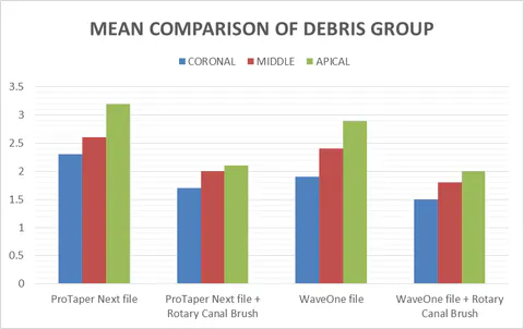
In the present study, when the canals were instrumented with ProTaper Next and Wave One (Group 1 and Group 3, respectively), ProTaper Next showed significantly lower () debris and smear layer scores at coronal, middle, and apical thirds of the canals [Fig. 1, 2 and Table 3].
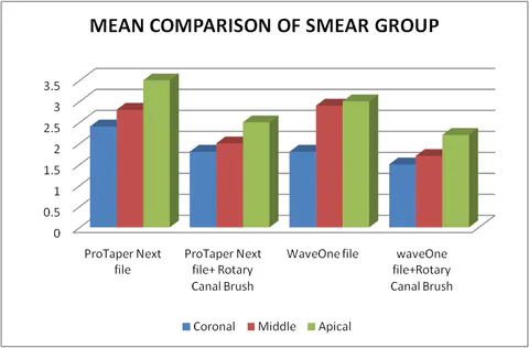
Comparison of debris scores between Group 1 and Group 3.
Comparison of smear layer scores between Group 1 and Group 3.
| Parameter | ProTaper Next | Wave One |
|---|---|---|
| Debris score | ||
| Coronal | ||
| Middle | ||
| Apical | ||
| Smear layer | ||
| Coronal | ||
| Middle | ||
| Apical |
Mean and standard deviation of Groups 1 and 3 at coronal, middle, and apical thirds of the canals for debris and smear layer scores.
Mean values represented with different superscript upper letters or lower-case letters are significantly different ().
When the canals were instrumented with ProTaper Next and Wave One, along with QMix (Group 2 and 4 respectively), ProTaper Next + QMix showed significantly lower () debris and smear layer scores at coronal, middle, and apical thirds of the canals [Fig. 1, 2 and Table 4].
| Parameter | ProTaper Next with brush | Wave One with brush |
|---|---|---|
| Debris score | ||
| Coronal | ||
| Middle | ||
| Apical | ||
| Smear layer | ||
| Coronal | ||
| Middle | ||
| Apical |
Mean and standard deviation of Groups 2 and 4 at coronal, middle, and apical thirds of the canals for debris and smear layer scores.
Mean values represented with different superscript upper case letters or lower letters are significantly different ().
SEM images of ProTaper Next and Wave One without brush, showing smear layer and debris layer.
SEM images of ProTaper Next with brush, showing smear layer and debris layer removal.
Discussion
In general, the results showed that, regardless of the final irrigation regimen used, the canal walls were cleaner in specimens prepared with Wave One than those prepared with the ProTaper Next. This may be attributed to the fact that the size 25 Wave One file has an 8% taper and the larger size 40 Wave One file has a large fixed taper of 8% from D1 to D3, whereas from D4 to D16 they have a unique progressively decreasing tapered design [8].
The larger apical taper of Wave One allows effective shaping contact, which will create a continuous, smooth, wide, and funnel-shaped canal. This allows better penetration of needle and irrigating solutions, and this shape will also allow more removal of debris and smear layer during flushing. This was in agreement with the study of Usman et al. [9] that showed an increased efficacy of canal debridement with increased apical size preparations and an increased taper of instruments. It has been found that the reciprocating motion leads to a more centred preparation when compared with continuous rotating motion.
All endodontic instruments create debris and smear layer as a consequence of their action on canal walls. [10, 11, 12] In this study, SEM evaluation revealed that partially uninstrumented areas with remaining debris and smear layer were found in all sections of all groups, which is consistent with other studies [13]. Results showed that regardless of the instruments and final irrigation regimen used, the coronal and middle thirds were significantly cleaner when compared with the apical third. This may be attributed to the larger diameter in these regions, exposing dentin to a higher volume of irrigants and making smear layer removal more effective, in agreement with results of various studies [14].
Regardless of instruments used, agitation of the QMix with rotary canal brush increased the efficacy of debris and smear layer removal. In the current study, irrespective of the instrumentation system used, the coronal thirds of the root canal showed more cleanliness when compared to the middle and apical thirds of the canals [15]. In both the instrumentation groups, the apical thirds of the canals were observed to have less cleanliness when compared to the middle and coronal parts. The ProTaper Next file system showed significantly lower debris and smear layer scores when compared to Wave One instruments.
The above-mentioned findings were in accordance with previous reports [16, 17] that also showed ProTaper Next to be superior in cleaning efficacy. The cleaning efficacy of ProTaper Next could be attributed to its offset design. The swaggering effect helps in lesser file engagement, more cross-sectional space for improved cutting, loading and auguring debris [18].
The Wave One had significantly higher debris and smear layer scores at all the three levels of the canal. The irregular geometry of the file design could be responsible for creating a non-uniform shape of root canals, which in turn could have prevented the flushing of debris and smear layer adequately [19].
Groups 2 and 4, which used ProTaper Next and Wave One systems along with QMix, showed significant reduction in debris and smear layer scores. The shape memory principles of the NiTi alloy and its inherent metallurgy contributed to the efficacy of the QMix finisher. In the cooled state, the file remained straight in its martensitic phase. The shape memory property of the NiTi alloy allowed the file to take on the austenitic phase and convert to a sickle shape when subjected to the body temperature (in the canal), thereby increasing its cleaning and contact areas during the rotation mode [20, 21, 22].
The continuous rotary motion results in an increased enlargement on the external side of the canal, especially in the apical third, leaving the inner curvature relatively untouched. Hence, it can be concluded that reciprocating motion results in superior cleaning efficacy [6].
Conclusion
The results of this study seem to point out that QMix can be considered as a necessary adjuvant with all contemporary rotary systems to significantly increase the cleaning efficacy of the root canals.
Within the limitations of this study, it is concluded that following biomechanical preparation, smear layer and debris are bound to form in the root canals. ProTaper Next and Wave One rotary files with canal brush produced cleaner canal walls than without canal brush, and Wave One rotary file gave superior results compared to ProTaper Next in removal of smear layer and debris. Further investigation is required on the efficacy of this introduced instrument.