I. INTRODUCTION
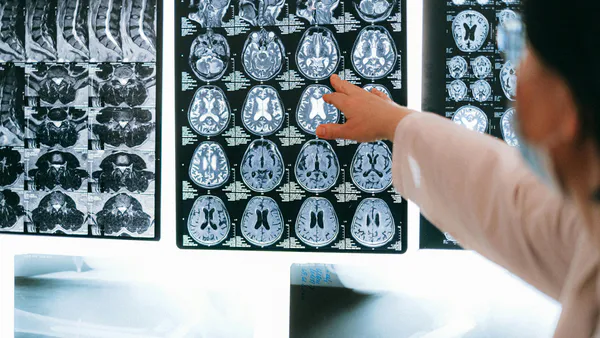
Scientific and diagnostic imaging is medical imaging used to create images of parts or the entire human/animal body for various clinical purposes, such as: B. Medical procedures and diagnostics or medicine, including the study of normal anatomy and function. Medical imaging in a broader sense is the subset of biological imaging that includes photography, microscopy, ultrasound, radiography, computed tomography (CT), magnetic resonance imaging (MRI), and positron emission tomography (PET) included. Detailed anatomical and physiological images of various Medical organs and tissues of the body for research, diagnostic and therapeutic purposes are used.
This post provides a detailed overview of medical imaging to outline past, present, and future aspects of the field.
II. METHODOLOGY
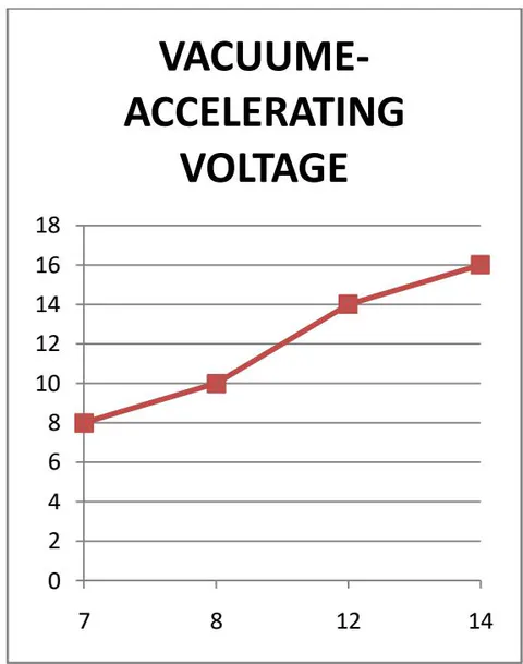
Acceleration Voltage is determined from 8 KV to 16 KV at different vacuum conditions for Advanced Materials
| Sl.No | Vacuume (Kv) | Accelerating Voltage (Kv) |
| 1 | 7 | 8 |
| 2 | 8 | 10 |
| 3 | 12 | 14 |
| 4 | 14 | 16 |
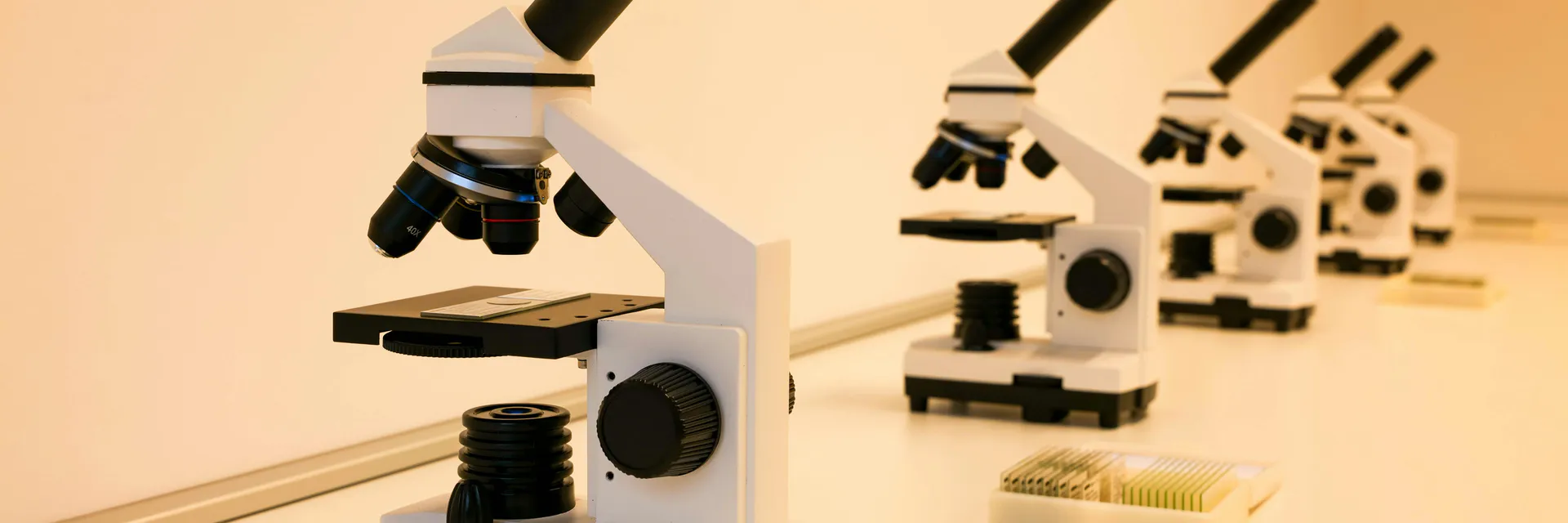
The overall structure of an electron microscope is similar to that of an optical microscope.
| Sl. No | Vacuume (Kv) | Accelerating Voltage (Kv) |
| 1 | 7 | 8 |
| 2 | 8 | 10 |
| 3 | 12 | 14 |
| 4 | 14 | 16 |
Light is replaced by electrons, and glass lenses are replaced by electromagnetic and electrostatic lenses.
Electron microscopes have an electron optical lens system similar to the glass lens in an optical microscope.
Mainly he has two types of electron microscopes. Between Transmission Electron Microscopy (TEM) and Scanning Electron Microscopy (SEM), TEM is the most commonly used. Electron microscopy allows the visualization of structures that are normally invisible to light microscopy. Electron microscopy can be used to visualize microorganisms, cells, large molecules, biopsy specimens, metals, and crystals. Modern electron microscopes use special digital cameras and frame grabbers to take electron micrographs and capture images.
Transmission electron microscopy is a technique developed to obtain much greater magnification, or detail, of specimens than conventional light microscopy. At the specimen it passes through, an image is formed from the interaction of electrons transferred through the sample. The image is magnified and focused onto an imaging device such as a fluorescent screen, a sheet of photographic film, or a sensor such as a CCD camera., which is roughly analogous to a biological optical microscope.
TEM consists of an illumination system, a sample stage, an imaging system, and a vacuum system. Transmission electron microscopy is an important analytical method in physical, chemical and biological sciences.
TEM has applications in cancer research, virology, materials science, pollution, nanotechnology, and semiconductor research.
The seven most commonly used medical imaging modalities today are X-ray (that is, conventional radiography), CT, PET, SPECT, OI, US, and MRI. The first two (X-ray and CT) use high-energy photons to create 2D or 3D image sets of biological anatomy.
In contrast, nuclear medicine procedures such as PET and SPECT use small amounts of radiotracers involved in metabolic and signaling pathways in vivo and their distribution and abundance can be measured by the emitted radiation.
Finally, MRI, OI, and US use non-ionizing radiation for diagnostic purposes. H. Mechanical waves (US) in the MHz range, optical (OI), or magnetic fields (MRI) oscillating in the MHz range.
In general, most of the imaging modalities mentioned above, as well as therapeutic approaches, use electromagnetic radiation over a wide range of frequencies or energies. These parameters partially determine penetration depth, spatial resolution, specific absorption rate, etc. This affects the sensitivity and specificity of the medical imaging modalities used.
III. CONCLUSION
Optical imaging (OI) often uses light from lasers or LEDs and allows imaging with high spatial resolution and good contrast, but the depth of tissue penetration is very limited. For these reasons, many optical imaging applications target cultured or fixed cell samples.
light microscopy is not only the method of choice in histopathology, but also the study of cell development and cell fate, gene expression analysis, cell-pathogen.
Interactions, cellular and intracellular signaling, metabolism, intercellular Also used for interaction analysis. Routine medical applications of optical imaging of target surfaces and transparencies of the human body (eg, dermatology, ophthalmology, various endoscopic procedures and dentistry).
Nevertheless, a number of additional optical imaging modalities have been developed to image normal and diseased patients and animals in clinical and pre-hospital settings. The Ol is primarily used to image human skin, eyes, and other accessible parts of the body such as teeth, mucus, throat, and colon.
For this purpose, multi photon imaging and optical coherence tomography (OCT) are the most commonly used OI techniques. Moreover, the use of highly specific markers such as fluorescent tags and novel imaging probes facilitates the adoption of His OI for in vivo imaging.
3D optical imaging techniques include two-photon microscopy, OCT, light field microscopy, diffuse optical tomography, optical projection tomography, light sheet microscopy, and optical imaging. Acoustics is included. An approach that uses laser light for illumination and contrast is used in conjunction with ultrasonic detection. Super-resolution microscopy of Oil was recently proposed, allowing non-invasive interrogation with a spatial resolution of less than .