I. CASE PRESENTATION
An 81-year-old woman was admitted to our cardiac surgery department because of a severe dyspnoea of one month duration also at rest and an episode of acute pulmonary oedema. At the admission she had mild dyspnoea, a blood pressure of and a long standing persistent a trial fibrillation rhythm at the electrocardiogram. Blood test were almost normal: haemoglobin , transaminases , creatinine , brain natriuretic peptide . Chest radiograph indicated a significant increase of the vascular network in both the lungs. Transthoracic echocardiography showed a normal left ventricular systolic function, with a ejection fraction, a moderately dilated left ventricle (tele-diastolic volume: ), and a huge left a trial chamber (area: , volume/BSA: ). At the doppler examination, a severe and symptomatic mitral valve regurgitation was diagnosed (vena contracta: 7 mm, EROA , regurgitant volume: 72 ml). The aortic valve appeared to be normal and there was a minimal physiologic tricuspid regurgitation. The heart team opted for a surgical approach and then she underwent mitral valve replacement and a porcine bioprosthesis (Carpentier Edwards Perimount Magna Mitral Ease, 29 mm) was implanted. Transoesophageal echocardiography (TEE) was used in a conventional intraoperative setting (at baseline, for de-iring and surgical result evaluation) and the probe was inserted without any resistance. On the fourth postoperative day, there was evidence of a yellowish material from the right thoracic drainage, while she was drinking. She was asymptomatic, a febrile, with modest leucocytosis and increase of inflammatory indexes.
Suspecting the existence of an esophageal pleural fistula, Methylene blue was given orally, and it coloured the chest tube drainage. Parenteral nutrition was started immediately. Antibiotic prophylaxis was then started and medications were given intravenously.
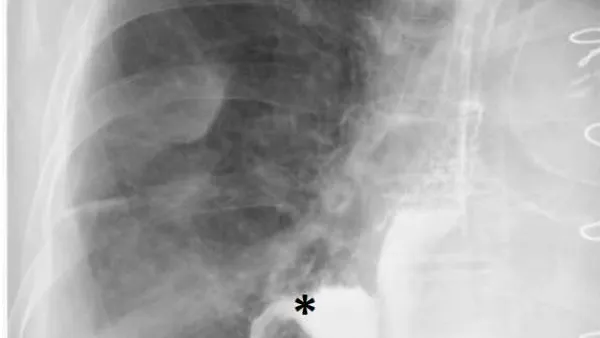
Oral Gastrografin radiography confirmed a communication between the esophagus and the right pleura (Fig. 1).
Thoracic computed tomography (CT) scans clearly indicated the fistula location (Fig. 2A) and esophagoscopy showed a wide opening - on the right surface of the distal esophagus (Fig. 2B). No other anomalies were found. We speculated that perforation could be due to ischemia of the esophagus resulting from the combination of TEE probe compression and non-pulsatile flow during a lengthy on pump procedure, in a fragile patient, as it is already well reported in the literature[^1]. Moreover, in our patient, CT images clearly showed a giant left atrium - indexed volume - compressing and displacing the oesophagus, right in the fistulated region. Several studies reported cases of esophagus compression and distortion by enlarged left atrium related to mitral valve disease[^2]: this anatomic feature should be recognized as a risk factor for TEE-related esophageal complications.
Finally, a 15 x 2 cm auto expandable esophageal stent was placed endoscopically. No residual communication was observed, and a new CTscan confirmed the good result (Fig.3).
II. DISCUSSION
TEE is used routinely during cardiac surgery to monitor cardiac haemodynamic, weaning off cardiopulmonary bypass, air removal, and valve function, butal though it is a relatively safe exam, it may result in some complications[^3]. During cardiac surgery, the insertion, manipulation, and removal of the probe may increase those complications[^4]. Moreover, when cardiopulmonary bypass (CBP) is necessary as in this case, patient's temperature is lowered to , there is a significant inflammation reaction and the blood flow provided has a continuous pattern, not a pulsatile one. All these conditions can cause micro-ischemia, facilitate tissue damage and weak the oesophageal wall, even if any movement or manipulation of the probe was gentle and TEE examination was discontinued intermittently and automatically to decrease the probe temperature and the risk of tissue damage. In addition, a huge left atrium might chronically compress the anterior oesophageal wall, causing local ischemia, and might displace it, generating dangerous bends which can make easier a TEE-mediated damage. However it has been reported that the majority of iatrogenic esophageal damages occurs in patients with an unknown esophageal or gastric pathology[^5].
In this case, our hypothesis was that the lesion was induced by transoesophageal echocardiography probe in a favourable setting: huge left atrium which displaced the thoracic oesophagus, in a very frail patient who underwent a quite long surgical procedure, using CBP with long-time perfusion with continuous blood flow.
III. CONCLUSION
TEE is a fundamental tools during cardiac surgery, however for patient safety, comprehensive intraoperative TEE guidelines should always be followed. And an enlarged left atrium should be recognized as a main risk factor for TEE-induced esophageal perforation, especially in fragile patients, with marked esophagus distortion.
The authors have no conflict of interest to declare and no founding.