I. INTRODUCTION
Neurological disorders in case of anomalies of the craniovertebral junction are extremely diverse and consist of both clinical signs of brainstem compression by lowered cerebellar tonsils and liquorodynamic disorders [1,3,7].
Analysis of the dynamics of neurological disorders does not always provide objective information, and a more informative functional quantitative scale for assessing neurological symptoms, proposed by Egorov et al., 2002 [3] determines the dynamics of neurological disorders in the pre- and postoperative period in Arnold Chiari type I (AK type I). A more objective picture of the functional state of the structures of the craniovertebral transition is provided by a comparison of clinical and neurophysiological research data [6,7,10]
Unfortunately, there is still no single system for assessing the degree of functional deficiency in this pathology. At the same time, the high proportion of subjective neurological disorders makes it difficult to determine the degree of compensation of the disease [4,9,10]. The question - to observe or to operate? - is extremely important in the modern practice of neurologists and neurosurgeons. Modern neurophysiological diagnostic methods make it possible to absolutely accurately objectify the level of pathological disorders and determine the degree of functional involvement of certain anatomical structures. Clinical manifestations of Chiari type I anomaly are described in detail in the literature. Many authors identify from 3 to 6 variants of clinical syndromes [3,4,7,10]. Most often, cerebellar, bulbar, pyramidal and syringomyelia variants are distinguished, and almost all researchers of this pathology point to a high proportion of patients with cerebellar disorders in AK type I [8,10]. Comparative analysis of subjective, objective symptoms in AK type I and neurophysiological data of ASEP, SSEP and ENMG in various AK type I syndromes greatly facilitates the decision on the issue of conservative and surgical tactics, however, a small amount of information about such results in the literature determined the purpose of our study.
The purpose of the study is to determine the dependence of the formation of the cerebellar syndrome on the degree of ectopia of the cerebellar tonsils and to identify significant predictors according to evoked potentials in determining conservative or surgical tactics in patients with this syndrome in Chiari type 1 anomaly.
II. MATERIALS AND METHODS
We studied 207 patients with AK type I who were treated at the Republican Specialized Scientific-Practical Medical Centre of Neurosurgery from 2015 to 2018, aged 18 to 57 years, the number of men was 82 observations, and women were 125 observations. Among them, 63 patients were identified with neurological disorders characteristic of the cerebellar syndrome in AK type I. All patients were divided into groups depending on the degree of displacement of the cerebellar tonsils below the Chamberlain line. The first group consisted of 17 patients with ectopia of , the second had 75 patients with ectopia of , the third was 84 patients with ectopia of , in the fourth group consisted of 31 patients with the displacement and ectopia was more than according to MRI study. The control group consisted of 30 healthy individuals.
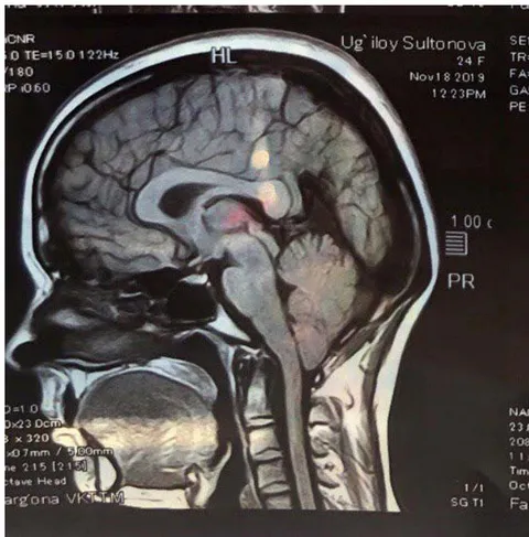
The standard for determining the degree of descent of the cerebellar tonsil in Chiari anomalies was the Chamberlain line, passing from the hard palate to the Opistion (a point located in the centre of the posterior edge of the BSO) [2,8,9]. We considered the displacement of the cerebellar tonsils beyond the Chamberlain line up to to be acceptable. In our studies, we used the Chamberlain line to guide the anatomical anomalies of the craniovertebral junction and the degree of cerebellar tonsil ectopia (Fig. 1).
All patients underwent a multimodal neurophysiological protocol, including acoustic stem evoked potentials (ASEPs), somatosensory evoked potentials (SSEPs), and motor evoked potentials (ENMG) [7]. The studies were carried out on a 4-channel Synopsis complex (Neurotech, Russia) with computer data processing.
For ASEp, the standard vertex-mastoid lead (M1-Cz, M2-Cz) was used.
During SSEP, the electrodes were installed according to the standard C4-Fz method - with stimulation of n.medianus S C3-Fz- with stimulation of n.medianus D. Stimulation was carried out with electrical impulses in the projection of the median nerve at the level of the wrist with a current of , a frequency of .
Stimulation EMG was performed for n.glossopharyngeus with the establishment of recording electrodes in accordance with muscle innervation. If necessary, we supplemented the studied nerves based on the neurological deficit.
Statistical data analysis was carried out using IBM SPSS Statistics 26 version. Differences in the distribution of quantitative values were assessed using Student's parametric test. To assess the likelihood of surgical treatment, the method of discriminant analysis was used, followed by the construction of a prognostic function, and an assessment of the specificity and sensitivity of the model. Statistically significant differences in the group of operated and non-operated patients were assessed using the Wilks coefficient .
III. RESULTS AND DISCUSSION
Patients with cerebellar disorders had the highest representation among the examined patients - 63 cases .
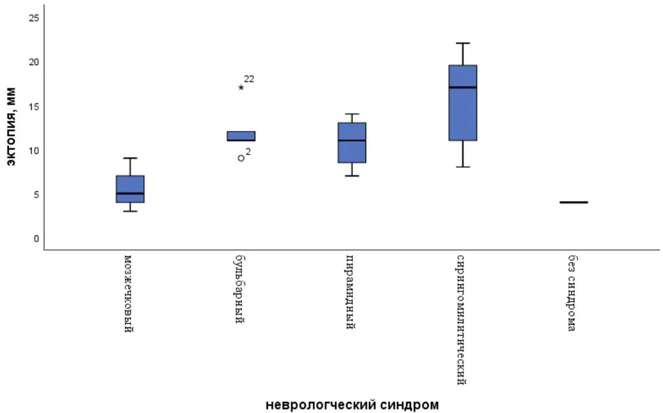
After analyzing the above complex of subjective and objective neurological symptoms in patients with type I Chiari anomaly, we identified 4 types of clinical syndromes - cerebellar, bulbar, pyramidal, syringomyelitic, which were most clearly formed in groups of patients. The distribution of these syndromes in patients with AK type I, depending on the degree of displacement of the cerebellar tonsils below the Chamberlain line, is proposed in Table 1 (Fig. 2).
| I group 0-4 mm (n=17) | II group 5-9 mm (n=75) | III group 10-15 mm (n=84) | IV group >15 mm (n=31) | |||||
| Syndromes | # | % | # | % | # | % | # | % |
| Cerebellar | 10 | 58.8 | 39 | 52.0 | 12 | 14.3 | 2 | 6.45 |
| Bulbar | - | - | 10 | 13.3 | 20 | 23.8 | 9 | 29.0 |
| Pyramidal | - | - | 5 | 6.67 | 21 | 25.0 | 1 | 3.23 |
| Syringomyelitic | - | - | 18 | 24.0 | 31 | 36.9 | 19 | 61.3 |
| Without syndromes | 7 | 41.2 | 3 | 4.0 | - | - | - | - |
As can be seen from the presented data, cerebellar syndrome with clinical symptoms of dizziness, tinnitus, various types of nystagmus, static and dynamic ataxia phenomena most often occurred in patients of the I and II groups with tonsil descent to 5-9 mm and accounted for more than half of the cases (58.8% and 52.0% respectively). The representation of cerebellar syndrome in patients of the III and IV groups was significantly less. So, with severe tonsil ectopia of 10-15 mm, the cerebellar syndrome was observed in 12 (14.3%) patients, and in the group with ectopia of more than 15 mm, it was observed in only 2 examined individuals. Such a low number of patients in the III and IV groups with manifestations of the cerebellar syndrome, in our opinion, is associated with the intactness of the cerebellar pathways with a pronounced displacement of the cerebellar tonsils.
Next, we analyzed data from neurophysiological studies in patients with Chiari 1 anomaly.
The data obtained from ASEP studies in patients with cerebellar syndrome are presented in Table 2. It was found that in all examined patients, the latent periods of peaks III and V were extended bilaterally with significant differences compared to healthy individuals. The average values of the latencies of the remaining components - I, II, IV were unchanged compared to the results of the control group. The amplitude indices of the III and V peaks were significantly increased relative to the control values, which dissociated from the general idea of depression of the amplitude indices in ASEP in patients with pathology of stem structures. In our opinion, an increase in the amplitudes of the III components in patients with cerebellar syndrome indicated functional irritation of the stem structures at the level of the upper olivary complex. An analysis of the average values of the interpeak intervals showed a slight delay in III-V and I-V in the examined group with significant differences with control individuals, which indicated a slowdown in conduction at the pontomesencephalic level. Peak-to-peak intervals I-III were preserved compared to the control group,
which can be explained by the intactness of the peripheral portion of the auditory analyzer.
| Control group (n=30) | PI | PII | PIII | PIV | PV |
| S | 1.79± 0.16 | 2.95 ± 0.18 | 3.94 ± 0.24 | 5.06 ± 0.22 | 5.97 ±0.25 |
| D | 1.72 ± 0.17 | 2.98 ± 0.19 | 3.92 ± 0.22 | 5.13 ± 0.20 | 6.02± 0.25 |
| Cerebellar syndrome (n=63) | |||||
| S | 1.74 ± 0.18 | 2.96 ± 0.17 | 4.25 ± 0.25 | 5.25± 0.21 | 6.55± 0.22* |
| D | 1.68 ± 0.16 | 3.02 ± 0.19 | 4.25 ± 0.21* | 5.38± 0.19 | 6.70 ± 0.24* |
Amplitude,
| Control group (n=30) | PI | PIII | PV |
| S | 0.286± 0.05 | 0.262± 0.04 | 0.368 ±0.06 |
| D | 0.282± 0.04 | 0.265± 0.06 | 0.338± 0.08 |
| Cerebellar syndrome (n=63) | |||
| S | 0.348± 0.03 | 0.370± 0.03** | 0.375 ± 0.05* |
| D | 0.340± 0.04 | 0.372± 0.05** | 0.380 ± 0.07* |
Peak intervals, ms Significant differences in identical indicators between the control group and the group of patients (Student t-test)
| Control group (n=30) | PI-PIII | PIII-PV | PI-PV |
| S | 2.19± 0.16 | 2.06± 0.18 | 4.38± 0.22 |
| D | 2.24± 0.18 | 2.08± 0.22 | 4.46± 0.24 |
| Cerebellar syndrome (n=63) | |||
| S | 2.56± 0.15 | 2.52± 0.14** | 4.90±0.21* |
| D | 2.88± 0.17 | 2.60± 0.18** | 4.82±0.20* |
We analyzed the data of somatosensory EPs in 63 patients with clinical manifestations of cerebellar AK type I syndrome. Registration of SSEP was performed during stimulation of the median and tibial nerves from 2 sides, the average SSEP values were compared with the values in the control group. The results of the SSEP study in cerebellar syndrome are presented in Table 3.
| Latency, ms | Control group (n=30) | Cerebellar Syndrome (n=63) |
| N9 Erb | 9.6±0.7 | 9.4±0.7 |
| N13 Neck | 13.2±0.8 | 14.5±0.7* |
| N20 Cortex | 18.8±1.0 | 18.9±1.2 |
| Amplitude, μV | ||
| N9 Erb | 5.4±2.5 | 5.6±2.2 |
| N13 Neck | 2.9±1.3 | 2.7±1.2 |
| N20 Cortex | 2.8±1.6 | 2.9±1.5 |
| Peak intervals, ms | ||
| N9-N13 | 3.5±0.4 | 3.2±0.3 |
| N13-N20 | 5.8±0.5 | 6.9±0.2* |
| N9-N20 | 9.2±0.5 | 8.8±0.7 |
Significant differences in identical indicators between the control group and the group of patients (Student t-test)
As can be seen from the above data, in the group of patients with cerebellar syndrome, there was a significantly significant increase in the latency of the N13 component up to 14.5 ms compared with the control group, which was more often symmetrical bilateral (84% of cases). The amplitude indicators of all SSEP components were preserved relative to healthy individuals. The expansion of the N13-N20 interpeak intervals up to 6.9 ms was isolated in the group of patients with cerebellar AK type I; the parameters of the N9-N13 and N9-N20 interpeak intervals were unchanged compared to the control values.
When analyzing the data on SSEP indicators for stimulation of the tibial nerve, shown in Table 4, a significant extension of the latent period of the N30 component to 38.1 ms was determined in patients with cerebellar syndrome relative to the control group. Changes in the amplitudes of the components N22, N30, P37 in the study group of patients were not recorded. The N30-P37 peak-to-peak interval was moderately extended to 12.5 ms in the majority of cases (68%) with cerebellar syndrome compared to healthy individuals, the N22-N30, N22-P37 peak-to-peak latencies were consistent with the control group.
| Latency, ms | Control group (n=30) | Cerebellar Syndrome (n=63) |
| N22 lumbar | 23.6±1.9 | 23.2±1.6 |
| N30 cervical | 30.6±2.5 | 38.1±1.2** |
| P37 Cortex | 37.5±3.4 | 36.±3.0 |
| Amplitude, μV | ||
| N22 lumbar | 1.3±0.5 | 1.7±0.3* |
| N 30 cervical | 0.9±0.3 | 1.1±0.2 |
| P37 Cortex | 2.6±1.5 | 2.9±1.5 |
| Peak intervals, ms | ||
| N22-N30 | 7.62±1.14 | 7.86±1.07 |
| N30-P37 | 8.05±1.32 | 12.5±1.54* |
| N22-P37 | 15.7±1.65 | 16.9±1.35 |
Significant differences in identical indicators between the control group and the group of patients (Student t-test)
Thus, the analysis of SSEP data during stimulation of the median and tibial nerves revealed an increase in the latency of the N13, N30 components in patients with cerebellar AK type 1 syndrome in the predominant number of cases combined with an expansion of the interpeak intervals N13-N20 (64% of patients) and N30-P37 (55% of patients), which indicated a slowdown in afferentation at the level of the cervical spinal cord and then the medulla oblongata - the thalamus cortex with a tendency to reduce postsynaptic activation of the nuclei of the medulla oblongata.
We analyzed the ENMG data obtained by stimulation of the oculomotor, facial, glossopharyngeal nerves, as well as the median and tibial nerves in a group of patients with cerebellar disorders in AK type 1. As follows from Table 5 below, motor conduction velocity SPI eff was slightly reduced in the facial and glossopharyngeal nerves with significant differences from the control group. In the oculomotor nerve, the efferent velocity in the study group was preserved relative to the control. The parameters of the conduction velocity of the SPI eff impulse along the nerves of the upper and lower extremities were unchanged in comparison with healthy individuals. Also, we did not register significant deviations in the Amax values of the M-response amplitudes for all the studied nerves in the group of patients. However, after stimulation, in of patients with cerebellar syndrome, pathological waves along the facial nerve were noted, while in the group of healthy individuals such a phenomenon was not recorded.
| Control group (n=30) | SPI eff, m/s | Amax, μV | Additional pathological waves |
| Oculomotor nerve | 29.4±2.2 | 1080±105.5 | - |
| Facial nerve | 39.5±1.8 | 1235±126.3 | - |
| Glossopharyngeal nerve | 42.6±2.0 | 1860±164.0 | - |
| Median nerve | 61.0±1.7 | 6254±267.0 | - |
| Tibial nerve | 49.6±2.1 | 7125±745.5 | - |
| Cerebellar Syndrome (n=63) | |||
| Oculomotor nerve | 29.1±2.0 | 1072±105.8 | |
| Facial nerve | 34.8±1.6** | 1130±138.0* | + |
| Glossopharyngeal nerve | 39.2±1.4** | 1851±170.5 | |
| Median nerve | 60.4±1.5 | 6158±245.6 | |
| Tibial nerve | 48.3±1.9 | 7245±760.8 |
In our opinion, small downward deviations of the SPI eff values in the facial and glossopharyngeal nerves against the background of relatively intact values of the M-response amplitudes indicated the functional involvement of the structures of the pons and medulla oblongata in cerebellar syndrome. Pathological waves along the facial nerve may have corresponded to irritable disorders at the cerebellopontine level. Unchanged parameters of SPI eff and amplitudes of muscle responses during stimulation of the median and tibial nerves in the group of patients with AK type 1 indicated the absence of dysfunction of the segmental apparatus in cerebellar disorders.
Taking into account the above data, we tried to use the discriminant analysis method to identify the likelihood of an operative outcome of treatment in patients with neurological manifestations of the cerebellar syndrome in AK type 1, depending on the degree of tonsil ectopia, as well as quantitative indicators - the amplitudes and latencies of the components of evoked potentials - ASEP, SSEP and ENMG, followed by the construction of a predictive model.
As a result of discriminant analysis by the method of stepwise selection, the following model was obtained (1):
where is a discriminant function that characterizes the likelihood of surgery in patients with cerebellar syndrome with AK type 1, is the degree of tonsil ectopia (mm), is the latency of the inter-peak interval P3-P5 in ASVP.
The discrimination constant separating patients into operated and non-operated was determined as the value of the function equidistant from the centroids, which amounted to 1.259 in the group of operated patients, and 2.698 in the group of non-operated patients.
The discrimination constant for this model was calculated by the formula - arithmetic mean of centroids (sum/2), or
when comparing the mean values of the discriminant function in both groups of operated and non-operated patients with AK type 1 using the Wilks coefficient , statistically significant differences were established .
Given the calculated values of the prognostic discriminant function, it is possible to determine a high or low probability of the risk of surgical intervention in patients with cerebellar syndrome with Chiari anomaly type 1: with a function value of more than -0.719, the patient belonged to the group with a high probability of surgical intervention, and with a function value of less than -0.719 The risk of surgery was significantly lower.
The sensitivity of the model was , the specificity was .
Thus, as a result of discriminant analysis, we obtained a function that allows us to predict the likelihood of a surgical outcome of treatment in patients with cerebellar syndrome in AK type 1.
In conclusion, it should be noted that the proposed prognostic model of a conservative or surgical outcome of the disease in patients with AK type 1 has a significant relationship with the degree of cerebellar tonsil ectopia and changes in neurophysiological parameters according to evoked potentials. At the same time, registration of the expansion of interpeak latencies P3 and P5 according to the ASEp study is especially important in addressing the issue of surgical intervention in patients with manifestations of cerebellar syndrome in AK type 1.