The use of nanoparticles in the health area is a research topic that has been increasing in recent years, from that perspective this work focused on making a characterization of nanoparticles, their evolution and interaction with blood, aspect addressed through the description of the biomagnetic fluid, focusing on characteristics such as viscosity and geometry. Also, the evolution of the applications or techniques in which nanoparticles have been used is presented, focusing the review on cancer treatments, for which the four progressive generations of this research field were considered, as well as the use of nanoparticles in diagnostic imaging. Finally, some fields of implementation and study in Colombia were identified. The review carried out allows concluding that the evolution of the use of nanoparticles.
## I. INTRODUCTION
The term nanotechnology refers to a multidisciplinary field that deals with the research, design, synthesis, application of materials and functional systems by controlling substances at the nanometer level. The interest of nanotechnology is not only to manipulate matter on a small scale, but also to study the unique physical and chemical properties of nanostructures (e.g., surface properties, electrical conductivity and magnetic properties). In recent years, nanotechnology has had a major impact in areas such as biology and medicine (Rojas, Aguado, & González, 2016). With the aim of advancing in this field, a bibliographic review was carried out, oriented towards three topics: characterizing nanoparticles (np) and their evolution; describing the medium in which they move and therefore their interaction with it, and identifying the applications or techniques of nanotechnology in health sciences, specifically in cancer treatments (tumors), and the obtaining of diagnostic images through methods that use nanoscopic contrast agents or markers.
## II. CHARACTERIZATION, DESCRIPTION AND EVOLUTION OF THE NP
The np are particles with dimensions of the order of $1 \, \text{nm} = 1 \times 10^{-9} \, \text{m}$, which facilitates their application to different fields of nanotechnology, in medicine for example, they are used for the purpose of monitoring, control, construction or repair, defending or improving the human biological system at the molecular level (Wakaskar, 2018). Some of the np used in cancer treatments have been dentrimers, being np with three-dimensional tree-like structures in the range of 1-100 nm, they can host a variety of carrier molecules, both hydrophobic and hydrophilic and are useful delivery agents for genes, drugs and anticancer agents; thanks to their size and geometry they can be specifically controlled in groups, in order to possess pre-designed and specific physical and chemical properties (Alfonso & Casado, 2016).
On the other hand, micelles are hydrophobic spherical structures that are grouped to form the central core of the sphere in a liquid environment (Haley & Frenkel, 2008), so they are useful for the administration of water-insoluble drugs with sizes in the range of 10 - 50 nm (Urrejola et al., 2018). Nanospheres, on the other hand, are spherical structures composed of a matrix system where the surface can be modified by adding polymers and also biological materials, have a size in the range of 10 - 100 nm (Haley & Frenkel, 2008). From the perspective of their use as carriers, nanocapsules are vesicular systems with a central cavity or core to which it is possible to confine a drug, their size is in the range of 10 - 500 nm (Chávez, Olvera, Ganem, & Quintanar, 2002). Finally, we find the magnetic ones, on which we will go deeper, because their characteristics have made possible advances in the transport through the blood as a biomagnetic fluid.
### a) Magnetic Nanoparticles (MNP)
They are np that are iron-based, therefore they can be manipulated by employing an external magnetic field (B) (Aval et al., 2020). Magnetite (Fe3O4) is a black ferromagnetic iron oxide of Fe(II) and Fe(III), which has been the most studied, due to the potential to act as an electron donor (Mohammed, Gomaa, Ragab, & Zhu, 2017).
### b) Ferrofluids
When talking about ferrofluids, we refer to a colloidal dispersion made by a special multidomain particles based on iron oxide and iron hydroxide by a wet chemical method, which facilitates the steering capability under the influence of a B (Lübbe, Bergemann, Riess, et al., 1996). Additionally, these colloidal magnetic np have unique surface properties that allow biocompatibility and biodegradability in addition to having minimal toxicity, they are suitable as drug-delivery vehicles that have excellent magnetic saturation (Liu, Xu, Wang, & Ke, 2008).
### c) Superparamagnetic
They are a unique type of MNP, because they have many desirable properties, from the point of view of biomedical applications, such as: biocompatibility, biodegradability and ease of synthesis, to which we must add their superparamagnetic nature. On the other hand, they do not produce hysteresis, since they leave a zero residual magnetization after an external B is removed, this feature helps to prevent coagulation, so compared to other MNP, it reduces the possibility of agglomeration in the body (Mohammed, Gomaa, Ragab, & Zhu, 2017).
The size of these particles influences both their physicochemical and pharmacokinetic properties, so two groups are classified: Spions: (superparamagnetic iron oxides) are np that in particular are generally based on inorganic iron oxide coated with hydrophilic polymers, whose size is larger than $50~\mathrm{nm}$ (including the coating). USpions: (ultra-small superparamagnetic iron oxides) are np that have a size smaller than $50~\mathrm{nm}$, being blood pooling agents they could be used for perfusion imaging enabling the diagnosis of cerebral or myocardial ischemic diseases (Zhang et al., 2020).
## III. CHARACTERIZATION AND INTERACTION OF THE ENVIRONMENT
Currently there is a field of research associated with the interaction of B with living beings, and in particular it has gained relevance in nanomedicine, from this perspective we can consider two basic areas: magnetobiology and biomagnetism. The former deals with the effects produced by magnetic fields on organisms, ranging from the orientation capacity of some animals, to the controversial damage to health caused by exposure to low-frequency electromagnetic waves. Biomagnetism, on the other hand, focuses on the study of the B associated to the organism itself, in particular, we refer to the biomagnetic fluid, which is found in organisms and reacts to the presence of a B. The results of these experimental fields are useful to obtain information that does not allow us to understand biophysical systems, to implement new clinical diagnostic techniques and to create new therapies centered on the use of np (Sosa, Alvarado, & Gonz, 2002).
### a) Blood as a Transport Medium for Nanoparticle
The various applications of np as a means of transporting drugs or contrast chemicals, have led researchers to deepen both the knowledge of the blood, as the fluid through which these particles move, the interaction with the components thereof, and the incidence of external magnetic fields applied, in this sense, Bose & Banerjee, (2015) mention measurements made to estimate the magnetic susceptibility of blood and reported that this is between $3.5 \times 10^{-6}$ and $6.6 \times 10^{-7}$ for venous and arterial blood, respectively, also Bartoszek & Drzazga, (1999) show an experimental study of the magnetic anisotropy of blood cells at a B of up to $1.8 \mathrm{~T}$ for a temperature range between $75 - 295 \mathrm{~K}$, employing torsional magnetometry.
Ichioka & Ueno, (2000) conducted in vivo experiments using rats as subjects, which were subjected to magnetic fields of $8\mathrm{T}$, showing a reduction in blood flow and temperature of the animal. In the same direction, the in vitro experiments conducted by Haik, Pai & Chen (1999), in which they used human blood samples subjected locally to a B of the same intensity, reduced by $30\%$, additionally established that as the biofluid enters and exits the gradient of B, In relation to the biomagnetic flux in narrow channels, Tzirtzilakis in 2005 found that these are affected by a constant and local B (Bose & Banerjee, 2015).
### b) Simulating Biomagnetic Fluid (Blood)
Liu, Zhu, Rao, Clausen & Aidun, (2018) simulate the biotransport of np under a complex cell flow environment using a multiscale method based on the Lattice Boltzmann method (LBM), the basic components of which include liquid-phase LBM processing, the red blood cell spectrum linkage method (SLM), and the Langevin dynamics (LD) method to capture pauses of np motion. In addition, extensive bidirectional coupling schemes are established to capture precise interactions between each component and thus simulate np transport in cellular blood flow with high efficiency.
In turn, Lee, Ferrari & Decuzzi, (2009) present a general mathematical model to predict the transport behavior of particles with different physical properties: size (nano-microparticles) and shape, as well as the material in which they are immersed, considering a linear laminar flow. Their results show that non-spherical particles, under the concurrent action of inertial and hydrodynamic forces, can deflect laterally, an effect known as hydrodynamic margination, increasing the probability of interaction with the wall surface. Thus, in blood, np will periodically oscillate around their trajectory, thus reducing their distance from the vessel wall, while the particles may actually separate with a net lateral deflection.
On the other hand, Duncan & Bevan, (2015) generated a Monte Carlo simulation, measuring the net interactions between np "decorated" by ligands, which possess distinct chemical structures, providing multiple interactions in self-assembly, and membrane proteins on the surfaces of healthy and diseased cells. From their analysis, they identify that these ligand-functionalized np are able to selectively bind to populations of diseased cells rather than healthy cells, proving attractive for improving the efficacy of drug therapies by using lower affinity ligands to target cancer cells with targeted membrane proteins.
Along the same lines, Müller, Fedosov & Gompper, (2014) performed simulations and attempted to study the marginal characteristics of carriers of different shapes and sizes using mesoscopic hydrodynamic simulations to explain the related physical mechanisms, finding that the properties of particle edges increase with increasing carrier size. The above results lead to the conclusion that addressing the various problems associated with drug delivery is a complex issue; its solution requires an interdisciplinary effort, including in vitro and in vivo experiments and realistic numerical simulations.
### c) Viscosity
Blood viscosity and plasma viscosity are the best known parameters characterizing the properties of blood flow. Haik, Pai & Chen, (2001) conduct an investigation on the behavior of viscosity due to magnetic discharges in human blood, for which they carried out flow experiments on oxygenated blood in vitro and which is subjected to a B of up to 10 T. Additionally, they use a mathematical model to simulate biomagnetic fluid dynamics under similar conditions, which allows them to identify that the magnetization action will introduce a rotational motion to orient the magnetic fluid particles with the B (Afkhami & Renardy, 2017); however, the behavior and characteristics of the blood are unique to each patient. As a complement to the aforementioned computational experiments, Rukshin, Mohrenweiser, Yue & Afkhami, (2017) proposed a mathematical model describing the behavior of equations to visualize particle trajectories and calculate capture rates to assess the impact of various physical conditions on the success of magnetic drug targeting.
### d) Geometry of the Medium
Another element to take into account in the characterization of the medium is the geometry, muscle arteries have three major geometrical differences with capillaries, for example in microvessels, the anti-slip boundary condition reduces the velocity of blood near the wall relative to the centerline and improves the ability to trap particles by weaker magnetic force due to lower resistance compared to millimeter vessels (Aviles et al., 2005). In this case, the force required is well below the maximum allowable exposure, however, due to the higher blood flow rate in muscles and blood vessels, the particles require a higher magnetic force to resist the "tenacity" that occurs in the opposite direction of particle motion.
## IV. EVOLUTION OF APPLICATIONS IN CANCER TREATMENTS, USING NP
Cancer is a disease in which the cells of the human body acquire the ability to divide and multiply uncontrollably a (Sharma, Sharma, Punj, & Priya, 2019), this condition acquired by the diseased cells through mutations of the genome, as the cancer cells divide, leads to the fact that the new cells will inherit the same growth capacity and increase the number of cancer cells (Miller, 2018). The treatments for this disease are diverse and in any case, side effects must be considered, therefore, targeted treatments are an important option for patients. In this sense, the evolution in the use of np is presented, which is approached through four generations, identifying the main investigations and their results.
### a) First Generation
The first experiment was performed "in vivo" by Lübbe et al, (1996), who developed a magnetic fluid to which drugs, cytokines and other molecules can chemically bind to allow these agents to be directed into an organism by an external high-energy B. They used male rats and mice, kept in a controlled environment. For which they used male rats and mice, kept in a controlled environment. For the design of the external B, using high-energy permanent magnets made of rare earths (neodymium), they consisted of disks or blocks with variable thickness, configured in the shape of a column or a block (see Figure 1). In this way, the magnets could be arranged more closely around the individual tumor configuration, with a B between $0.2 - 0.5\mathrm{T}$ (depending on tumor size). They tested two forms of therapy with the magnetic fluid: treatment of tumors by mechanical occlusion with the ferrofluid in high concentrations; and magnetic therapy, using small amounts of the ferrofluid as a drug carrier vehicle, which allowed epirubicin to be concentrated locally in the tumors.
The first part of the study focused on tolerance to ferrofluid and magnetically bound epirubicin. The results show that hematological and blood chemistry values did not change from baseline after injection of different amounts of ferrofluid. On the other hand, epirubicin caused changes in hematological parameters. Histological data showed that the magnetic particles accumulated in the liver and spleen, without causing significant hepatosplenomegaly, the latter two results were within predicted. After the sixth week of observation, one animal in each high-dose group died, possibly due to cardiovascular failure caused by sepsis. In those groups in which the highdose epirubicin was administered, the animals died quickly; in the low-dose groups, they died somewhat later, around 4 - 6 weeks. In all other groups, including those receiving low levels of epirubicin, the animals survived the observation period (Lübbe, Bergemann, Huhnt, Fricke, & Riess, 1996).
The second part of this investigation focused on the mechanical embolization by the ferrofluid after its injection and concentration in the tumor by means of an external B, regardless of the type of tumor, there was a rapid and constant decrease in tumor volume within 14 days after treatment. It was impossible to reproduce this tumor response in the animals given only epirubicin, although the tumors responded to the high dose of this drug, this was only for a brief period and most of the animals in this group died shortly thereafter.
Subsequent experiments by Lübbe led to the conclusion that magnetic fluid is a good agent to decrease tumor volume and with further studies, it can be used in different forms of local cancer treatment in conjunction with high-energy magnetic fields, avoiding mortality of the subject (Lübbe., Bergemann, Brock, & McClure, 1999).
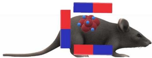
Source: own Fig. 1: Graphic Illustration of Placement of Rare Earth Magnets in Block form on the Tumor to be Treated
Thus, in the second experiment, Lübbe et al., (1999) chose their subjects with an eligibility criterion among them, seven patients with breast cancer, two with chondrosarcoma, two with parotid squamous cell carcinoma, one with Ewing's sarcoma and two with malignant tumor; with a life expectancy of at least three months and renal preservation. Continuing their line of research, they employed anticancer drugs that would reversibly bind to magnetic fluids, which were focused on locally advanced tumors through a B disposed on the tumor surface outside the body.
They tested for magnetite concentration in the tumor, and 10 of the 14 patients, had intact skin covering the tumors, the other four showed wound healing and open superficial wounds. In the first four cases, since the B will obscure the shape of the magnetic block attached to the tumor, it is easy to observe magnetite absorption into the tumor, this discoloration lasted for 24 - 36 hours and then disappeared completely, these areas were not locally toxic and ensured that the discoloration could not be removed to rule out the possibility of iron deposition from the magnetic lumps in the superficial layer of the skin. So the targeted magnetic drug with epirubicin was well tolerated and mild tumor reductions were achieved at day 10 and some small responses at day 40 (Lübbe et al., 1999) (Lübbe, Bergemann, Riess, et al., 1996).
### b) Second Generation
Based on the results described above, Alexiou et al. (2000) used mitoxantrone-linked ferrofluid (FF-MTX) to treat rabbits with squamous cell carcinoma and concentrated it under a B. When the tumor reaches a volume of $3500~\mathrm{mm}^3$, FF-MTX is injected intra-arterially or intravenously. When an external B is focused on the tumor, it is activated by an electromagnet with a maximum flux density of 1.7 T, producing a non-uniform B, both in direction and magnitude, a feature that is crucial for the use of magnetic drugs. Magnetic drug targeting is a means of keeping the chemotherapeutic agent at the desired site of activity, thus increasing efficacy and decreasing systemic toxicity.
Only when the MTX dose was increased to $75\%$ and $100\%$, tumor remission was observed, but this resulted in severe side effects (hair loss, ulcers and leukopenia). However, this "magnetically targeted drug" provides a unique opportunity to locally treat malignant tumors without systemic toxicity. In addition, it is possible to use these magnetic particles as "carrier systems" for various anticancer agents such as radionuclides, antibodies and cancer-specific genes (Alexiou et al., 2002).
### c) Third Generation
Under another line of research, Gitter & Odenbac (2011) presented experimental results based on systematic "in vitro" quantitative measurements of a tube model simulating a Y-branched artery, in which they conclude that the success of particle orientation towards branching depends largely on the crossing point and the magnetic force at the site, elements that as previously mentioned must be evaluated.
For their part, Krukemeyer, Krenn, Jakobs & Wagner, (2011) performed a verification method on the effectiveness of cytostatic drugs coupled to ferromagnetic np and extracorporeal magnets, using 42 adult rats that were transfected with rhabdomyosarcoma. In the biodistribution assay, concentrations of mitoxantrone iron oxide and conventional mitoxantrone with and without $0.6\mathrm{T}$ magnets were measured in vitro in plasma and tumor tissue for one and two doses. During magnetic drug treatment, iron particles are rapidly removed and remain in the area where the tumor remained.
### d) Fourth Generation
From the perspective of assessing patient safety, Attar et al., (2016) propose a configuration which investigates the thermal effect of superparamagnetic np on human cells, present a study considering general details on the design and construction of the configuration needed to generate a safe B to examine the thermal effect of superparamagnetic np on human cancer cells, then performed a series of experimental tests to study the effect of B on the cells for 30 minutes, which allowed them to calculate the temperature rise and specific absorption. While it is true, hyperthermia treatment (Eivazzadeh-Keihan et al., 2019) is a mechanism to destroy malignant cells by increasing tissue temperature up to a range of $42 - 45^{\circ}\mathrm{C}$, temperatures above $45 - 56^{\circ}\mathrm{C}$, can cause necrotizing damage and subsequent tissue inflammation (Shabestari Khiabani, Farshbaf, Akbarzadeh, & Davaran, 2017). In this regard, ferromagnetic materials are commonly used to treat hyperthermia and the general procedure involves the allocation of magnetic particles of various sizes depending on the type of treatment to the tissue, then, when the particles generate heat through two mechanisms (including hysteresis and eddy currents), the tissue is exposed to an alternating B.
Among the advances in this fourth generation, it is important to mention the work of Al-Jamal et al., (2018), who performed in vivo experimentation based on a solid theoretical foundation for the design of a magnetic nanocarrier, capable of magnetizing uptake after intravenous administration, in order to elucidate the parameters necessary for the detection of magnetic tumors. Because long-circulating polymeric magnetic nanocarriers are capable of encapsulating increasing amounts of superparamagnetic iron oxide np (SPIONs) in a biocompatible oil carrier, they were able to study the effects of SPION loading and applied B intensity on magnetic tumor targeting in tumor-bearing mice.
Another important element is the fact that the high loading of SPIONs eliminates the need to use highly magnetized np and the oil core promotes high hydrophobic drug loading, compared to polymer-coated SPIONs. The objective of this experiment was to evaluate the key factors influencing magnetic targeting efficiency, including the loadings of SPIONs on m-NC (polymeric oil-core magnetic nanocapsules) and the magnitude of the magnetic force applied at a distance. Under controlled conditions, they quantified magnetic targeting in vivo and found that it was directly proportional to SPIONs loading and B intensity, however, higher SPIONs loading resulted in reduced blood circulation time and stabilization of magnetic targeting.
## V. DIAGNOSTIC IMAGING WITH NP
Due to the physicochemical properties presented by nanomaterials, the development of nanodevices as contrast agents in medical imaging has clear advantages over traditional agents used in the diagnosis of diseases, among which we can mention: better optical dispersion (absorption of light in the material, with a clearer visual spectrum), increased biocompatibility, decreased probability of denaturation and especially, their ability to bind to ligands, which turns them into devices with multiple functions that bind to cells, simultaneously allowing imaging for diagnosis and transport of drugs to specific sites, thus, achieving targeted and efficient treatment (Minbashi, Kordbacheh, Ghobadi, & Tuchin, 2020).
Nan, Suciu, Ardelean, Senila & Turcu (2020), report a simple reaction strategy for the synthesis of magnetic iron oxide np's stabilized with ethylenediaminetetraacetic acid (EDTA) followed by the chelation reaction of gadolinium (Gd) ions. These results show that these magnetic nanosystems represent a promising dual-mode contrast in agents for MRI applications with biomedical applications in mind.
Another type of contrast agent used for feature detection are SPIONs, due to their long half-life and small diameter, they provide a variety of possibilities to visualize intracellular targets, they can also be coupled with fluorescent dyes so that these particles can be detected in vitro and in vivo by optical fluorescence methods.
In this technique, SPIONs are inhibited by the binding of polyethylene glycol (PEG) chains that are anchored by peptide substrates shed by proteases, in diagnostic imaging, dextran-coated SPIONs provide stability for imaging, such as magnetic resonance imaging, computed tomography and optical fluorescence (Cicha, Lyer, Alexiou, & Garlichs, 2013).
In this direction, Nahrendorf et al., (2014) performed a study, where single-crystalline fluorochromelabeled SPIONs in the infrared were chelated with DTPA (diethylenetriaminepentaacetic acid) to allow binding of the PET radiotracer 64 Cu. While the iron oxide core provided the MRI contrast, the fluorochrome served for fluorescence imaging (fluorescence microscopy, flow cytometry and fluorescence mediated tomography), and the 64Cu radiotracer allowed PET (positron emission tomography) imaging, while the iron oxide core provided the MRI contrast.
The reported results show a trend towards the increasing use of SPIONs in various biomendicine applications.
## VI. IMPLEMENTATION IN COLOMBIA
Jaimez, Gonzales, Granados, Álvarez & Espitia, (2012) of the Pontificia Universidad Javeriana carried out a review article to see what advances and expectations there are in surgery to date, explaining what nanotechnology consists, its basic principles and some utilities in the field of surgery. On the other hand, Mendez and Muñoz [43] of the National University of Colombia wrote an article describing the clinical and molecular characteristics of premalignant lesions and oral cancer, as well as diagnostic methods using nanotechnology (nanochips, nanosensors, etc.) as an effective method for the early detection of cancer. Rodriguez, Moyano and Roa [44] from the Universidad Distrital Francisco Jose de Caldas, obtained a mathematical model and a computational simulation describing the trajectory of magnetic np injected near the target tissue. The magnetic np propagate along the blood vessel in the Z-direction and point to the target area through a cylindrical magnet located outside the body generating a constant B.
Likewise, Gallo and Ossa [45] from the University of Antioquia carried out a study where they evaluated two silver np synthesis processes, using, in addition, a biofunctionalization process with polyethylene glycol (PEG) to improve the anchoring properties and biocompatibility of the np, for possible treatments against skin cancer.
In the master's thesis in engineering of Pantoja, (2020) of the Universidad Distrital Francisco José de Caldas, he has focused on proposing a mathematical model that can estimate the trajectory of NPM through the action of an external B and the blood flow is obtained through computational simulation. The model includes forces that significantly affect NPM dynamics, including magnetic fields generated by magnets, scattering forces, and drag. Molecular dynamics results show that NPM under the action of a B will be captured and attracted by it, so that they can be directed to the proposed target.
The reported results show that although there is no defined line of research on the use of np in Colombia, they nevertheless highlight the possibility of joining efforts to strengthen this field of knowledge.
## VII. CONCLUSION
The np seen as drug nanocarriers play a leading role and it is in this direction in which research has been carried out, from this perspective to characterize the np and evaluate its evolution, it is identified that currently the work is focused on superparamagnetic np. The review carried out provides clarity regarding the evolution of np, as well as the importance of understanding how their kinematics are through the blood, seen as a biomagnetic fluid, a characteristic that has allowed the evaluation of strategies for directing nanoparticles that move through this medium, in this direction the main advances are associated with SPIONs for their biomedical application. There are many challenges from the treatment of diseases, starting from an accurate diagnosis to achieve an effective treatment, which involves a reduction of the adverse effects that these may have on the organism of the treated subject, in this sense the np offer a viable possibility both in diagnosis and therapy with low adverse effects, due to the possibility of targeting the treatment.
1. Afkhami, S., & Renardy, Y. (2017). Ferrofluids and magnetically guided superparamagnetic particles in flows: a review of simulations and modeling. Journal
- of Engineering Mathematics, 107(1), 231-251.
https://doi.org/10.1007/s10665-017-9931-9
2. Al-Jamal, K. T., Bai, J., Wang, J. T. W., Protti, A., Southern, P., Bogart, L.,... Pankhurst, Q. A. (2018). Magnetic Drug Targeting: Preclinical in Vivo Studies, Mathematical Modeling, and Extrapolation to Humans. Nano Letters, 16(9), 5652-5660. https://doi.org/10.1021/acs.nanolett.6b02261
3. Alexiou, C., Arnold, W., Klein, R. J., Parak, F. G., Hulin, P., Bergemann, C.,... Lubbe, A. S. (2000). Locoregional cancer treatment with magnetic drug targeting. Cancer Research, 60(23), 6641-6648.
4. Alexiou, Ch., Schmidt, A., Klein, R., Hulin, P., Bergemann, C., & Arnold, W. (2002). Magnetic drug targeting: Biodistribution and dependency on magnetic field strength. Journal of Magnetism and Magnetic Materials, 252(1-3 SPEC. ISS.), 363-366. https://doi.org/10.1016/S0304-8853(02)00605-4
5. Alfonso, B., & Casado, C. (2016). DENDRIMEROS: MACROMOLECULAS VERSATILES CON INTERES INTERDISCIPLINAR. Journal of Chemical Information and Modeling, 01(01), 1689-1699.
6. Attar, M. M., Amanpour, S., Haghpanahi, M., Haddadi, M., Rezaei, G., Muhammadnejad, S.,... Javadi, S. (2016). Thermal analysis of magnetic nanoparticle in alternating magnetic field on human HCT-116 colon cancer cell line. International Journal of Hyperthermia, 32(8), 858-867. https://doi.org/10.1080/02656736.2016.1204667
7. Aviles, M. O., Ebner, A. D., Chen, H., Rosengart, A. J., Kaminski, M. D., & Ritter, J. A. (2005). Theoretical analysis of a transdermal ferromagnetic implant for retention of magnetic drug carrier particles. Journal of Magnetism and Magnetic Materials, 293(1), 605-615. https://doi.org/10.1016/j.jmmm.2005.01.089
8. Awval, Z. M., Malekpour, L., Raeisi, F., Babapoor, A., Mousavi, S. M., Hashemi, S. A., & Salari, M. (2020). Introduction of magnetic and supermagnetic nanoparticles in new approach of targeting drug delivery and cancer therapy application. Drug Metabolism Reviews, 52(1), 157-184. https://doi.org/10.1080/03602532.2019.1697282
9. Bartoszek, M., & Drzazga, Z. (1999).; A study of magnetic anisotropy of blood cells. 197, 573-575.
10. Bose, S., & Banerjee, M. (2015). Magnetic particle capture for biomagnetic fluid flow in stenosed aortic bifurcation considering particle-fluid coupling. Journal of Magnetism and Magnetic Materials, 385, 32-46. https://doi.org/10.1016/j.jmmm.2015.02.060
11. Chávez, F., Olvera, B. I., Ganem, A., & Quintanar, D. (2002). Liberación de sustancías lipofílicas a partir de nanocápsulas poliméricas. Journal of the Mexican Chemical Society, 46(4), 349-356.
12. Cicha, I., Lyer, S., Alexiou, C., & Garlics, C. D. (2013). Nanomedicine in diagnostics and therapy of cardiovascular diseases: Beyond atherosclerotic
- plaque imaging. Nanotechnology Reviews, 2(4), 449-472. https://doi.org/10.1515/ntrev-2013-0009
13. Duncan, G. A., & Bevan, M. A. (2015). Computational design of nanoparticle drug delivery systems for selective targeting. Nanoscale, 7(37), 15332-15340. https://doi.org/10.1039/c5nr03691g
14. Eivazzadeh-Keihan, R., Radinekiyan, F., Maleki, A., Salimi Bani, M., Hajizadeh, Z., & Asgharnasl, S. (2019). A novel biocompatible core-shell magnetic nanocomposite based on cross-linked chitosan hydrogels for in vitro hyperthermia of cancer therapy. International Journal of Biological Macromolecules, 140, 407-414. https://doi.org/10.1016/j.ijbiomac.2019.08.031
Generating HTML Viewer...
References
44 Cites in Article
Shahriar Afkhami,Yuriko Renardy (2017). Ferrofluids and magnetically guided superparamagnetic particles in flows: a review of simulations and modeling.
Khuloud Al-Jamal,Jie Bai,Julie Tzu-Wen Wang,Andrea Protti,Paul Southern,Lara Bogart,Hamed Heidari,Xinjia Li,Andrew Cakebread,Dan Asker,Wafa Al-Jamal,Ajay Shah,Sara Bals,Jane Sosabowski,Quentin Pankhurst (2018). Magnetic Drug Targeting: Preclinical in Vivo Studies, Mathematical Modeling, and Extrapolation to Humans.
C Alexiou,P Hulin,R Klein,A Schmidt,C Bergemann,F Parak,W Arnold (2000). Magnetic drug targeting: biokinetic study and therapeutic efficacy.
Ch Alexiou,A Schmidt,R Klein,P Hulin,Ch Bergemann,W Arnold (2002). Magnetic drug targeting: biodistribution and dependency on magnetic field strength.
B Alfonso,C Casado (2016). DENDRÍMEROS: MACROMOLÉCULAS VERSÁTILES CON INTERÉS INTERDISCIPLINAR.
Mohammad Attar,Saeid Amanpour,Mohammad Haghpanahi,Mahnaz Haddadi,Gita Rezaei,Samad Muhammadnejad,Mehran Hajiakhoundzadeh,Tahereh Barati,Fatemeh Sadeghi,Saba Javadi (2016). Thermal analysis of magnetic nanoparticle in alternating magnetic field on human HCT-116 colon cancer cell line.
Misael Avilés,Armin Ebner,Haitao Chen,Axel Rosengart,Michael Kaminski,James Ritter (2005). Theoretical analysis of a transdermal ferromagnetic implant for retention of magnetic drug carrier particles.
Zhila Avval,Leila Malekpour,Farzad Raeisi,Aziz Babapoor,Seyyed Mousavi,Seyyed Hashemi,Marjan Salari (2020). Introduction of magnetic and supermagnetic nanoparticles in new approach of targeting drug delivery and cancer therapy application.
M Bartoszek,Z Drzazga (1999). A study of magnetic anisotropy of blood cells.
S Bose,M Banerjee (2015). Magnetic particle capture for biomagnetic fluid flow in stenosed aortic bifurcation considering particle-fluid coupling.
F Chávez,B Olvera,A Ganem,D Quintanar (2002). Liberación de sustancias lipofílicas a partir de nanocápsulas poliméricas.
Iwona Cicha,Stefan Lyer,Christoph Alexiou,Christoph Garlichs (2013). Nanomedicine in diagnostics and therapy of cardiovascular diseases: beyond atherosclerotic plaque imaging.
G Duncan,M Bevan (2015). Computational design of nanoparticle drug delivery systems for selective targeting.
R Eivazzadeh-Keihan,F Radinekiyan,A Maleki,M Salimi Bani,Z Hajizadeh,S Asgharnasl (2019). A novel biocompatible core-shell magnetic nanocomposite based on cross-linked chitosan hydrogels for in vitro hyperthermia of cancer therapy.
J Gallo,C Ossa (2019). Fabricación y caracterización de nanopartículas de plata con potencial uso en el tratamiento del cáncer de piel.
K Gitter,S Odenbach (2011). Experimental investigations on a branched tube model in magnetic drug targeting.
Yousef Haik,Vinay Pai,Ching-Jen Chen (1999). Development of magnetic device for cell separation.
Amirhossein Hajiaghajani,Soheil Hashemi,Ali Abdolali (2017). Adaptable setups for magnetic drug targeting in human muscular arteries: Design and implementation.
Barbara Haley,Eugene Frenkel (2008). Nanoparticles for drug delivery in cancer treatment.
S Jaimes,A Gonzáles,C Granados,D Álvarez,E Espitia (2012). Redalyc.Nanotecnología: avances y expectativas en cirugía.
Manfred Krukemeyer,Veit Krenn,Martin Jakobs,Wolfgang Wagner (2012). Mitoxantrone-Iron Oxide Biodistribution in Blood, Tumor, Spleen, and Liver—Magnetic Nanoparticles in Cancer Treatment.
Sei-Young Lee,Mauro Ferrari,Paolo Decuzzi (2009). Shaping nano-/micro-particles for enhanced vascular interaction in laminar flows.
H Liu,W Xu,S Wang,Z Ke (2008). Hydrodynamic modeling of ferrofluid flow in magnetic targeting drug delivery.
Zixiang Liu,Yuanzheng Zhu,Rekha Rao,Jonathan Clausen,Cyrus Aidun (2018). Nanoparticle transport in cellular blood flow.
Andreas Lübbe,Christian Bergemann,Jeffery Brock,David Mcclure (1999). Physiological aspects in magnetic drug-targeting.
Andreas Lübbe,Christian Bergemann (1996). Selected Preclinical and First Clinical Experiences with Magnetically Targeted 4’-Epidoxorubicin in Patients with Advanced Solid Tumors.
A Lübbe,C Bergemann,H Riess,F Schriever,P Reichardt,K Possinger,D Huhn (1996). Clinical experiences with magnetic drug targeting: A phase I study with 4'-epidoxorubicin in 14 patients with advanced solid tumors.
B Méndez,C Muñoz (2012). Nanochips y nanosensores para eldiagnóstico temprano de cáncer oral: una revisión.
M Miller (2018). Kernis Meets the New York Philharmonic.
Leena Mohammed,Hassan Gomaa,Doaa Ragab,Jesse Zhu (2017). Magnetic nanoparticles for environmental and biomedical applications: A review.
Kathrin Müller,Dmitry Fedosov,Gerhard Gompper (2014). Margination of micro- and nano-particles in blood flow and its effect on drug delivery.
Matthias Nahrendorf,Hanwen Zhang,Sheena Hembrador,Peter Panizzi,David Sosnovik,Elena Aikawa,Peter Libby,Filip Swirski,Ralph Weissleder (2008). Nanoparticle PET-CT Imaging of Macrophages in Inflammatory Atherosclerosis.
Alexandrina Nan,Maria Suciu,Ioan Ardelean,Marin Şenilă,Rodica Turcu (2020). Characterization of the Nuclear Magnetic Resonance Relaxivity of Gadolinium Functionalized Magnetic Nanoparticles.
León Karen Liliana,Gina Rey Ardila,Juan Rodríguez Miranda (2020). Ecuaciones econométricas para los costos de inversión en plantas de tratamiento de agua potable en Colombia.
D Rodriguez,J Moyano,L Roa (2018). Estudio por dinámica molecular browniana de nanopartículas bajo efectos de campo magnéticos externos.
Y Rojas,K Aguado,I Gonz´alez (2016). La nanomedicina y los sistemas de liberación de fármacos: ¿la revolución de la terapia contra el cáncer?.
Iris Rukshin,Josef Mohrenweiser,Pengtao Yue,Shahriar Afkhami (2017). Modeling Superparamagnetic Particles in Blood Flow for Applications in Magnetic Drug Targeting.
Saeid Shabestari Khiabani,Masoud Farshbaf,Abolfazl Akbarzadeh,Soodabeh Davaran (2017). Magnetic nanoparticles: preparation methods, applications in cancer diagnosis and cancer therapy.
Varruchi Sharma,Anil Sharma,Vasu Punj,Panneerselvam Priya (2019). Recent nanotechnological interventions targeting PI3K/Akt/mTOR pathway: A focus on breast cancer.
M Sosa,J Alvarado,J Gonz (2002). Los metodos de generacion de casos de prueba y su automatizacion.
Sheng Tong,Haibao Zhu,Gang Bao (2019). Magnetic iron oxide nanoparticles for disease detection and therapy.
Madelein Urrejola,Liliam Soto,Consuelo Zumarán,Juan Peñaloza,Beatriz Álvarez,Ignacio Fuentevilla,Ziyad Haidar (2018). Sistemas de Nanopartículas Poliméricas II: Estructura, Métodos de Elaboración, Características, Propiedades, Biofuncionalización y Tecnologías de Auto-Ensamblaje Capa por Capa (Layer-by-Layer Self-Assembly).
Rajesh Wakaskar (2018). General overview of lipid–polymer hybrid nanoparticles, dendrimers, micelles, liposomes, spongosomes and cubosomes.
Guilong Zhang,Li Zhang,Yuanchun Si,Qingdong Li,Jianmin Xiao,Bin Wang,Chaozhao Liang,Zhengyan Wu,Geng Tian (2020). Oxygen-enriched Fe3O4/Gd2O3 nanopeanuts for tumor-targeting MRI and ROS-triggered dual-modal cancer therapy through platinum (IV) prodrugs delivery.
No ethics committee approval was required for this article type.
Data Availability
Not applicable for this article.
How to Cite This Article
Camila Andrea Gualdría Sandoval. 2026. \u201cEvolution of the use of Nanoparticles in Cancer Diagnosis and Treatment\u201d. Global Journal of Research in Engineering - J: General Engineering GJRE-J Volume 23 (GJRE Volume 23 Issue J4): .
Explore published articles in an immersive Augmented Reality environment. Our platform converts research papers into interactive 3D books, allowing readers to view and interact with content using AR and VR compatible devices.
Your published article is automatically converted into a realistic 3D book. Flip through pages and read research papers in a more engaging and interactive format.
The use of nanoparticles in the health area is a research topic that has been increasing in recent years, from that perspective this work focused on making a characterization of nanoparticles, their evolution and interaction with blood, aspect addressed through the description of the biomagnetic fluid, focusing on characteristics such as viscosity and geometry. Also, the evolution of the applications or techniques in which nanoparticles have been used is presented, focusing the review on cancer treatments, for which the four progressive generations of this research field were considered, as well as the use of nanoparticles in diagnostic imaging. Finally, some fields of implementation and study in Colombia were identified. The review carried out allows concluding that the evolution of the use of nanoparticles.
Our website is actively being updated, and changes may occur frequently. Please clear your browser cache if needed. For feedback or error reporting, please email [email protected]
Thank you for connecting with us. We will respond to you shortly.