I. INTRODUCTION
The genus Eimeria comprises obligate intracellular protozoan parasites belonging to the phylum Apicomplexa. Members of this genus cause enteric disease in a wide range of vertebrate hosts, including fish, reptiles, birds and mammals. These parasites complete their development in a single host species and their sporocysts can be recognised by the presence of a Stieda body, an organelle through which the sporozoites exist. Duszynski and Wilber 1997 several species cause high levels of morbidity and/or mortality in certain hosts, resulting in economic losses in various animal production industries (Daugschies and Najdrowski 2005; Aarthi et al. 2010; Sharma et al. 2018).
A total of 157 species of fish-parasitic Eimeria have been described on the basis of sporulating oocyst morphology, host specificity, pathology and geographical distribution (Belova and Krylov 2000). Although these characteristics have traditionally been used to identify Eimeria species (Duszynski and Wilber
1997), they are often insufficient for reliable differentiation between species due to overlapping morphometric and biological characteristics (Long et al. 1984; Zhao and Duszynski 2001). A combination of morphological and molecular analyses is therefore necessary to delimit species and determine phylogenetic relationships between them.
The development of molecular tools has allowed not only diagnosis but also the study of genetic variability of pathogens from small quantities of oocysts using molecular markers (Schnitzler et al, 1998; Costa et al, 2001). Fernandez et al (2003) identified species-specific markers for Eimeria spp from a cluster of SCAR (Sequence-Characterized Amplified Region) markers. This allowed the use of the polymerase chain reaction (PCR) technique as an efficient and integrated diagnostic method, capable of detecting Eimeria species individually or simultaneously in a single reaction (Fernandez et al, 2003; Lien et al, 2007).
Of the forty-two species of Coccidia described in marine fishes (Dykova and Lom 1983), sixteen exist in Mediterranean fishes and are divided into four genera: Crystallospora Labbe 1896, Eimeria Schneider 1875, Epieimeria Dykova and Lom, 1981, and Goussa Labbe 1896. Little research has been carried out on these parasites since the end of the last century; the main works are those of Thélohan (1892), Labbe (1896), Léger and Hollande (1922) and finally Lom and Dykova (1981, 1982).
Molecular information on the diversity of Eimeria species infecting fish is scarce. Thus, only a few species of Eimeria isolated from different marine, estuarine and freshwater fish have been genetically characterised: Eimeria percae from perch (Perca fluviatilis); Eimeria anguillae from the European eel (Anguilla anguilla); Eimeria variabilis, from the long-billed bullhead (Taurulus bubalis); Eimeria daviesae on gudgeon (Gobius fluviatilis); Eimeria rutilus on roach (Rutilus rutilus); and Eimeria nemethi on bleak (Alburnus alburnus). (Molnár et al. 2012).
This work constitutes the first study of Coccidia Eimeriidae in the Mugilidae of the Algerian coast. The objective of the present study was to molecularly characterise, at the small subunit ribosomal RNA (rRNA-
US) locus using the primer pair ESSP841 CRP999, the Eimeria isolates obtained from the mullet (Mugil cephalus), and to develop a quantitative PCR for rapid detection.
II. MATERIALS AND METHODS
a) Collection and Processing of Samples
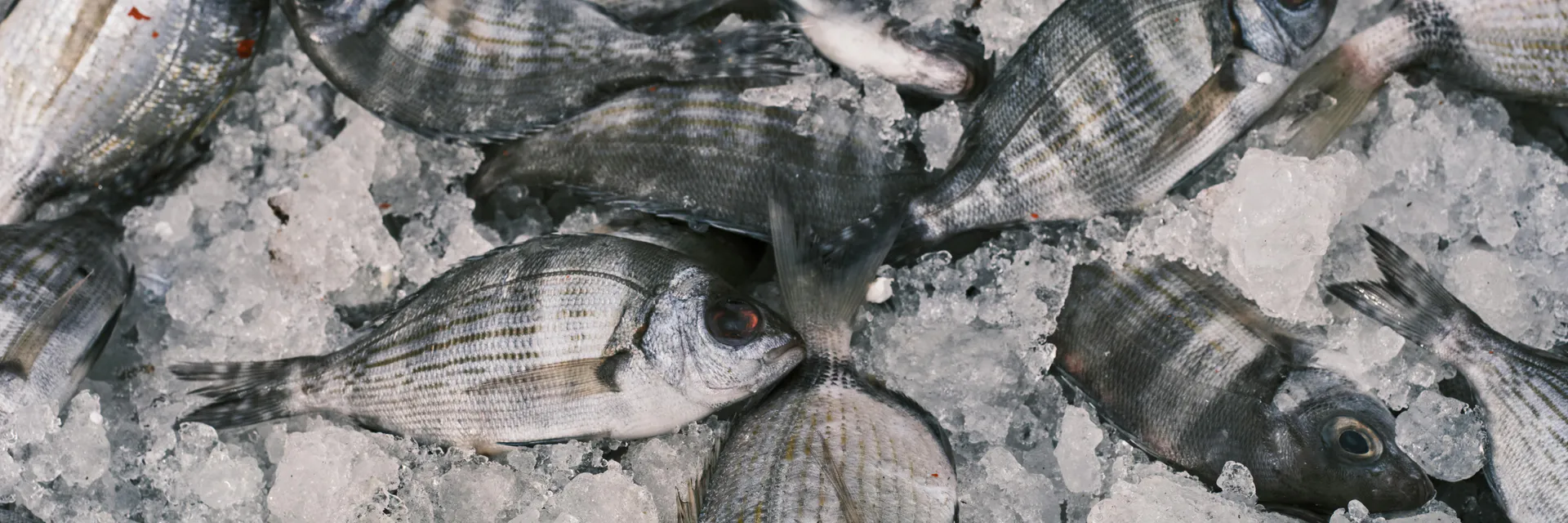
From February 2017 to March 2018 a total of 816 Mugil cephalus were caught by fishermen in the east coast of Algeria.
The first step of the present study is to identify positive samples from whole fish samples and examine them under the microscope for oocysts, either by direct methods where samples are examined either freshly, using a concentrated natural formalin buffer, or by staining the samples with iodine or Giemsa to make the internal components clearer.
- The gastrointestinal tract was differentiated into the pyloric cecum and the intestine. The pyloric caeca were homogenised using an Ultra-Turrax® T10 homogeniser (Ika®-Werke GmbH and Co., KG, Staufen, Germany). The intestinal contents were removed by scraping with a scalpel blade and then ground in a mortar with phosphate buffered saline (PBS) pH 7.2. The resulting homogenates were filtered through a set of two sieves (mesh size, 150 and ) before being subjected to a two-phase concentration of PBS pH 7.2/ diethyl ether (2:1) by centrifugation at , , for 15 min. The supernatants were carefully discarded and the concentration step was repeated until lipid-free sediments were obtained. Finally, the pellets were resuspended in of PBS and stored at .
Aliquots of of sediment were examined under brightfield microscopy to detect Eimeria oocysts magnification), which were confirmed by appearance, presence of four sporocysts and thin wall. A total of 50 oocysts from several fish specimens were observed under differential interface contrast (DIC) microscopy magnification) and measured under a light microscope (AX70 Olympus Optical Co., Ltd., Tokyo, Japan) using a micrometer eyepiece and DP Controller 2.1.1.183 software (©2001-2004 Olympus Optical Co., Ltd.).
b) DNA Extraction
Genomic DNA was extracted from the samples using the Qiamp Stools Quiagen DNA extraction kit.
c) DNA Profile
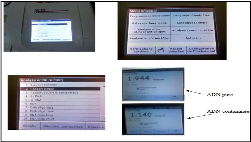
For the detection of DNA that is extracted from stool samples by using a Nanodrop spectrophotometer (THERMO. USA) for the detection and measurement of the concentration of nuclear acids (DNA and RNA), where the concentration of DNA is detected and the measurement of the purity of the DNA by reading the absorbance at a wavelength between (280-260 nm) Figure 1.
Wavelength 260 nm: represents the area of maximum absorbance of nucleic acids.
The 280 nm wavelength: is used to establish the ratio and to control the purity of the extraction.
d) Real-Time PCR Protocols
Real-time PCR performed for the detection of Eimeria species from Mugil cephalus using primers and TaqMan probe specific to the ITS1 region of the DNA that code for ribosomal RNA. The technique performed as described by Ogedengbe et al. 2011.
e) Real-Time PCR Master Mix Preparation
Real-Time PCR master mix prepared by one-step Reverse Transcription and Real-Time PCR detection kit (Accu Power Rocket Script RT-qPCR Pre Mix, Bioneer. Korea), and done according to company instructions as following Table (1):
| qRT-PCR Master mix | Volume |
| 2X Green star master mix | 25 μL |
| DNA template | 5μL |
| ITS1 forward primer 10pmol | 1μL |
| ITS1 reverse primer10pmol | 1μL |
| DEPC water | 18μL |
| Total | 50μL |
Primer
Primers were designed in this study using the complete sequence of the ITS1 region in the rDNA using the NCBI Gene-Bank and Primer 3 plus online and provided by (Bioneer company, Korea) as shown in Table (2):
| Real-Time Primer | Sequence (ESSP841-CRP999) | PCR SIZE |
| Eimeria spp | 5 GTTCTATTTTGTTGGTTTCTAGGACCA-35-CGTCTTCAAACCCCCTACTGTCG-3 | 174 bp |
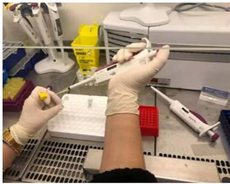
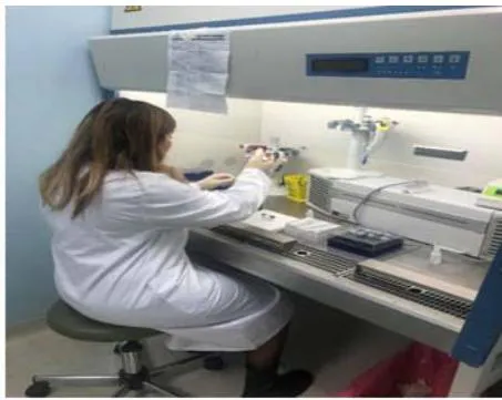
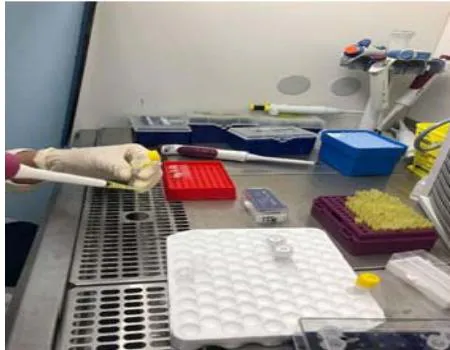
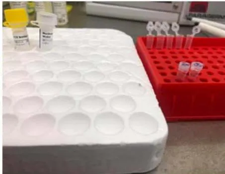
The reaction components of the qRT-PCR mix listed in Table 1 were added to a standard qPCR tube (8-well strip tubes containing Rocket Script Reverse Transcriptase and TaqMan probe pre-mix) (Fig. 2). Next, all strip tubes were vortexed and centrifuged at 3000 rpm for 3 minutes in an Exispin centrifuge and transferred to a real-time PCR thermal cycler.
f) Real-Time PCR Thermocycler Conditions
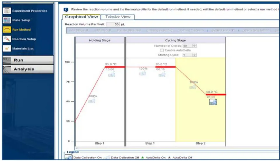
Real-Time PCR thermocycler conditions was set up according to primer annealing temperature and RT-qPCR TaqMan kit instructions as following Table (3):
| Step | Condition | Cycle |
| Reverse transcriptase | 95°C 15 min | 1 |
| Pre-Denaturation | 95°C 5 min | 1 |
| Denaturation | 95°C 20 sec | 45 |
| Annealing/Extension | 60°C 30 sec | |
| Detection (Scan) |
Thermal cycles were applied to inspect the Real-Time PCR and relying on instructions AccuPower® 2X Green-StarTM qPCR Master Mix as well as by calculating the degree Tm prefixes using the device MiniOpticon Real-Time PCR system BioRad/USA as in Figure (3) below:
g) Real-Time PCR Data Analysis
qRT-PCR data analysis was performed by calculation the threshold cycle number (CT value) that presented the positive amplification of gene in Real-time cycle number.
III. RESULTS
a) Direct Examination and Staining
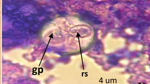
In the present study, Eimeria oocysts were detected in 378 of 816 (46.3%) gastrointestinal tracts of Mugil cephalus examined. This coccidia produces equally spherical oocysts containing sporoblasts and sporocysts (Fig. 4). The oocysts measure um in diameter. The oocyst residue is absent but three polar granules of um diameter each are present (Fig. 4). Each mature oocyst contains four pyriform sporocysts um long and um wide (Fig. 4). At one end of the sporocysts there is a conspicuous projection corresponding to the body of Stieda (Fig. 4). Each sporocyst contains two vermiform sporozoites between which the sporocystic residue is present as three or four refractive granules (Fig. 4).
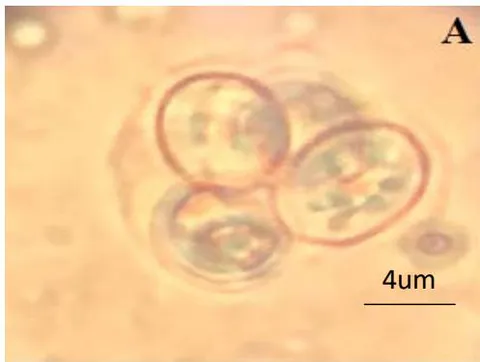
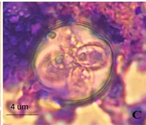
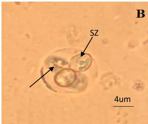
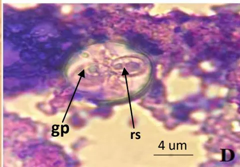
(A) Oocyst containing sporoblasts (bar = 4 um). - (B) Sporulated oocyst. Sporozoites (sz) are visible inside the sporocysts. The arrow indicates the Stieda body (bar = 4 um) - (C) Sporulated oocyst showing three polar granules (gp) and (D) sporocystic residue (rs) in one of the sporocysts (bar = 4 um).
b) Results of Molecular Examination by qRT-PCR
In the present study, Eimeria oocysts were detected in 378 of 816 (46.3%) gastrointestinal tracts of Mugil cephalus. Measurements of sporulated oocysts, sporocysts and other morphological characteristics identified the oocysts as Eimeria sp. We confirmed by molecular analysis of the small ribosomal RNA subunit (rRNA-SSU) gene, a single sequence of bp was obtained for all positive samples. The results of the molecular examination using qRT-PCR revealed that of 378 samples collected, 378 (100%) were positive. This complemented and confirmed the results of our microscopic examinations.
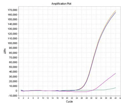
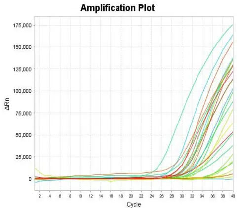
The use of qRT-PCR techniques in the specific detection of Eimeria sp. showed a fluorescence of the SYBER green dye which was most clearly seen through the formation of an amplification pattern for positive samples from cycle 22 onwards as shown in Figure 5.


IV. DISCUSSION
The genus Eimeria comprises obligate intracellular protozoan parasites belonging to the phylum Apicomplexa. Members of this genus cause enteric disease in a wide range of vertebrate hosts, including fish, reptiles, birds and mammals. A total of 157 species of Eimeria that parasitise fish have been described; however, molecular information on these fish parasites is scarce.
In the present study, Eimeria oocysts were detected in 378 of 816 (46.3%) gastrointestinal tracts of Mugil cephalus in the eastern coast of Algeria. Measurements of sporulated oocysts, sporocysts and other morphological characteristics identified the oocysts as Eimeria sp. By molecular analysis of the small ribosomal RNA subunit gene (rRNA-SSU), by quantitative PCR all direct positive samples came back positive with different Ct's from 22. This confirmed the presence of Eimeria sp and complemented the direct examination.
Coccidia of the genus Eimeria Schneider, 1875 produce tetrasporidoocysts and dizoicsporocysts. The sporocysts have a Stieda body and sometimes a Stiedasubbody at one end (Lom and Dykova, 1992). The coccidia described here has these characteristics.
Several studies have used the PCR technique targeting different regions of the Eimeria genome, such as the 5S rRNA (the small rRNA subunit (Mushattat and Sukayna (2013), Ogedengbe et al., 2011), the sporozoite antigen gene EASZ240/160 (Qvarnstrom et al., 2005) and the genomic regions ITS-1 (Long and Reid, 1982, Williams, 1998, Lew et al., 2003) and ITS-2 (Lien et al., 2007; Shirley et al., 2005). As the ITS regions are less conserved than the rRNA genes, the wide variation in this region of the DNA sequence between Eimeria species makes primer design straightforward and reduces the risk of cross-reactions between different species (Morris and Gasser, 2006). The REAL-TIME test has been shown to be directly comparable in sensitivity and robustness, capable of detecting 10 parasite genomes but not a single one, without being affected by the presence of DNA derived from the host or other species tested (Kirs and Smith, 2007). Each sporulated oocyst contains eight eimerial genomes, suggesting that the DNA equivalent of a single oocyst will be consistently detectable given normal experimental replication (between one and 10 genomes detected per reaction). Mature intracellular stages represent in the order of 10 to 100 eimerial genomes (depending on species and stage (Johnston et al., 2001). This suggests that even a fraction of one can be counted (Damer et al., 2008).
V. CONCLUSION
This study is the first to characterise Eimeria sp in Mugil cephalus from the Algerian east coast. Although routine tests such as macroscopic and microscopic diagnosis are important, they are unable to establish a qualitative diagnosis of the Eimeria causing the infection in Mugil cephalus. The use of molecular methods such as real-time PCR which is characterised by high accuracy, but these methods are expensive compared to routine methods. The use of specific primers for the diagnosis of the ITS1 region is important for the molecular detection of Eimeria species that are isolated from the intestines of mules.