I. INTRODUCTION
Every year, around 700,000 patients succumb to antimicrobial resistance (AMR) globally. Projections indicate that this mortality rate could rise to 10 million by the year 2050 (O'Neill, 2014).
The emergence of superbugs, which are pathogenic bacteria resistant to the majority or entirety of available antibiotics, presents a significant challenge. The World Health Organization has cautioned that these multidrug-resistant pathogens could potentially revert global health to conditions reminiscent of the preantibiotic era (WHO, 2017).
There is a pressing necessity to identify novel alternatives to address the issue of antimicrobial resistance. Potential solutions include bacteriophages, antibodies, probiotics, lysins, and antimicrobial peptides (AMPs), which have shown promise in tackling this critical challenge (Ghosh, Sarkar, Issa, & Haldar, 2019).
AMPs are short oligopeptides, comprising up to 50 amino acids, that are present in all living organisms. Plant-derived AMPs represent a valuable natural alternative to synthetic antibiotics for applications in human healthcare and agriculture, particularly for protection and healing (Almeida et al., 2020; Tang, Prodhan, Biswas, Le, & Sekaran, 2018).
These peptides exhibit a wide range of activities, including antibacterial, antifungal, antiviral, and even anticancer properties (R. E. W. Hancock, 2001; Kamysz, Okrój, & Lukasiak, 2003). Their mechanisms of action are varied and closely linked to their structural characteristics; AMPs interact electrostatically with bacterial cell walls, leading to a disruption of membrane integrity (Tzong-Hsien, Kristopher, & Marie-Isabel, 2016). Additionally, these peptides possess the ability to penetrate biological membranes, allowing them to exert effects intracellularly (Cardoso et al., 2019). Beyond their antimicrobial functions, AMPs also play a significant role in immunomodulation (Haney & Hancock, 2013).
The broad activity spectrum, straightforward synthesis, and specific mechanisms of action associated with antimicrobial peptides (AMPs) significantly mitigate the risk of bacterial resistance. This makes them attractive candidates for the development of new therapeutic agents to address the pressing issue of bacterial resistance (Baltzer & Brown, 2011). There is a critical need to identify new AMPs that can either replace or improve the effectiveness of current antibiotics. Moreover, AMPs can be easily obtained from natural sources, such as plants, for applications in human healthcare, agriculture, and the food industry.
This research focused on screening a variety of plant species to identify novel antimicrobial peptides. We achieved this by extracting protein and peptide fractions from the plants and evaluating their antimicrobial efficacy against several standard bacterial strains. The study involved determining the minimum inhibitory concentration of the extracts exhibiting antibacterial properties, isolating the plant protein mixture into distinct proteins and peptide fractions, and localizing the active compounds present in the extracts with antibacterial activity.
II. MATERIALS AND METHODS
a) Plant Material
Seeds and leaves of various plants screened for the presence of antimicrobial peptides (AMPs) are listed in Table 1.
| No. | Binomial Name | Family | Part tested | Protein Concentration (μg/ml) |
| 1. | Punica granatum | Lythraceae* | seeds | 261 |
| 2. | Vitis labrusca 'Fragola' | Vitaceae* | seeds | 4064 |
| 3. | Annona | Annonaceae* | seeds | 4285 |
| 4. | Ricinus communis | Euphorbiaceae* | seeds | 1797 |
| 5. | Ocimum basilicum | Lamiaceae* | seeds | 4561 |
| 6. | Foeniculum vulgare | Apiaceae* | Grounded seeds | 2557 |
| 7. | Cucumis melo flexuosus | Cucurbitaceae* | seeds | 5806 |
| 8. | Ammi visnaga | Apiaceae* | seeds | 1037 |
| 9. | Brassica rapa subsp. Rapa | Brassicaceae* | seeds | 1659 |
| 10. | Matricaria recutita | Asteraceae* | Leaves, flowers | 2351 |
| 11. | Raphanus sativus | Brassicaceae* | seeds | 2156 |
| 12. | Coriandrum sativum | Apiaceae* | Leaves | 2557 |
| 13. | Lepidium sativum | Brassicaceae* | Leaves | 414 |
| 14. | Thymus vulgaris | Lamiaceae* | Leaves | 3253 |
| 15. | Olea europaea | Oleaceae* | Leaves | 4936 |
| 16. | Psidium guajava | Myrtaceae* | Leaves | 2323 |
| 17. | Zingiber officinale | Zingiberaceae* | roots | 2653 |
| 18. | Pimpinella anisum | Apiaceae* | Leaves | 3285 |
| 19. | Rosmarinus officinalis | Lamiaceae* | Leaves | 1543 |
| 20. | Trigonella foenum-graecum | Fabaceae* | Leaves | 4147 |
| 21. | Nigella sativa | Ranunculaceae * | seeds | 4117 |
| 22. | Rumex obtusifolius L. | Polygonaceae* | seeds | 1707 |
| 23. | Salvia hispanica | Lamiaceae* | seeds | 1102 |
| 24. | Cuminum cyminum | Apiaceae* | Grounded seeds | 1382 |
| 25. | Cinnamomum zeylanicum | Lauraceae* | Cinnamon sticks | 2599 |
| 26. | Prunus dulcis | Rosaceae* | seeds | 1974 |
| 27. | Hibiscus sabdariffa | Malvaceae* | flowers | 1828 |
| 28. | Brassica oleracea var. capitata f. rubra | Brassicaceae* | Leaves | 4212 |
b) Bacteria, Growth Media and Reagents
The isolated antimicrobial peptides (AMPs) were evaluated for their efficacy against various bacterial strains from the ATCC collection, which included Gram- positive species such as Staphylococcus aureus ATCC 12493, Streptococcus epidermidis ATCC 12228, and Bacillus subtilis ATCC 6633, as well as Gram-negative species including Escherichia coli NCTC 13846,
Klebsiella pneumoniae ATCC 2021, Pseudomonas aeruginosa ATCC 27853, and Acinetobacter baumannii ATCC 19606.
Muller Hinton Agar (MHA), Nutrient Agar (NA), Nutrient Broth (NB), Brain Heart Infusion Broth (BHIB), 50 mM phosphate buffer pH 7, 2 mM EDTA, Glycerol, 50 mM NaCl, distilled water (D.W), tap water, 0.1% 2,3,5 triphenyl tetrazolium chloride (TTC), n-butanol, acetone, acetic acid, 5% ammonia, ninhydrin, methylthiazol tetrazolium (MTT).
c) Total Water-Soluble Proteins Extraction
The plant material underwent a washing process with tap water followed by distilled water, after which it was dried under sunlight. For the extraction of water-soluble proteins, the milled plant material was combined with a cold extraction buffer at a ratio of 1:10 (w/v), consisting of phosphate buffer at pH 7, 2 mM EDTA, glycerol, and NaCl. Subsequently, the mixture was agitated on a shaker for two hours at a temperature of , followed by centrifugation at for 20 minutes at the same temperature. The clear solution was then filtered through sterile gauze and stored at (Aliahmadi, Roghanian, Emtiazi, & Ghassemi pour, 2011).
d) Total Protein Concentration Estimation
The concentration of the extracted water-soluble proteins was estimated using a nano-drop spectrophotometer.
e) Testing Extracts on Bacteria by Agar Well Diffusion Assay
Muller Hinton agar (MHA) plates were inoculated using a cotton swab that had been moistened with a McFarland standardized test organism. Using a sterile pipette blue tip, holes measuring 6 to in diameter were created and subsequently filled with of the plant extract. The petri dishes were then incubated at for a duration of 24 hours. The diameters of the zones of growth inhibition were measured. The inoculum was prepared in a sterile saline solution, with its turbidity adjusted to the 0.5 McFarland standard . A potassium iodide (KI) solution was utilized as a positive control (Abed, 2015; Nigussie, Davey, Legesse, Fekadu, & Makonnen, 2021).
f) Determination of Minimum Inhibitory Concentration (MIC) by Microtiter Broth Dilution Method
In a 96-well microtiter plate, of plant extract was introduced into the first well, while of BHIB media was allocated to each of the remaining wells. Subsequently, was extracted from the first well and transferred to the second well, continuing this process until the last well to achieve serial dilution for each extract. A volume of of bacterial suspension, calibrated to the 0.5 McFarland standard, was added to each well, with the exception of the last well designated as the "negative control." The plates were then covered and incubated at for 24 hours. Following this incubation, of 2,3,5 triphenyl tetrazolium chloride (TTC) was introduced into each well, and the plates were re-incubated for an additional 15 minutes. The plates were subsequently examined, and the final dilution that exhibited antibacterial activity was identified by noting the last well that did not display red coloration, as the presence of red color indicates bacterial growth (Abou-Elkhair, Fadda, & Abu-Mohsen, 2010).
g) Protein and Peptide Separation by Thin Layer Chromatography (TLC)
This technique was derived from the work of Jaskiewicz et al. (2016), utilizing Silica gel 60 RP-18 F254S (Merck) thin-layer chromatography (TLC) plates as the stationary phase. A volume of from each plant extract was applied to the TLC plate. The separation process employed a mobile phase composed of n-butanol, acetone, acetic acid, ammonia, and distilled water in a ratio of 4.5:1.5:1:1:2 (v/v/v/v/v) within a sealed chamber. The separation was allowed to proceed until the solvent front reached the end line, at which point the plates were removed and air-dried to facilitate the evaporation of the eluents. Subsequently, the dried plates were treated with a (w/v) ninhydrin solution in a mixture of acetone and glacial acetic acid (25:1, v/v). Following this, the plates were left to dry for several minutes at ambient temperature before being heated in an oven at until the peptide zones became visible (Gwarda, Tomczyszyn, Misicka, & Dzido, 2013; Sharma, Abid, & Sajgotra, 2017). For subsequent antibacterial assays, the TLC plates were sterilized under ultraviolet light for 15 minutes. The identification of specific compounds was constrained by the lack of available reference standards.
h) TLC-Agar-Overlay Bioautography
Developed TLC plates were placed within a sterile Petri dish. A suspension of representative bacterial strains, measuring one milliliter, was combined with of melted Mueller-Hinton agar, which was then poured as a thin layer over the developed TLC plate. Following the solidification of the medium, the TLC plate was incubated for 24 hours at a temperature of . Subsequently, the TLC-bioautography plates were sprayed with an aqueous solution of methylthiazol tetrazolium (MTT) at a concentration of (Sigma, USA). Clear zones of inhibition were noted against a purple background (Valle, Puzon, Cabrera, & Rivera, 2016).
III. RESULTS
a) Total Protein Concentration Estimation
The plant protein extracts examined in this study exhibited a range of protein concentrations between 261 and . Among these, the protein extract from
Punica granatum displayed the lowest protein content, while the extract from Cucumis melo flexuosus demonstrated the highest protein concentration. Detailed protein concentrations for each plant extract can be found in Table (1).
b) Testing Plants Protein Extracts on Bacteria by Agar Well Diffusion Assay
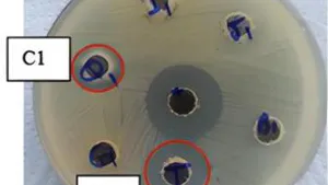
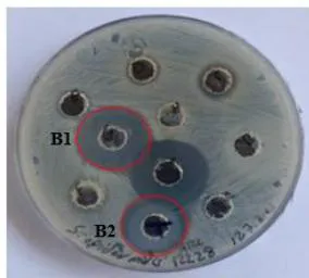
The protein extracts derived from five different plant species exhibited varying levels of antibacterial activity against a range of both Gram-positive and Gram-negative bacteria, with inhibition zones measuring between 11 and . Extracts from Ricinus communis and Vitis labrusca demonstrated antibacterial effects against the Gram-positive bacteria S. aureus and S. epidermidis. The extract from Raphanus sativus displayed a more extensive antibacterial spectrum, affecting both Gram-positive (S. aureus and S. epidermidis) and Gram-negative bacteria (E. coli and A. baumannii). Notably, none of the protein extracts exhibited antibacterial activity against B. subtilis or K. pneumoniae. Antibacterial effects were recorded against S.aureus for the extracts of R. sativus, R. communis, V. labrusca, and Prunus dulcis, with inhibition zones measuring 16, 15, 18, and , respectively (Figure 1).
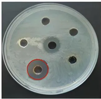
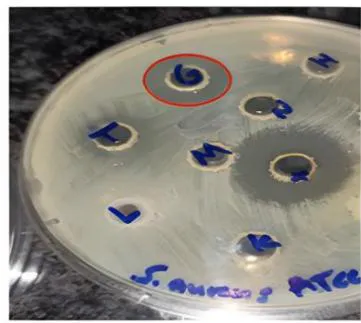
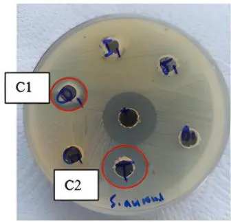
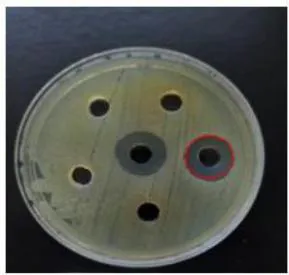
Figure 2 illustrates that the extracts of sativus, communis, and labrusca exhibit antibacterial properties against S. epidermidis, resulting in zones of inhibition measuring 20 mm, 22 mm, and 11 mm, respectively.
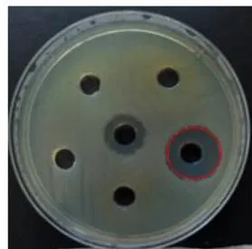
R. sativus has demonstrated antibacterial properties against E. coli, exhibiting an inhibition zone of and against A. baumannii, with an inhibition zone of , as illustrated in Figure 3.
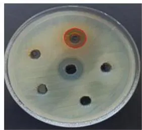
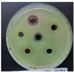
The growth of aeruginosa was suppressed by the protein extract of sabdariffa, resulting in a modest zone of inhibition measuring , as illustrated in Figure 4.
c) Determination of Minimum Inhibitory Concentration (MIC) by Microtiter Broth Dilution Method
The minimum concentration that resulted in the inhibition of bacterial growth by plant protein extracts was determined through the observation of color changes in the wells of a 96 microtiter plate, utilizing the micro broth dilution assay alongside TTC as a growth indicator. The protein extracts from V. labrusca, R. communis, R. sativus, and P. dulcis exhibited growth inhibition against S. aureus at concentrations of 2.03, 0.45, 0.54, and , respectively. Conversely, the protein extracts from V. labrusca, R. communis, and R. sativus demonstrated inhibitory effects on the growth of S. epidermidis at concentrations of 4.06, 0.45, and . Additionally, the growth of A. baumannii and E. coli was inhibited at concentrations of and for R. sativus, respectively. Furthermore, the growth of P. aeruginosa was inhibited by a concentration of of sabdariffa protein extract.
d) Protein and peptide separation by Thin Layer Chromatography (TLC)
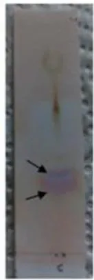
Antibacterial activity from plant protein extracts was isolated using the thin-layer chromatography (TLC) technique, and the resulting fractions were analyzed with ninhydrin spray. The separation of extracts from V. labrusca (Fig. 5-A), P. dulcis (Fig. 5-D), and P. guajava (Fig. 5-E) yielded two distinct bands. In contrast, the separation of R. communis resulted in a continuous line featuring two distinct bands, one positioned at the beginning and the other at the end of the migration line (Fig. 5-B). The separation of Raphanus sativus displayed two intertwined bands (Fig. 5-C). Additionally, the separation of H. sabdariffa resulted in three distinct bands.




e) TLC-Agar-Overlay Bioautography
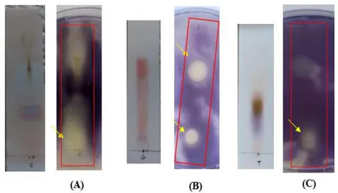
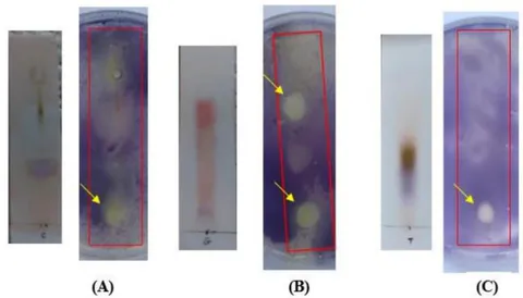
The antibacterial activity of the compounds separated on TLC was further evaluated using thin-layer chromatography (TLC) agar overlay bioautography. The antibacterial properties of V. labrusca, R. communis, R. sativus, Prunus dulcis, Psidium guajava, and H. sabdariffa were evidenced by the presence of clear zones of inhibition against the respective bacteria on a purple background. Notably, V. labrusca, R. communis, and R. sativus exhibited inhibitory effects against both S. aureus and S.epidermidis. Additionally, R. sativus demonstrated inhibitory activity against E. coli and A. baumannii, while H. sabdariffa showed effectiveness against P. aeruginosa. Conversely, Prunus dulcis did not
produce any zones of inhibition against S. aureus. The antibacterial effects of V. labrusca (Fig. 6-A) and R. sativus (Fig. 6-C) against S.aureus are illustrated by clear zones near the origin line, with the absence of stained bands on the corresponding TLC plates indicating that the inhibitory compounds are non-protein in nature. Two stained compounds on the TLC plates of communis (Fig. 6-B) resulted in two zones of inhibition, one located near the origin line ( ) and the other positioned in the middle of the TLC plate ( ).
Comparable outcomes were observed in agar overlay bioautography using S. epidermidis, where V. labrusca (Fig. 7-A) and R. sativus (Fig. 7-C) exhibited minor inhibitory zones near the origin line. In contrast, two stained compounds on the R. communis TLC plate (Fig. 7-B) revealed two distinct zones: one located close to the origin line ( ) and the other positioned in the middle of the TLC plate ( ).
Non-stained fraction on the line of origin of . sativus TLC plate exhibited a large inhibitory zone against E. coli (Fig. 8-A) and A. baumannii (Fig. 8-B).

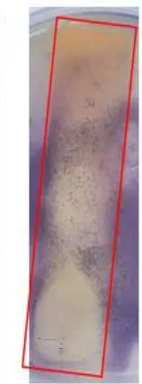

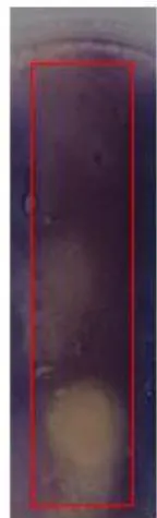
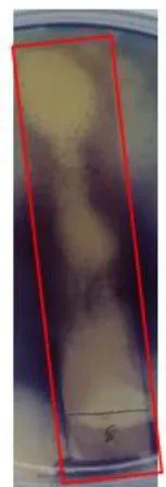
Figure 9 illustrates that the TLC plate of sabdariffa exhibited three inhibitory zones against aeruginosa. The first zone had an value of 0.11, the second zone had an value of 0.46, and the third, which was a non-stained compound, generated a zone close to the end line.

IV. DISCUSSION
The rise in antimicrobial resistance to existing antibiotics is a major global health issue, limiting treatment options. This problem is worsened by the scarcity of newly developed antibiotics. Research shows that antibiotic resistance has increased worldwide over the past twenty years, mainly due to overprescription and misuse of antibiotics. Finding new antimicrobial agents is crucial, yet the development of new antibiotics has slowed down, stressing the need for innovative solutions (Klein et al., 2018; O'Neill, 2016).
Plants have been used for medicinal purposes for a long time and are important in discovering new antimicrobial agents. Many plants contain natural compounds that fight infections. Research emphasizes the value of plants as a source of new antimicrobial compounds due to their diverse secondary metabolites and cost-effectiveness (Cowan, 1999b; Newman & Cragg, 2012; Newman & Cragg, 2020; Atanasov et al., 2015). The extraction of antimicrobial compounds from plants has advantages over synthetic methods, such as lower toxicity risks and unique mechanisms of action. Therefore, exploring plants for new antimicrobial compounds can lead to effective treatments for drug-resistant infections (Anand et al., 2022; Akinyemi, Oladapo, Okwara, Ibe, & Fasure, 2005; Djeussi et al., 2013; Jubair, Rajagopal, Chinnappan, Abdullah, & Fatima, 2021).
Plants contain a diverse range of antimicrobial peptides that can be extracted and studied for their potential therapeutic applications (Broekaert et al., 1997;
Lei et al., 2019; Tam, Wang, Wong, & Tan, 2015). Several studies found that antimicrobial peptides were effective against drug-resistant bacteria, suggesting their potential as a therapeutic option for these infections (Chung & Khanum, 2017; S.-C. Park, Park, & Hahm, 2011).
This research was designed to evaluate various plant species in order to identify novel antimicrobial peptides through the extraction of protein and peptide fractions. The antimicrobial activity of these extracts was tested against a range of standard bacterial strains, and the peptide nature of the isolated substances was confirmed following their separation and subsequent activity testing. This study builds upon previous investigations that have highlighted the potential of peptides derived from plants as a source of new antibiotics (da Silva et al., 2012; Gully et al., 2019; Lage et al., 2018; Ljoljić Bilić et al., 2022).
During the study, twenty-eight plant seeds, leaves, or flowers were collected from local markets. A phosphate buffer was used to extract water-soluble proteins and peptides, followed by purification through TLC chromatography (Melnikova, Mineev, Finkina, Arseniev, & Ovchinnikova, 2016; Osborn et al., 1995; Taveira et al., 2014). The total protein concentration of the extracts was measured using a NanoDrop instrument, which has been documented in various studies (Jafari, Khavari Nejad, Vaziri, and Siadat 2017; Najib 2017; Desjardins and Conklin 2010).
a) Antibacterial Activity of Plant Protein Extracts Against Several Standard Bacteria
As part of this study, Ricinus communis seeds protein extract has been studied for its activity against several Gram-positive and Gram-negative standard bacteria. The findings of this study indicate that the protein extract from R. communis demonstrates antibacterial activity against S. aureus and S. epidermidis. The observed antibacterial properties of the protein extract may be linked to the presence of lectins, such as ricin, which are recognized for their antibacterial effects (Al-Mamun et al., 2016), as well as the presence of antimicrobial peptides (Al-Mamun et al., 2016; Boldbaatar, Gunasekera, El-Seedi, & Goransson, 2015).
Numerous studies have evaluated the antimicrobial properties of communis seed protein extract against various bacteria. For example, research conducted by Al-Mamun et al. (2016) assessed the antibacterial efficacy of the protein extract against coli, aeruginosa, and S. aureus through agar well diffusion and MIC assays, yielding MIC values of 250, 125, and , respectively. Additionally, a study by Afzal, Bakhsh, Ahmad, Manzoor, and Liaquat (2011) utilized the disc diffusion method alongside MIC assays to explore the antibacterial effects of communisseed protein extract against S. aureus, revealing a MIC value of 15.29 mg/ml. Conversely, research by Patil and Bhise (2015) examined the antimicrobial activity of R.communis seed aqueous extract against several bacteria, including S. aureus and P.aeruginosa, using the agar well diffusion method, and found significant antibacterial activity solely against P. aeruginosa. Furthermore, a study by Abd-Ulgadir, Suliman, Zakria, and Hassan (2015) concluded that the seed protein extract exhibited no notable antimicrobial activity against a variety of Gram-negative and Gram-positive bacteria.
The variations noted in the results of different studies could be attributed to the diverse methodologies used in the protein extraction from seeds (Ben Brahim et al. 2022; Djeussi et al. 2013). Additionally, differences in bacterial strains and the specific experimental culture conditions applied may also contribute to these discrepancies (Ben Brahim et al. 2022). Furthermore, the exact mechanism through which the protein extract functions has not been sufficiently clarified (Worbs et al. 2011).
This study demonstrates that the extract derived from Raphanus sativus seeds exhibited antibacterial properties S. aureus, S. epidermidis, E. coli, and A. baumannii, with concentrations ranging from 0.537 to . The antibacterial effects observed may be linked to the presence of compounds such as alkaloids, flavonoids, and saponins, which are likely to be extracted using an aqueous cold extraction method (Ahmad, Hasan, Chishti, & Ahmad, 2012).
Two investigations conducted by Ahmad et al. (2012) and Khamees (2017) assessed the antimicrobial properties of . sativus seed extract against various bacterial strains, including S. aureus, K. pneumoniae, P. aeruginosa, and E. coli. The findings from both studies indicated that the . sativus seed extract demonstrated considerable antibacterial efficacy against all bacterial strains examined. Additionally, both studies revealed the presence of various bioactive compounds, such as alkaloids, flavonoids, and saponins, within the extract, which are believed to play a role in its antibacterial effects. In a related study by Jadoun, Yazbak, Rushrush, Rudy, and Azaizeh (2016), the antibacterial activity of . sativus seed extract was tested against several standard bacterial strains, including S. aureus, Escherichia coli, and K. pneumoniae, with minimum inhibitory concentrations ranging from 0.5 to . The study also identified a novel sulfur compound that contributes to the antibacterial activity. Collectively, these studies indicate that . sativus extract holds promise as a natural antibacterial agent, with potential applications in food preservation and various other industries.
A study conducted by Törün, Çoban, Biyik, and Barişik (2017) examined the antimicrobial properties of methanolic extracts from R. sativus against ten distinct pathogenic microorganisms, such as S. aureus, E. coli, and K. pneumoniae. Utilizing the Disc diffusion method, the findings revealed that the extract exhibited antibacterial activity solely against S. aureus.
In the current study, the growth of S. aureus was found to be inhibited by of Vitis labrusca seeds protein extract. Additionally, the extract was also observed to inhibit the growth of S. epidermidis at a concentration of . Few studies have investigated the antibacterial properties of V. labrusca seed extracts. Junior et al. (2021) found that the extracts presented some inhibitory effect against E. coli (IAL 2064) and S. aureus.
In this investigation, the findings indicate that the protein extract derived from Prunus dulcis seeds exhibits a minimal antibacterial effect against S. aureus. Nevertheless, the extract failed to show any antibacterial properties against the other bacteria that were examined. Conversely, research conducted by Hifza (2018) on the antimicrobial characteristics of fatty acids extracted from P. dulcis seeds revealed a moderate inhibitory effect against three bacterial strains, including S. aureus, B. subtilis, and E.coli, which aligns partially with the results of the current study.
Additional research has explored the antibacterial properties of dulcis seed extract, suggesting that the presence of various bioactive compounds, such as phenolic compounds and flavonoids, may play a role in their antibacterial efficacy (Dhingra, Kar, Sharma, & Bhasin, 2017; Shelly, Shikha, & Narayan, 2015). Consequently, while the protein extract from dulcis seeds appears to have limited antibacterial effects against S. aureus, it is plausible that other compounds within the seeds possess more potent antibacterial properties, warranting further investigation to assess their potential applications.
The findings of this research indicate that Hibiscus sabdariffa effectively inhibited the growth of . aeruginosa. Numerous studies have investigated the antibacterial properties of . sabdariffa calyces extract. In 2011, a study found that the methanolic extract showed antibacterial activity against various bacteria (Elmanama et al., 2011). In 2022, Khalil et al. found that the . sabdariffa extract had bactericidal effects against multidrug-resistant . aeruginosa from burn wound exudates. Al-Hashimi's 2012 study found significant inhibition zones for . coli and S. aureus. Vargas-Sanchez et al.'s 2018 study found moderate to high antibacterial activity against various Gram-positive and Gram-negative bacteria.
b) Thin-layer chromatography (TLC) and TLC agar overlay bioautography for plant protein extracts
During the course of this research, the use of TLC chromatography with agar overlay bioautography to investigate Ricinus communis produced noteworthy findings, demonstrating two separate zones of inhibition on the agar plate against S. aureus and S. epidermidis. One zone corresponded to a fraction with an Rf value of
0.06, while the other was linked to a fraction with an Rf value of 0.73. These zones of inhibition may be attributed to the presence of lectins, such as ricin, known for their antibacterial properties, as well as antimicrobial peptides, as indicated by Al-Mamun et al. (2016). Further investigations are necessary to isolate and characterize the chemical nature of each fraction. In a separate study conducted by Lekganyane (2015), TLC chromatography was employed to separate phytochemical compounds from various leaf extracts, including that of communis. The resulting fractions were subjected to bioautography to evaluate their antimicrobial efficacy against different bacterial strains. The study revealed that a fraction with an Rf value of 0.77 demonstrated significant antimicrobial activity against S. aureus.
The antibacterial activity of V. labrusca seed extract was assessed using TLC plates and a TLC agar overlay bioautography assay. The results showed two stained fractions but no antibacterial effects against S. aureus and S. epidermidis. A zone of inhibition was noted near the sample application line, suggesting that non-proteinaceous compounds may be responsible for the antibacterial effects. Previous studies identified bioactive polyphenols like anthocyanins, flavonoids, and resveratrol in V. labrusca, which are known for their antioxidant, anti-inflammatory, and antimicrobial properties (Xia, Deng, Guo, & Li, 2010; Yu & Ahmedna, 2013). These compounds likely contribute to the antibacterial activity observed in the extract.
In the context of this study, the thin-layer chromatography (TLC) analysis of Raphanus sativus seed extract revealed two overlapping bands, neither of which demonstrated any antibacterial properties. Nevertheless, a notable zone of inhibition was detected adjacent to the sample application line on the TLC plate when tested against S. aureus, S. epidermidis, E. coli, and A. baumannii. The findings suggest that the antibacterial effects of sativus extract are unlikely to be linked to the proteins or peptides present, as evidenced by the lack of antibacterial activity in the two protein bands observed. Instead, the inhibition zone implies that other constituents within the extract may be responsible for the antibacterial effects. Research has pointed to sulfur compounds, particularly isothiocyanates, as significant contributors to the antibacterial properties of sativus extract, with sulfuraphane being a prominent isothiocyanate known for its efficacy against various bacterial strains (Gutiérrez & Perez, 2004; Jadoun et al., 2016; Lim, Han, & Kim, 2016). The presence of these sulfur compounds may elucidate the antibacterial activity observed in sativus extract, underscoring its potential as a natural source of antimicrobial agents In this research, the antibacterial properties of the extract derived from Prunus dulcis seeds were assessed. Interestingly, although the agar well diffusion
method indicated minimal antibacterial effects against S. aureus, the agar overlay bioautography did not reveal any significant antibacterial activity from the extract. This discrepancy may suggest that the concentration of antibacterial compounds present in the extract was insufficient to be detected by the agar overlay bioautography technique (Bieleski & Turner, 1966).
In the course of this investigation, the protein extract from the calyces of Hibiscussabdariffa was subjected to thin-layer chromatography (TLC), which successfully identified three distinct stained bands on the chromatographic plate. Subsequent evaluation of the sabdariffa extract against aeruginosa through agar overlay bioautography demonstrated the presence of several large zones of inhibition, a stark contrast to the smaller zone noted in the agar well diffusion assay. This discrepancy in findings may be attributed to the potential antagonistic interactions among the extract's components during the agar well diffusion assay; however, following separation via TLC, these compounds likely ceased to interact, thereby allowing for individual antimicrobial activity and resulting in the observed larger inhibition zones in the agar overlay bioautography. The phenomenon of antagonism among constituents within a single plant extract has also been documented in research conducted by Vaou et al. (2022) regarding artemisinins, which are antimalarial compounds derived from Artemisia annua. Additionally, another plausible reason for the variation in outcomes between the agar well diffusion assay and the agar overlay bioautography could be that the TLC process eliminated certain interfering substances that were present in the original extract, as noted by Bieleski and Turner (1966).
The identification of several zones of inhibition against aeruginosa indicates that the plant extract is likely composed of various bioactive compounds with antibacterial properties. Notably, only one of these inhibition zones was linked to the protein fraction, while the remaining two zones were located close to the sample application line and the edge of the TLC plate. This observation implies that a limited aspect of the antibacterial effect may be due to the presence of antimicrobial peptides. Furthermore, there is a lack of prior research that has successfully isolated or evaluated antimicrobial peptides derived from the extract of sabdariffa calyx.
In the present work, the antibacterial properties of several plant extracts, including Vitis labrusca, Ricinus communis, Raphanus sativus, Prunus dulcis, and Hibiscus sabdariffa, were examined through various methodologies such as agar well diffusion assay, micro broth dilution, bioautography, and thin-layer chromatography (TLC). The findings indicated that the antibacterial effects of these extracts could not be exclusively linked to the presence of proteins or peptides; rather, other phytochemicals, including polyphenols, flavonoids, tannins, and alkaloids, may play a significant role in their efficacy. The specific active compounds were not identified, which points to the necessity for additional investigations employing advanced techniques like mass spectrometry and nuclear magnetic resonance (NMR) spectroscopy. The study also highlighted the intricate nature of plant extracts and the difficulties associated with pinpointing their active constituents. Future research endeavors may focus on the isolation and characterization of individual compounds, as well as exploring their mechanisms of action against bacterial strains. Overall, this investigation offers valuable insights into the potential of plant extracts as antibacterial agents and emphasizes the critical need for continued research in this field.
Disclosure statement: The authors declare no conflict of interest.