INTRODUCTION
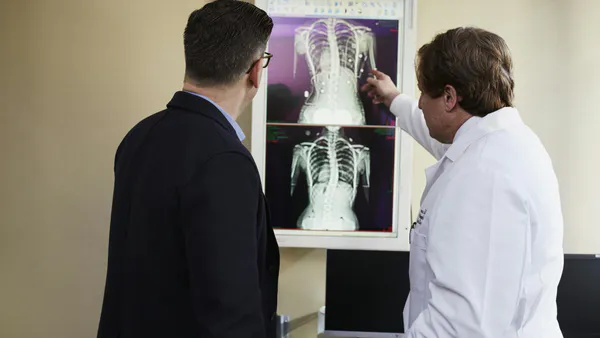
Having had some experience in various medical disciplines including diagnostic radiology (primarily), radiotherapy, nuclear medicine, surgery and primary care and after several visits to Central America, I have come to the following conclusions concerning medical care in the developing world: The basic routine history and physical examination remains the solid bedrock foundation for good medical care for the patient. One-on-one with the patient is irreplaceable. Vital well-proven adjuncts are simple imaging studies, mainly x-ray and ultrasound, in addition of course to laboratory studies[^10]. More sophisticated imaging procedures like CT, MRI, PET and nuclear medicine studies are inappropriate as first-line studies for the developing world because of cost, inconvenience and limited relevance. The World Health Organization, recognizing this state of affairs, designed a simple, easily used and serviced, highly efficient, low-cost x-ray unit, which is highly suitable for developing nations with climate and energy challenges15,9. The unit was designed as the WHIS-RAD, the World Health Imaging System-Radiology. The unit was designed by WHO radiologists and physicists: Dr. Philip Palmer (University of California), Dr. Thure Holm (University of Lund, Sweden) and Dr. Gerald Hanson (Pan American Health Organization). Over 1,000 units are now in use around the world, providing a service which was not available previously. The simple chest x-ray, a hallmark examination, had been a distinct luxury, but no longer. As the COVID-19 virus is primarily a respiratory disease, a routine chest x-ray is more important than ever. Naysayers over the years have disparaged the WHIS-RAD as too simple and low-tech to meet the need. However, the great success of the unit has proven otherwise.
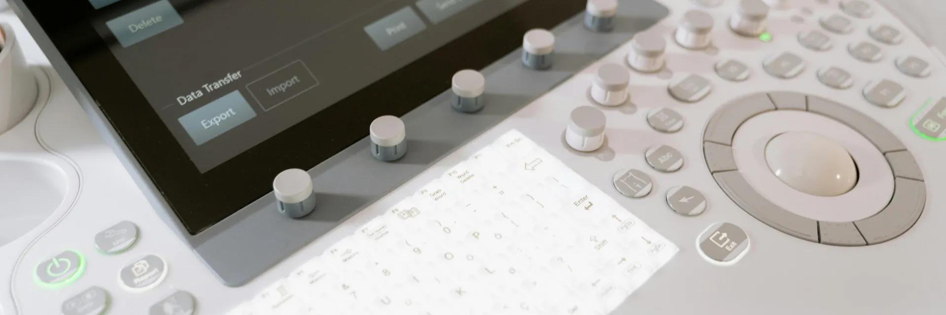
It is important to keep in mind and to stress that about of medical conditions in the developing world can be diagnosed with just two imaging modalities: WFUS-RAD and a portable ultrasound unit like Lumify by Philips. The range of conditions is extensive, including tuberculosis, pneumonia, COVID pneumonia, pulmonary masses, chronic obstructive pulmonary disease (COPD), cardiac failure, air, fluid or blood in the pleural space, bowel obstruction, genitourinary abnormalities, fractures, abdominal and thoracic and skull trauma. This is quite a list, but not complete. Obstetrical and gynecological pathology, including ectopic pregnancy, breech presentation, multiple pregnancies, placental abnormalities, among others, will be diagnosed by ultrasound. Maternal and infant mortality remain monumental issues in the developing world. Beyond the tactical challenges encountered in the installation of WHIS-RAD and ultrasound, there are the issues of interpretation, appropriate training, storage, and transmission of the images to distant locations. None of these issues is insurmountable, as evidenced by successes, for example, in Guatemala[^718], Nepal, Uganda and Tanzania. This represents a small beginning for a project that will stretch out over the coming decades. Success in this endeavor can only be achieved by partnering with organizations such as: World Health Organization; Pan American Health Organization; American College of Radiology; Rotary International; Radiological Society of North America; manufacturers, e.g. Sedecal and Carestream and Philips; academic institutions; and international organizations, e.g. Imaging the World arid RAD-AID. An excellent example of partnering is practiced by Diagnostic Imaging Informatics Support Team or DIMIST (See: w5vw.dimist.org)?
Hopefully, the health worker toiling in the developing world will now have the necessary technical tools to facilitate his or her mission.