I. INTRODUCTION
Grading of Meningiomas using Meningiomas are relatively common dura-based tumors that constitute about 25-30% of primary brain tumors in Saudi Arabia (1,2). Grading of meningiomas has an essential role in patient management, including classification, treatment and prognosis prediction. Tumors with a higher grade have more chances of increased recurrence and mortality rates (3). The treatment of atypical and anaplastic meningioma is based on surgery and radiation therapy (4). The updated 2016 edition of the World Health Organization (WHO) Classification of Tumours of the Central Nervous system (5) provides the criteria used in the classification of meningiomas into three grades (from grade I to grade III). These criteria include the subtype of the tumor, brain invasion or mitotic counts, in addition to the presence of an overtly malignant high-grade morphology (i.e. sarcomatoid, carcinomatoid or melanoma). If three out of specific five criteria are present in a given meningioma, it should be graded as grade II. These five criteria are increased cellularity, small cells change with high N/C ration, large and prominent nucleoli, patternless or sheet-like growth and foci of spontaneous or geographic necrosis. These criteria do not clarify a quantitative definition. This paper explores the responses of ten pathologists who examined ten cases of meningiomas, where grading variability may occur due to different interpretations of these five criteria.
II. METHOD
sheet was used, expecting each pathologist to follow the WHO (2016) criteria for Meningioma grading. The results obtained from each pathologist were independently documented as meningioma (grade I), atypical meningioma (grade II) or anaplastic meningioma (grade III). A Neuropathologist recorded the morphological features of the ten cases. The results were tabulated and analyzed by the multiple-reader Cohen kappa statistical analysis method using a website based calculator (6) and a free-marginal multi-rater kappa. The aim was to assess the precision of agreement between the various observers (interobserver agreement).
III. RESULTS
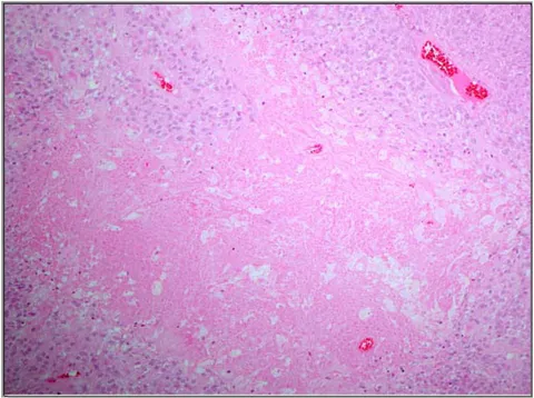
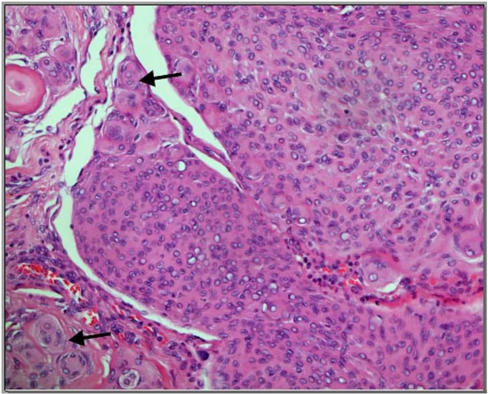
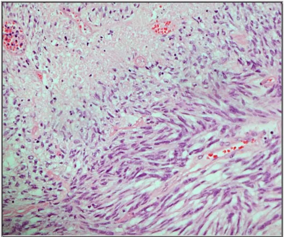
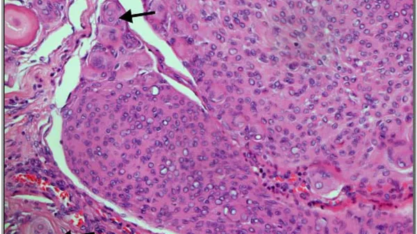
The results obtained by the ten participating pathologists and the two neuropathologists are summarized in Table 1. The kappa score for interobserver agreement between the ten anatomical pathologists was 0.53 (95% CI for free-marginal kappa [0.32, 0.74]) with an overall agreement percent of 68.44%. The kappa score for interobserver agreement between the two neuropathologists was 0.55 (95% CI for free-marginal kappa 0.1, and 1) with an overall agreement percent of 70%. Both kappa scores are in keeping with above chance "intermediate to good" agreement (7). The cases that showed 100% agreement were three cases. Two cases were WHO grade I. The third case was WHO grade II (Case 2, Figure 1). The latter was the only meningioma from the selected cases that showed focal necrosis. Another five cases were graded as either WHO grade I or WHO grade II by the reviewers. These cases were showing a variable degree of small cell change, lack of pattern, and cellularity (Figure 2). These cases did not show features of necrosis or prominent nucleoli. Two cases (Cases 8 and 9) were labelled as WHO grade II by the majority of the reviewers. However, in each one of them, two reviewers labelled them as WHO grade III (Figure 3). In their opinion, the reason for such designation was the focal presence of sarcomatoid morphology.
IV. DISCUSSION
Meningiomas grading has an essential role in patients management and related research studies (8). The risk of recurrence further increases with WHO grade. In one study, patients with benign, atypical, and malignant meningiomas had a 10-year cumulative incidence of recurrence of 6%, 17%, and 30%, respectively. The 10-year relative survival of patients with WHO grade I, II, and III meningiomas were 97%, 90%, and 30%, respectively. These numbers demonstrate the significant increase in tumor-related mortality based upon the WHO grade (3). There is no clear recommendation about the use of radiation therapy in meningiomas (9). However, the higher grade the tumor, the more chances that the patient will receive adjuvant therapy (e.g., External Beam Radiotherapy). Surgical resection extent is the most important prognostic factor among malignant meningioma patients (3, 10). The extended safety margins are necessary to achieve a favorable local control for high-grade meningiomas (11). 15 to 80% improvement of the 5-year progression-free survival was reported when RT was added to surgical resection for malignant meningioma. Atypical meningiomas appear to be more frequently diagnosed under the WHO classification system updates (12). No consensus exists for "atypical" meningiomas treatment, and radiation therapy has mostly been reserved for recurrence and progression (13,14). Gross total resection and adjuvant radiation therapy appear to be highly associated with improved survival, independent of other factors, in patients with atypical meningiomas (15). Overall, the grading of meningiomas is essential and has a significant impact on both the clinical research studies and the treatment of these tumors.
Among the ten practicing pathologists and the two neuropathologists, this study's findings show that the inter-observer agreement on the grading of meningioma that is based upon the presence of the specified three out five features is "intermediate to good" above chance. Clinicians and researchers should be aware of this issue and the subjectivity element in the grading criteria. Three cases out of ten had a perfect agreement. Seven cases had discrepancies, while five cases were graded either as grade I or grade II, and two cases were essentially graded as grade II or grade III.
This paper does not incorporate grade II and grade III meningiomas that were classified based on relatively more objective criteria, including the brain invasion or the specific tumor subtype Morphology. Similar studies are limited in this field. In one study, the mitotic count is considered an objective method of grading, but variation in the grading using mitoses has been reported between pathologists based upon the number of fields examined (16). Another study (17) using previous WHO versions showed high concordance between the pathologists for brain invasion, mitoses/10 high-powered field and spontaneous necrosis. The concordance was lowest for small cells, sheeting and mitoses/10 HPF. For atypical meningioma, the criteria of diagnosis include the presence of the three out of five morphological features (3 out 5), as mentioned earlier. The case that displayed necrosis was the only one that had a perfect agreement as grade II. It appears that necrosis presence was a feature that prompted all the pathologists to look for more needed features to label the tumor as grade II during the case screening. However, the rest of the five features appears to be more problematic. The WHO criteria do not state the percentage of the tumor area, showing the features that are needed to apply criteria. Furthermore, the features are needed to apply criteria. Furthermore, the features do not have a quantitative definition. For example, regarding "increased cellularity", the criteria do not mention how many cells are needed per space unit to consider the tumor cellular. For "small cell change", the N:C ratio that should be present to consider a tumor cell as a small cell is not mentioned. "Prominent nucleoli" is left for the pathologist's judgments. In other body systems, the magnification power is used as a quantitative method for defining visible, prominent nucleoli (18).
For grade III tumors, one of the defining grading methods is the presence of sarcomatoid or carcinomatoid morphology. No statement of the volume of the tumor that should show this feature is clarified in the WHO criteria. Besides, these patterns may have a room for personal interpretation and opinions diversity. This explains the two cases where two pathologists labeled them as grade III, while the majority of the remaining pathologists graded them as grade II.
V. CONCLUSION
Atypical and anaplastic meningiomas can be challenging diseases, not only from a treatment perspective but also from a diagnosis perspective. As demonstrated, the current Meningioma grading system provided by the WHO book does not draw a sharp line between the different grade categories in a significant subset of meningiomas, and more precise criteria and definitions can help. This issue is particularly applicable to the following features: cellularity, lack of pattern, small cell change, prominent nucleoli and sarcomatoid or carcinomatoid morphology. A significant difference in interpretations may make it difficult to establish a definitive cut off that would translate accurately from one laboratory to another. Hence, clinicians and researchers should be aware of this concern. Understanding the grading criteria and the pathology report and communication with the pathologist are an essential element of meningiomas management. These tumors are signed out by general Anatomical Pathologists in many places. Hence, they should also be aware of the meningioma grading criteria and related-concerns. The pathology reports should include the basis of the grading and any difficulty that is associated with it. Besides, intradepartmental consultations, Pathologist's education and joint reporting by two experienced pathologists can help to maintain a high level of grading concordance. The biological signature of meningiomas is likely to play a significant role in the evolution of the grading system strategy. Hopefully, studies focusing on the immunohistochemical and genetic features of these complex tumors and relating these features to the treatment response and the prognosis will provide a more reproducible system with better concordance between pathologists and laboratories.
ACKNOWLEDGMENTS
The author would like to express his gratitude to all the pathologists who kindly participated in this study.
| Cases | NP | NP | AP | AP | AP | AP | AP | AP | AP | AP |
| 1 | 1 | 1 | 1 | 1 | 1 | 1 | 1 | 1 | 1 | 1 |
| 2 | 2 | 2 | 2 | 2 | 2 | 2 | 2 | 2 | 2 | 2 |
| 3 | 1 | 2 | 1 | 1 | 2 | 2 | 1 | 2 | 1 | 1 |
| 4 | 1 | 1 | 1 | 1 | 1 | 1 | 1 | 1 | 1 | 1 |
| 5 | 2 | 2 | 2 | 2 | 1 | 1 | 2 | 2 | 1 | 2 |
| 6 | 1 | 1 | 1 | 1 | 1 | 1 | 2 | 2 | 1 | 1 |
| 7 | 1 | 1 | 1 | 1 | 1 | 1 | 2 | 2 | 1 | 2 |
| 8 | 2 | 2 | 2 | 2 | 2 | 2 | 2 | 3 | 2 | 3 |
| 9 | 3 | 2 | 2 | 2 | 1 | 2 | 2 | 3 | 2 | 2 |
| 10 | 1 | 2 | 1 | 1 | 1 | 1 | 2 | 2 | 1 | 1 |