I. INTRODUCTION
Mangroves are saline resistant forest ecosystems found in tropical and sub – tropical intertidal regions around the world (Ottoni et al., 2015). Mangrove soils have a unique character for the growth of various kinds of microorganisms that play an important role in degrading the soil. Several factors that affect mangrove ecosystems cause microorganisms to adapt by producing unique metabolites, namely primary and secondary metabolites (Thatoi et al., 2013). Mangrove soils provide a unique ecological niche for the growth of diversified microorganisms which find use in recycling environmental nutrients and production of exclusive secondary metabolites of pharmaceutical importance. The total microbial community of tropical mangrove forest comprise 91% of bacteria and fungi, 7% of algae and 2% of protozoa. (Pallaa et al., 2018).
Actinomycetes are group of aerobic, branched, unicellular Grampositive bacteria with high percentage of G+C (70%) in their genetic material. This bacterium has many important roles in various industries because of its ability to produce a number of diverse metabolite compounds. These metabolite compounds have benefits such as, antibiotics, antifungal, antiviral, anticancer, enzymes, immune suppressants and other compounds that are beneficial in industry (Xu et al., 2014). Actinomycetes particularly Streptomyces are well known as major sources of secondary metabolites particularly antibiotics (Berdy, 2005).
Many researchers discovered novel actinobacteria from poorly explored mangrove habitats such as isolation of Asanoa iriomotensis (Tamura and Sakane, 2005), Nonomuraea maheshkhaliensis (Ara et al., 2008) and Streptomyces xiamenensis (Xu et al., 2009). Aquatic actinomysetes have been shown to have an important role in the discovery of several new bioactive compounds such as research conducted by Huang et al. (Huang et al., 2008) on rifamycin from Micromonospora, Fehling et al. (Feling et al., 2003) found salinosporamide-A as an anticancer metabolite of the Salinispora strain, Marinomisin from Marinophilus sp. and much more. According to Anzai et al. (2008) out of 22,500 biologically active compounds, 45% are derived from actinomycetes.
Actinobacterial diversity from these ecosystems has been studied worldwide for their unique biochemical processes. The present study includes isolation, morphological characterization and identification of rhizospheric actinobacteria using biochemical and molecular biology techniques (Brinda and Mathew, 2012; Page, 1997). Molecular biology techniques like 16S rRNA techniques are an important tool in final identification of bacteria sequencing this gene, and provide genus and species identification for isolates that do not fit any recognized biochemical profiles. It gives acceptable identification which otherwise according to conventional system of taxonomy is not possible (Malik et al., 2008). Zhao et al. (2011) recognized that antimicrobial activity and amplifying genes coding for PKS-I, PKS-II and NRPS from endophytic actinomycetes isolated from medicinal plants in Panxi plateau performed as valuable reservoirs of novel bioactive compounds therefore the method has been studied the distribution of PKS and NRPS biosynthetic systems in a collection of wild-type actinomycetes isolated from tropical soil samples (Ayuso et al., 2005).
This research was conducted to isolate and identify actinomycetes from Can Gio Mangrove ecosystem, HoChiMinh city, Vietnam. Actinomycetes in this ecosystem are thought to have the potential to produce primary and secondary metabolites. Actinomycetes will produce primary and secondary metabolites in extreme conditions like the Can Gio mangrove ecosystem, which is polluted by various types of waste from rivers passing through big city as HoChiMinh city. The results of isolation from this study can be used for further research such as screening for primary and secondary metabolite produced by actinomyces.
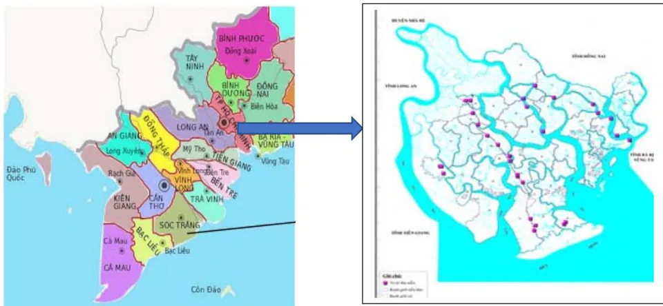
The Can Gio Mangrove (approximately 35,000 ha, extending from 10_220-10_440N and 106_460-107_010E (Tuan and Kuenzer,2012)) is located in the south of Vietnam, at the downstream part of the Sai Gon and Dong Nai Rivers watershed and in the SouthChina Sea coastal zone (Figure 1). Very little information is available about beneficial bacterial diversity (Thanh Nho et al., 2018) and their activity in mangrove soil of Vietnam. Therefore, the aims of this study were (i) to isolate and select together with identify actinobacterial isolates having good resistance to human pathogenic bacteria (ii) to obtain their characterization as analysing genes coding for PKS-I, PKS-II, and NRPS actinobacteria as well as to explore the potential use of these newly actinobacteria as a novel source of bioactives, CMC degradation and phosphate solubilization (iii) to identifying by 16S rDNA genes techniques and sequencing.
II. MATERIALS AND METHODS
a) Isolation
i. Collect of plant samples and rhizosphere soil
Plant soil samples were collected carefully from many species of mangroves viz. Bruguiera sexagula, Ceriops decandra, Sonnertia, Avicennia, Rhirophora..., from the 3 years-old plants in plantation site, Tam Thon Hiep – village, (soil pH = 4.22, salinity 10‰; Thanh An site, (soil pH= 6.18, salinity 7‰;An Thoi Đông village (soil pH= 4.16, salinity 8‰). (Lat. 10°68' 04" N; Long. 107° 02' 64" E) (Figure 1).
ii. Bacterial isolation
The samples were collected on in December, 2019. For isolation of bacterial rhizosphere, samples were collected during the low tide. Soil samples were collected by using a sterile spatula and stored in sterile polythene bags, and then were stored in ice-box (5°C) and transportation to Can Tho University as soon as possible; soil samples were stored in refrigerator (-10°C) in Microbiology Lab. until isolation. The soil samples were removed adherent particles and were superficially disinfected according to Araújo et al.(2014).
A known weight of soil (1 g) was aseptically weighed and transferred to a stoppered (150 mL) sterile conical flask containing 99 mL of sterile saline (0.9%) diluent. The sediment-diluent mixture was agitated by means of mechanical shaking for about 45 minutes. After the above time, the supernatant was collected and streaked on the Starch Casein Agar medium (Mohseni et al., 2013) was used for the isolation of actinobacteria. It was supplemented with Aginalxic (0.5 mg/L) and Nystatin (0.5 mg/L) to inhibit fungi and Gram-negative bacteria. The inoculated plates were incubated at 28°C for 3–6 weeks. The colonies bearing distinct morphological characteristics were picked up and transferred to freshly prepared media until pure cultures were obtained.
b) Morphological Characterization
The morphological characterization of the bacterial colonies were carried out according to on the basis of their shape, size, colour, margin, elevation on the media and Gram staining method was also performed to decide the further determinative protocol. All isolates were tested on CSA media with higher NaCl concentrations (4.50% NaCl).
c) Screening assays for antibacterial activity
The bioactivity of bacterial isolates was examined (Gobi et al., 2012; Manikandan et al., 2014). The pathogenic bacteria including Bacillus cereus, Escherichia coli, Staphylococcus aureus and Vibrio haemoparalyticus were provided by College of Aquaculture and Fisheries, Biotechnology Research and Development Institute (Can Tho University) and Can Tho Center for Technology, Standard, Quality (Department of Science and Technology, Can Tho City). The liquid cultures were grown with shaking at 150 rpm for 7–14 days depending on their growth rate at 30°C. The broth was centrifuged in 50 mL falcon tubes at 4,193xg for 15 min at room temperature (28-32°C); Megafuge 1.0R, Heraeus) and the supernatant was stored at 4°C. The bacterial organisms were plated in Mueller Hinton Agar medium. Antimicrobial extract was added to the wells, then the plates were incubated at 30°C for 2 – 3 weeks for diffusion of antimicrobial extract and observed for the zones of inhibition at 21 day after incubation.
d) The Agar well diffusion method
The active isolates were cultured by the method given in the previous step. The supernatants were used for testing extracellular antimicrobial activity by the agar well diffusion method. By using a sterile cork borer, the wells were punctured in appropriate agar medium previously seeded with one of the test organisms. One hundred microlitre of the culture supernatants were added to each well. The plates were then incubated at C for at least 2 h to allow the diffusion of crude extracts followed by incubation for 2-3 weeks at C for bacteria. The diameters of inhibition zones were monitored and measured (Rinaudo, 1992).
e) 16S rDNA Gene Amplification and Sequencing
The amplification of 16S rDNA by PCR was carried out using the universal primers 27F (Weisburg et al., 1991) and 1492R (Reysenbach et al., 1992). The 50 L reaction mixture consisted of 2.5 U Taq Polymerase (Fermentas), 50 M of each deoxynucleotide triphosphate, 500 nM of each primer (Fermentas) and 20 ng DNA. The thermocycling profile was carried out with an initial denaturation at 95°C (5 min) followed by 30 cycles of denaturation at 95°C (30 s), annealing at
55°C (30 s), extension at 72°C (90 s) and a final extension at 72°C (10 min) in C1000 Thermal Cycler (Bio-Rad). Aliquots (10 μL) of PCR products were electrophoresed and visualized in 1% agarose gels using standard electrophoresis procedures.
The following actinobacteria-specific primers were used for the amplification of actinobacterial 16S rRNA gene fragment (Stach et al., 2003). Cycling conditions were as follows: initial denaturation at 95°C for 4 min, 30 cycles of 95°C for 45 s, 68°C for 45 s, and 72°C for 1 min, and a final extension of 5 min at 72°C. S-C-Act-0235-a-S-20(5'-CGCGGCCTATCAGCTTGTTG 3'), and S-C-Act-0878-a-A-19 (5'-CCGTACTCCCCAGGC GGGG-3')
f) Sequence Analysis
The 16S rRNA gene sequences were compared with those from the type strains available in NCBI (http://www.ncbi.nlm.nih.gov/) using the Basic Local Alignment Search Tool (BLAST) (Altschul et al., 1990). For the phylogenetic analysis, multiple sequence alignment was performed using CLUSTALX, version 1.81. Phylogenetic tree was constructed using Mega 7.0 (Kumar et al., 2018). The consistency of the trees was verified by bootstrapping (1000 replicates) for Maximum Likelihood method.
Detection and Analysis of PKS-I, PKS-II, and NRPS
PCR primers and amplifications were carried out and analysed (Ayuso et al., 2005; Zhao et al., 2011).
g) Statistical analysis
The experimental results were analysed as a two-way ANOVA with the isolates and with levels of diameters of inhibition zones. All analyses were conducted using the programme MSTATC, Minitab 16. The data were considered significantly different at p<0.01. Duncan test at P = 0.01 was used to differentiate between average value statistically.
III. RESULTS AND DISCUSSION
a) Isolation
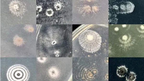
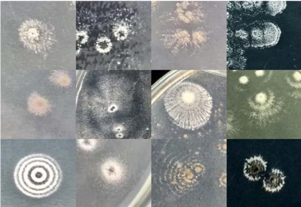
A total of 63 isolates of actinomycetes was purified from 25 soil samples collected at 9 sites of CanGio mangrove forest. Almost their colonies have round-shaped; milky, white clear and yellow, entire or loabate margin; diameter size of these colonies varied from 0.2 to 3.0 mm and all of them have Gram-positive. (Figure 2).

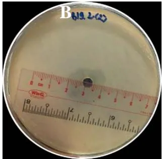
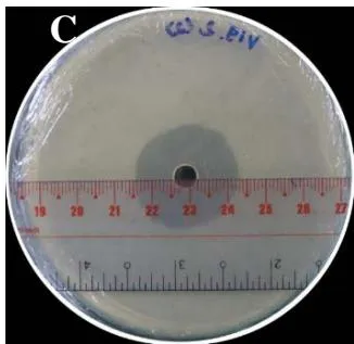
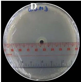
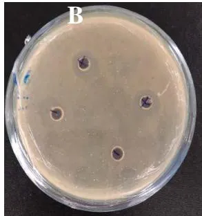
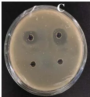
Twenty-nine of 75 tested isolates could produce antimicrobial active metabolites inhibiting at least one of the test pathogens (Figure 3). Over fifty percent isolates were capable of inhibiting the growth of Gram-positives, 23 isolates were actively against Gram-positive bacteria and 26 isolates showed activity against Gram-negative bacteria and 5 isolates were actively against following pathogens including Bacillus cereus, Staphylococcus aureus, Escherichia coli, Vibrio haemoparalyticus (Table 1).
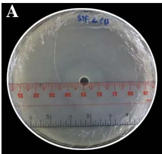
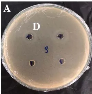
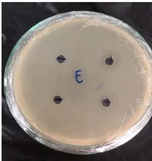
A: Staphylococcus areus (Streptomycin)
B: Bacillus cereus (Streptomycin)
C: Vibrio parahaemolyticus (Tetracyclin)
D: Escherichia coli (Tetracyclin)
Figure 3: Antimicrobial activity of ANTHOIDONG 3.1 isolate against Staphylococcus areus (A), Bacillus cereus (B), V. parahaemolyticus(C) and Escherichia coli (D) by observation of halo zone around colony (inhibition of ring) Table 1: Antimicrobial activity of isolated actinobacteria from Can Gio mangrove forest soil (HoChiMinhCity)
| No. | Actinobacterial name | B | E | V | S |
| 01 | TAMTHONHIEP 2.1 | ND | ND | ND | ND |
| 02 | TAMTHONHIEP 4.1 | ND | ND | ND | ND |
| 03 | TAMTHONHIEP 5.1 | ND | ND | ND | ND |
| 04 | THANHAN 5.1 | ND | ND | ND | ND |
| 05 | THANHAN 3.2 | ND | ND | ND | ND |
| 06 | THANHAN 3.3 | ND | ND | ND | ND |
| 07 | THANHAN 3.4 | ND | ND | ND | ND |
| 08 | THANHAN 4.1 | ND | ND | ND | ND |
| 09 | THANHAN 4.2 | ND | ND | ND | ND |
| 10 | THANHAN 4.3 | ND | ND | ND | ND |
| 11 | THANHAN 4.4 | 16 | ND | ND | 16 |
| 12 | THANHAN 4.5 | ND | ND | ND | 10 |
| 13 | THANHAN 5.1 | ND | ND | ND | ND |
| 14 | LYNHON 6.1 | ND | ND | ND | ND |
| 15 | LYNHON 6.2 | ND | ND | ND | ND |
| 16 | LYNHON 7.1 | ND | ND | ND | ND |
| 17 | LYNHON 7.2 | ND | ND | ND | ND |
| 18 | LYNHON 8.1 | ND | ND | ND | ND |
| 19 | LYNHON 8.2 | ND | ND | ND | ND |
| 20 | ANTHOIDONG 1.1 | ND | ND | ND | ND |
| 21 | ANTHOIDONG 1.2 | ND | ND | ND | ND |
| 22 | ANTHOIDONG 1.3 | ND | ND | ND | ND |
| 23 | ANTHOIDONG 1.4 | ND | ND | ND | ND |
| 24 | ANTHOIDONG 1.5 | ND | ND | ND | ND |
| 25 | ANTHOIDONG 1.6 | ND | ND | ND | ND |
| 26 | ANTHOIDONG 1.7 | ND | ND | ND | ND |
| 27 | ANTHOIDONG 3.1 | 10.7 | 07.7 | 17.3 | 21.7 |
| 28 | ANTHOIDONG 3.2 | 05.7 | ND | ND | ND |
| 29 | ANTHOIDONG 3.3 | ND | ND | ND | ND |
| 30 | ANTHOIDONG 3.4 | ND | 05 | ND | 05 |
| 31 | ANTHOIDONG 3.5 | ND | ND | ND | ND |
| 32 | ANTHOIDONG 3.6 | ND | ND | ND | ND |
| 33 | ANTHOIDONG 3.7 | ND | ND | ND | ND |
| 34 | ANTHOIDONG 3.8 | ND | ND | ND | ND |
| 35 | ANTHOIDONG 3.9 | ND | ND | ND | ND |
| 36 | ANTHOIDONG 4.1 | 12.3 | 11.7 | 21.7 | 15.7 |
| 37 | ANTHOIDONG 6.1 | 19.7 | ND | ND | ND |
| 38 | ANTHOIDONG 6.2 | ND | ND | ND | ND |
| 39 | ANTHOIDONG 7.1 | 27.7 | 05.6 | 17.7 | 35.1 |
| 40 | ANTHOIDONG 7.2 | ND | ND | ND | ND |
| 41 | LONGHOA 1.1 | ND | ND | ND | ND |
| 42 | LONGHOA 2.1 | ND | ND | ND | ND |
| 43 | LONGHOA 2.2 | ND | ND | ND | ND |
| 44 | LONGHOA 4.1 | ND | ND | ND | ND |
| 45 | LONGHOA 4.2 | 11.3 | ND | ND | ND |
| 46 | LONGHOA 4.3 | ND | ND | ND | ND |
| 47 | LONGHOA 4.4 | ND | ND | ND | ND |
| 48 | LONGHOA 5.1 | ND | ND | ND | ND |
| 49 | LONGHOA 5.2 | ND | ND | ND | ND |
| 50 | CANTHANH 1.1 | ND | ND | ND | ND |
| 51 | CANTHANH 1.2 | ND | ND | ND | ND |
| 52 | CANTHANH 2.1 | ND | ND | ND | ND |
| 53 | ANTHOIDONG 8.1 | ND | ND | ND | ND |
| 54 | ANTHOIDONG 9.1 | ND | ND | ND | ND |
| 55 | ANTHOIDONG 9.2 | ND | ND | ND | ND |
| 56 | ANTHOIDONG 10.1 | ND | ND | ND | ND |
| 57 | ANTHOIDONG 10.2 | ND | ND | ND | ND |
| 58 | ANTHOIDONG 10.3 | ND | ND | ND | ND |
| 59 | ANTHOIDONG 11.1 | 07.3 | ND | ND | ND |
| 60 | ANTHOIDONG 12.1 | ND | ND | ND | ND |
| 61 | ANTHOIDONG 13.1 | ND | ND | ND | ND |
| 62 | ANTHOIDONG 13.2 | ND | ND | ND | ND |
| 63 | ANTHOIDONG 13.3 | ND | ND | ND | ND |
| control | 08.7 streptomycin | 09.0 tetracyclin | 13.7 tetraclin | 12 streptomycin |
A total of 9 isolates was chosen for identification due to their highly antimicrobial activity. The fragments of 1050-1225 bp 16S rRNA were obtained from PCR and sequencing. Homology searches of 16S rRNA gene sequence of selected strains in GenBank by BLAST (Table 2) revealed that they had similarity to sequences of genus Streptomyces (8 isolates) and other genera including 1 isolates was Gram-negative bacteria (Stenotrophomonas) [not suitable for this study].
| Taxonomic group and strain | Closest species relative | Similarity (%) |
| LONGHOA 4.2 | MH472998 Streptomyces africanus strain E3SQ | 97.7 |
| MK368443 Streptomyces sp. strain MUM203J | 97.6 | |
| THANH AN 4 | HM594286 Streptomyces tendae strain M23 | 97.4 |
| MN339840 Streptomyces sp. strain MGB 2769 | 97.4 | |
| ANTHOIDONG 3.2 | U841673 Streptomyces tanashiensis strain HBUM174095 | 97.7 |
| EU593580 Streptomyces tanashiensis strain 173004 | 97.6 | |
| ANTHOIDONG 3.1 | MW217198 Streptomyces pavulus strain DSD2596 | 98.8 |
| MW217191 Streptomyces sp. strain DSD1692 | 98.8 | |
| ANTHOIDONG 4.1 | KP235209 Streptomyces celluloflavusstrain D4-17 | 99.6 |
| MN116554 Kitasatospora sp. strain SKW16 | 99.6 | |
| ANTHOIDONG 7.1 | HQ607433 Streptomyces albogriseolus strain 1168 | 99.4 |
| MK281547 Streptomyces albogriseolus strain SCAU-101 | 98.6 | |
| ANTHOIDONG 6.1 | U841673 Streptomyces tanashiensis strain HBUM174095 | 97.8 |
| MT505707 Streptomyces aegyptia strain 7 | 97.8 | |
| ANTHOIDONG 11.1 | MT072138 Streptomyces laurentii strain QT214 | 98.7 |
| LC497896 Streptomyces sp. 9R005 | 98.7 |
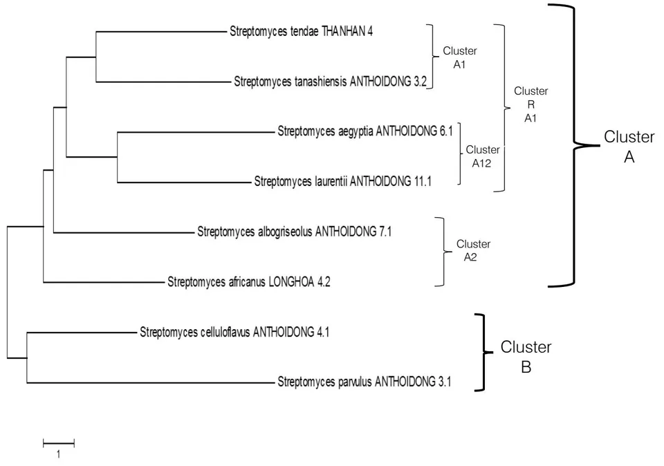
A Maximum Likelihood phylogenetic tree (Figure 4) of these isolates described the two clusters. Cluster A had 6 strains including 2 smaller clusters as cluster A1 with 4 strains divided to 2 smaller: cluster A11 composed of Streptomyces tendae THANHAN 4 and Streptomyces tanashiensis ANTHOIDONG 3.2 and cluster A12 with 2 strains: Streptomyces aegyptia
ANTHOIDONG 6.1 and Streptomyces laurentii ANTHOIDONG 11.1 while cluster A2 with 2 strains were Streptomyces albogriseolus ANTHOIDONG 7.1 and Streptomyces africanus LONGHOA 4.2. Cluster B had 2 strains: Streptomyces celluloflavus ANTHOIDONG 4.1 and Streptomyces pavulus ANTHOIDONG3.1.
Our results identified 8 strains of genus Streptomyces and all of them had the ability of antimicrobial activity, our results also conformed with the results of Qiu et al., (1994), when they determined that genus Streptomyces is the most resources of antibiotic production in comparison to other microbes.
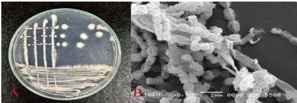
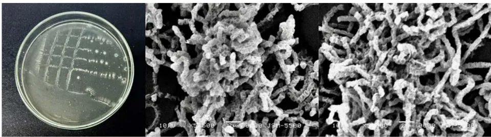
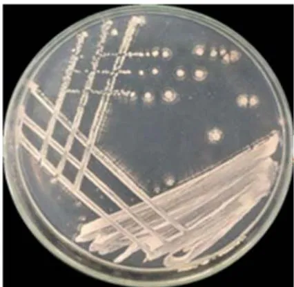
The species of genus Streptomyces, Streptomycescelluloflavus ANTHOIDONG 4.1(Figure 5A), their colonies had red white color, circular shape, many rays around colony on the ISP medium. The spores have many spines on the surface of spore and the cells connected to the string under the electric microscope X 8500 (Figure 5B).
Streptomyces celluloflavus first isolated at Japan soil, Streptomyces celluloflavussynthesized aureothricin and they have the ability of degrading-cellulose (Munray et al., 2008). In this study, Streptomyces celluloflavus has the ability of antibacterial activity to human pathogenic bacteria as Vibrio parahaemolyticus, Escherichia coli, Staphylococcus aureus and Bacillus cereus and this strain is the strongest phosphate solubilization and CMC degradation.
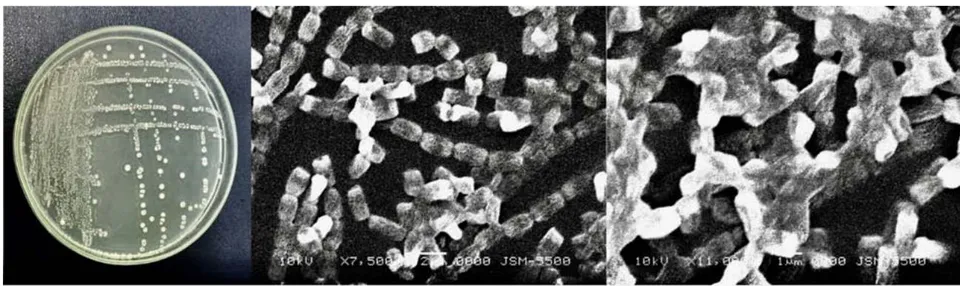
The second colonies of Streptomyces aegyptia ANTHOIDONG 6.1 strain have white color, circular shape, many rays around colony on the ISP medium (Figure 6A). Observation on SEM, Streptomyces aegyptia have smooth surface of spores, and connected to the string (Figure 6B and 6C).
Streptomyces aegyptia isolated from soil of Dakahliyah province, Egypt, they have the high ability of cellulose degradation (El-Naggar et al., 2011). Consequently, they produced cholesterol oxidase, anticancer in vitro, antimuscle infection, breast cancer and apoptosis in vivo (El-Naggar et al., 2018). Streptomyces aegyptia synthesized the nano-Ag seeds against bacteria as nanofactory friendly with the environment (Osama et al., 2014). In this study, Streptomyces aegyptia ANTHOIDONG 6.1 have the ability of against Bacillus cereus.
surface of spores, and connected to the string (Figure 7A).
The third strain, Streptomyces laurentii ANTHOIDONG 11.1 strain had colony with concentric circle surrounded by rays. Observation on SEM, Streptomyces laurentii ANTHOIDONG 11.1 have smooth
Streptomyces laurentii isolated from soil and they have the ability of Thiostrepton production (Trejo-Astreda et al., 1998). In this study, Streptomyces laurentii ANTHOIDONG 11.1 strain had the ability of agaisnt Bacillus cereus.
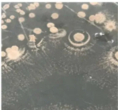
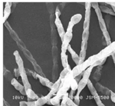
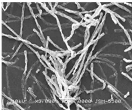
With the fourth strain, Streptomyces tanashiensis ANTHOIDONG 3.2 had characteristic of colonies as white color, circular shape, curled margin, pulvinate elevation, average diameter 6 mm; cells had circular, connected to short spring, no motile under observation of microscope; strait spores chain, smooth of surface of spore under SEM (Figure 8A, B, C).
Streptomyces tanashiensis isolated from mangrove forest soil Maowei, China, it had the ability of antibacterial activity to Bacillus cereus(Lu et al., 2019). According to Johnson and Dietz (1968), Streptomyces tanashiensis synthesized and secrete out to luteomycin (antifungal), mithramycin (anticancer), phosphoramidon (inhibition of enzyme thermolysin from Bacillus spp.) and kalafungin (antifunfal, anti-protozoa, and anti-gram-positive). The research 'Singh et al. (2009), showed that Streptomyces tanashiensis had the ability of against gram-positive bacteria and gram-negative bacteria together with it resistances to Candida albicans and Fusarium moniliform.
In the fifth strain, Streptomyces albogriseolus ANTHOIDONG 7.1 strain had characteristic of colonies as circular shape, one concentric ring, curled margin, pulvinate elevation, diameter of colony 5 mm; cells had circular and motile under light microscope; spiral spore chain, spiny surface of spore under observation of SEM (Figure 9).
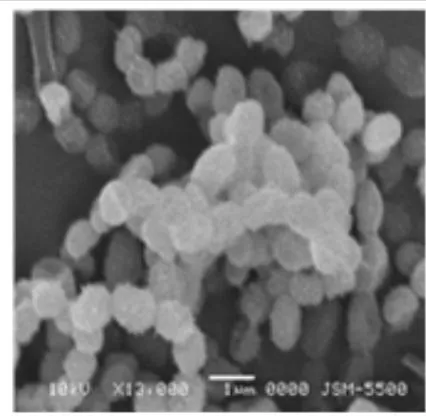
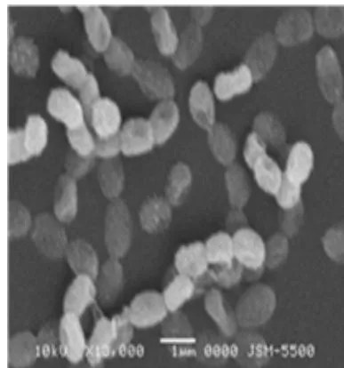
Streptomyces albogriseolus isolated from mangrove forest soil of Hainan, China. They had the ability of Macrocyclic Lactones production, this compound against Staphylococcus aureus and Escherichia coli(Xu et al., 2014). Qattan and Khattab (2019), Streptomyces albogriseolus had the ability of neomycin production, antibacterial compound, in the stress condition. Besides, Shao et al. (2019) determined Streptomyces albogriseolus using of polyethylene as carbon resource.
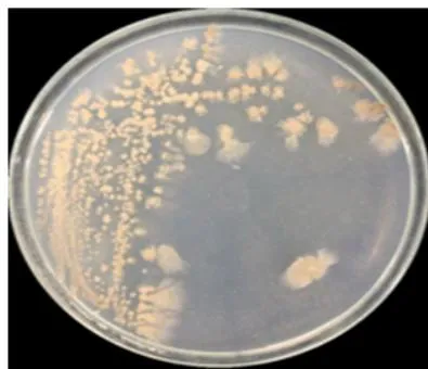
Streptomyces parvulus ANTHOIDONG3.1 strain had colonies had brown dark, circular shape and 1 long ray, curled margin, pulvinate elevation, diameter of colony 5 mm; cells had coccus, motile wken observation under light microscope, spores had long chain; Streptomyces parvulus had spiral spore chain, warty on surface of spore under SEM. (Figure 10A, B,C).
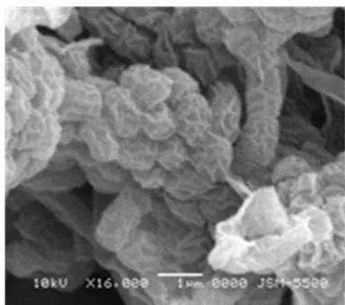
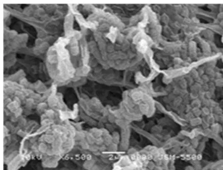
Streptomyces parvulus isolated from mangrove forest soil Visakhapatnam, Bengal by Prakasham et al. (2014), they had the ability of antibacterial activity as polypeptide (Actinomycin D) to agaisnt with 14 kinds of human pathogenic bacteria. Streptomyces parvulus had the ability of resistance to Bacillus subtilis, Staphylococcus aureus, Streptococcus fecalis, Pseudomonas aeruginosa, Proteus vulgaricus and Escherichia coli when test of antimicrobial activity by diffusion-well agar method(Usha et al. 2010),
The seventh strain, Streptomyces africanus LONGHOA 4.2 strain had white colonies color, circular shape, concentric circle on ISP medium. Under SEM (7,500 X), spores of Streptomyces africanus LONGHOA 4.2 strain had smooth surface, rods connected to string (Figure 11 A, B, C)
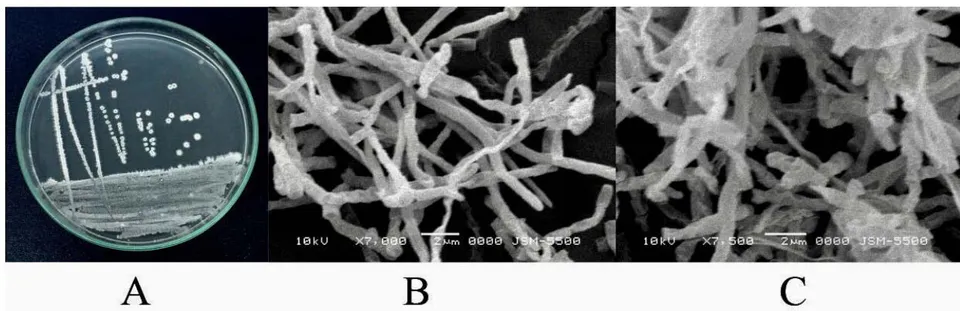
Streptomyces africanus isolated from soil at Cape Town, South Africa and they had low antibacterial activity to Enterococcus faecium (Paul et al., 2004). In this study, Streptomyces africanus LONGHOA 4.2 strain had good antibacterial activity to Bacillus cereus.
The final strain, Streptomyces tendae THANHAN 4 with the colonies had dark milk color, circular shape, curved margin, pulvinate elevation, diameter from 1.5 to 3 mm; cells had rods shape and motility. In this study, Streptomyces africanus LONGHOA 4.2 strain only had the ability of resistance to gram-positive bacteria as Bacillus cereus and Staphylococcus aureus at average level.
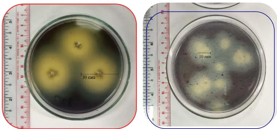
In order to test the other ability of Streptomyces isolates, 40 isolates were chosen to survey the ability of CMC (cellulose) degradation and phosphate solubilization. The isolates were grown on ISP medium in 15 days and supernatants were dropped into the wells on ISP added CMC agar (for degraded CMC test) and NBRIP (for phosphate solubilization test) and incubation in 21 days at room temperature. The results from Table 3 and Table 4 showed that 12/40 isolates had CMC degradation ability among two strains Streptomycescelluloflavus ANTHOIDONG 4.1 and Streptomyces africanus LONGHOA 4.2 had good cellulase synthesis ability and 12/40 isolates solubilized insolving phosphate among Streptomyces parvulus ANTHOIDONG 3.1, Streptomyces tanashiensis ANTHOIDONG 3.2 and Streptomyces albogriseolus ANTHOIDONG 7.1 solubilized phosphate. Therefore, five in twelve strains were sequenced and identified as species of genus Streptomyces. Actinobacteria from rhizosphere have the direct promotion of plant growth as nitrogen fixation, solubilization of Phosphorus and Potassium, IAA production and the indirect promotion of plant growth as Production of Antibiotics, Production of Lytic Enzymes, Production of Siderophores...(Yadav et al., 2018). Besides antibacterial compounds producing by Streptomycesspecies, our results also received 5/8 strains with capable of dissolving cellulose (CMC) and insoluble phosphate.
| No. | Isolate | Diameter of well (mm) | No. | Isolate | Diameter of well (mm) |
| 01 | LYNHON 6.1 | 24.33 bc | 07 | ANTHOIDONG 33 | 19.33 d |
| 02 | LYNHON 7.2 | 09.33 d | 08 | ANTHOIDONG 5.1 | 27.67 b |
| 03 | LYNHON 8.1 | 11.33 d | 09 | LONGHOA 4.1 | 5.45 cd |
| 04 | ANTHOIDONG 4.1 | 40.00 a | 10 | LONGHOA 4.2 | 06.22 c |
| 05 | LONGHOA 4.3 | 30.67 b | 11 | CANTHANH 1.1 | 03.93 d |
| 06 | LONGHOA 5.1 | 31.00 b | 12 | ANTHOIDONG 31 | 12.52 b |
| No. | Isolate | Diameter of well (mm) | No. | Isolate | Diameter of well (mm) |
| 01 | LYNHON 7.1 | 05.67 cd | 07 | ANTHOIDONG 3.4 | 02.03 d |
| 02 | LYNHON 7.2 | 05.01 d | 08 | ANTHOIDONG 3.5 | 05.67 cd |
| 03 | LYNHON 8.2 | 04.67 d | 09 | ANTHOIDONG 6.1 | 06.67 c |
| 04 | ANTHOIDONG 3.1 | 04.33 d | 10 | ANTHOIDONG 6.2 | 03.01 d |
| 05 | ANTHOIDONG 3.2 | 10.67 b | 11 | ANTHOIDONG 7.1 | 02.45 d |
| 06 | ANTHOIDONG 3.3 | 12.67 b | 12 | ANTHOIDONG 7.2 | 19.03 a |
The biosynthetic potential of 8 strains was investigated by the detection of polyketidesynthetase (PKS) and non-ribosomal polyketidesynthetase (NRPS) genes, the hall marks of secondary metabolites production. Results showed that many isolates were positive for PKS-I (2/8), without PKS-II (0), and NRPS (8/8) genes, indicating that mangrove Actinobacteria have significant biosynthetic potential. Our results highlighted that mangrove environment represented a rich reservoir for isolation of Actinobacteria, which are potential sources for discovery of antimicrobial secondary metabolites. Eight strains against Gram-positive bacteria (Bacillus cereus and Staphylococcus aureus) which were indicated with NPKS genes (8/8) while four strains resistant to Gram-negative bacteria (E. coli and Vibrio parahaemolyticus) which indicated with PKS-I genes (3/8) (3 strains: Streptomycescelluloflavus ANTHOIDONG 4.1, Streptomyces albogriseolus ANTHOI-DONG 7.1 and Streptomyces parvulus ANTHOIDONG 3.1) (Figure 13 and Table 5).
8.5 8.4 19.2 19.4 20.9 22.6 23.4 27.2 35.1 M
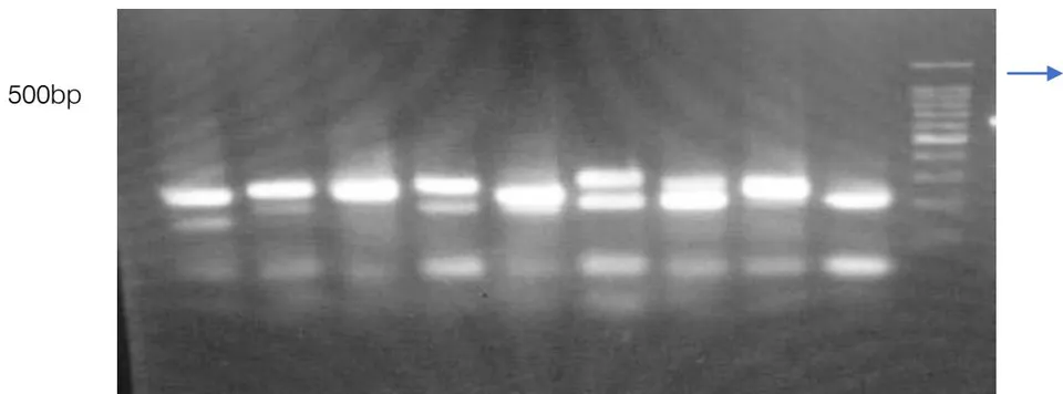
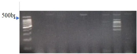
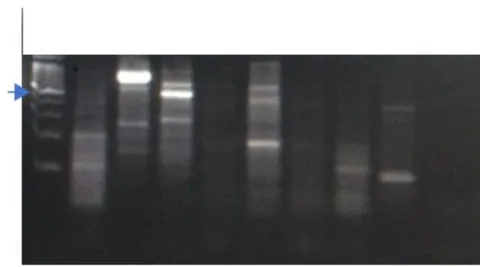
Figure 13: Agarose gel electrophoresis of PCR products from DNA isolated from representative actinomycete strains: (A) Selective amplification of 700–800 bp fragments using primers A3F/A7R specific for NRPS adenylation sequences; (B) Selective amplification of the 1200–1400 bp fragments using K1F/K2R and K1F/M6R; (C) Specific primers for PKS-I ketosynthase and methyl-malonyl-CoA transferase sequences Table 5: Distribution of NRPS, PKS-I, and PKS-II in 8 Streptomyces species or strains
| No | Taxa | NPRS (A3F/A7R) | PKS-I | PKS-II (KSα/ KSβ) | |
| (K1F/K2R) | (K1F/M6R) | ||||
| 01 | Streptomyces tendae THANHAN 4 | + | |||
| 02 | Streptomyces tanashiensis ANTHOIDONG 3.2 | + | + | ||
| 03 | Streptomyces pavulus ANTHOIDONG 3.1 | + | + | ||
| 04 | Streptomyces celluloflavus ANTHOIDONG 4.1 | + | + | ||
| 05 | Streptomyces aegyptia ANTHOIDONG 6.1 | + | |||
| 06 | Streptomyces africanus LONGHOA 4.2 | + | |||
| 07 | Streptomyces albogriseolus ANTHOIDONG 7.1 | + | + | ||
| 08 | Streptomyces laurentii ANTHOIDONG 11.1 | + | |||
The high rate of detection of PKS-I and NPKS genes in the isolates tested was mostly Streptomyces, providing strong evidence for the high potential of Streptomyces to produce high number of biologically active metabolites; the fact complies with other researches. Therefore, the molecular screening of Actinobacteria isolates for genes encoding biosynthesis of bioactive compounds is still an effective and valuable approach for pre-selecting isolates for useful secondary metabolites production (Ginolhac et al., 2004, Qui et al., 2009; Courtois et al., 2003; Hornung et al., 2007; Mets "a-Ketel" a et al., 1999; Schneemann, et al., 2010).
Can Gio mangrove forest located on the east coast of HoChiMinh city Vietnam is mostly unexplored the microbe resources; especially actinobacteria communnities. Therefore, this location is anticipated to be able to provide a rich source of Actinobacteria, the prolific producers of antimicrobial secondary metabolites. To date, no studies have reported the diversity and antimicrobial activities of Actinobacteria from Can Gio mangrove environment. For this reason, there is a high possibility to identify novel Actinobacteria and discover valuable antimicrobial secondary metabolites.
IV. CONCLUSION
Sixty-three actinomycetes were isolated from 25 soil samples of 9 different sites in mangrove forest in Can Gio, HoChiMinh city, Vietnam; Selected 29/63 isolates could produce antimicrobial active metabolites inhibiting at least one of the test pathogens and 8 best isolates were chosen to identify by 16S rRNA technique and sequencing. Eight strains belonged to genus Streptomyces among 5/8 strain had ability of dissolving cellulose (CMC) and insoluble phosphate, especially three strains: Streptomycescelluloflavus ANTHOIDONG 4.1, Streptomyces albogriseolus ANTHOIDONG 7.1 and Streptomyces parvulus ANTHOIDONG 3.1 had the ability of antibacterial activity with 4 human pathogenic bacteria.
ACKNOWLEDGEMENTS
The authors thank the helpfulness of Biotechnology BSc. students and technicians in the Environment Microbiology Laboratory, Biotechnology R&D Institute, Can Tho University, Vietnam. Drs. TRAN THI GIANG, Leader of the laboratory, Environment Microbiology Laboratory, Biotechnology R&D Institute, Can Tho University, Vietnam.