I. INTRODUCTION
Osteoarthritis (OA) is a localized and chronic joint disease that affects approximately one third of adults, with the prevalence of the disease increasing with advancing age[^1]. The economic impact of knee OA is also a growing problem for healthcare systems. Demographic predictions indicate that people over 65 will make up more than of the population by , so knee OA will only become more prevalent.
The knee is the most common site on the lower limb for OA, with the disease affecting the tibiofemoral and patellofemoral joints alone or in combination. The medial tibiofemoral compartment is the most commonly affected (67% medial versus 16% lateral)[^3]. Patients with knee OA often report symptoms of knee pain and difficulty with activities of daily living, such as walking, climbing stairs and household activities[^4]. Ultimately, the pain and disability associated with the disease lead to a loss of functional independence and a profound reduction in quality of life.
Knee OA treatment strategies can be considered as primary prevention (reduction of risk factors to reduce the incidence of the disease); secondary prevention (interventions to delay/prevent progression to serious illness) or; tertiary prevention (treatment of pain and disability) .
Given that there is currently no cure for knee OA and the only established treatment for end-stage OA is joint replacement, slowing the progression of structural disease is essential to help reduce the personal and social burden of knee OA. Traditionally, disease progression has been assessed by measuring joint space loss over time from serial radiographs. There is an increasing use of MRI to measure cartilage volume in the knee as it has proven to be a valid and reproducible technique that is more sensitive to change than x-rays .
Increased load on the joint is important in the pathogenesis of knee OA. Interventions that alter knee load can reduce symptoms and delay disease progression in patients with knee OA. The knee adduction moment determines the load distribution on the medial and lateral tibial plateaus, with a force in the medial compartment almost 2.5 times greater than the lateral[^7]. This may explain the much higher prevalence of medial OA compared to the lateral tibiofemoral joint.
The magnitude of this adduction moment is partially determined by the mechanical alignment of the knee. In medial knee OA, mechanical alignment becomes varus as the medial joint space narrows. Varus misalignment causes the ground reaction force vector to pass more medially to the center of the knee joint, resulting in a greater knee adduction moment. Cross-sectional studies demonstrate that patients with knee OA have a greater knee adduction moment during walking when compared to healthy controls of the same age[8,9].
Recent research has found that a longer adduction moment is associated with more intense pain in the knee and greater severity of radiographic disease . The severity of knee misalignment is also significantly associated with the intensity of knee pain and physical function . Longitudinal studies have shown that a minimum increase of one unit at the time of adduction is associated with an increase of up to 6.5 times in the risk of disease progression . Likewise, varus misalignment of the knee joint is also correlated with disease progression . Given the importance of knee adduction timing and joint alignment in relation to symptom severity and disease progression in knee OA, conservative strategies to alter these biomechanical factors constitute a logical rehabilitation approach.
Alternatives to reduce the adductor moment using lateral wedges in the heels for alignment and demonstrated that insoles statically aligned the knee in a more upright position, shifting the calcaneus to a valgus position in relation to the tibia. Studies that such changes would help to reduce the excessive load on the medial articular surface, leading to knee pain mitigation.
Since then, biomechanical studies have evaluated the effects of lateral wedge insoles on knee alignment and medial compartment load. However, despite its biomechanical effects, few randomized clinical trials have evaluated its clinical efficacy .
We classified varus knees as mild varus with mechanical femorotibial angle and moderate with mechanical femorotibial angle .
Treatments with osteotomies , much more for corrections of genu varus than in valgus with similar results even with moderate varus29.
Our objective is, focusing on knees with mild varus (femorotibial angle ), in addition to intra-articular corrections, perform ligamentopraxia (stretching by stretching by manipulation in valgus) of the medial complex, reducing the compression load on the medial compartment.
Evaluated preoperatively and postoperatively by X-Ray with monopodal support and Nuclear Magnetic Resonance, which qualified the femorotibial alignment and intra-articular lesions, as well as a specific and validated clinical questionnaire for the knee.
II. MATERIAL AND METHODS
16 knees of 13 patients with medial meniscal injuries and femoro-tibial alignment to mild genu varus (femoral-tibial diaphyseal mechanical angle ), (Figure 1 A and 1 B).
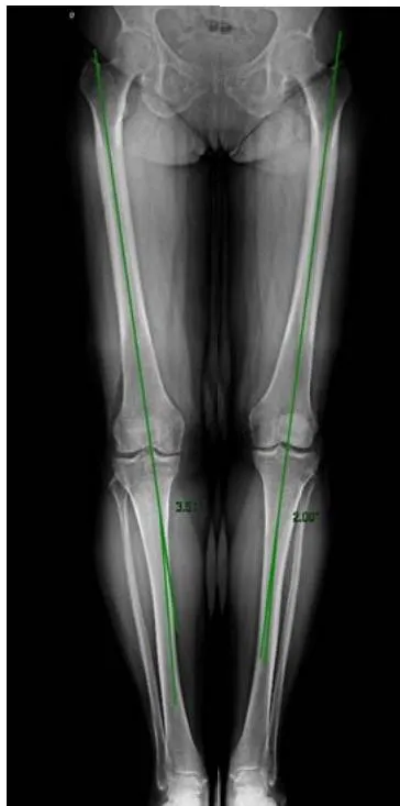
Fig. 1 A: Femorotibial angle of right knee varus
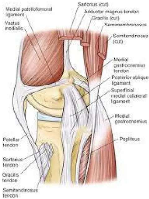
Surgically treated within one year (from 8/13/2020 to 8/13/2021) by the same orthopedist, undergoing in addition to video arthroscopy for resolution of intra-articular lesions, ligamentopraxia of the medial complex (medial collateral ligament and posterior oblique ligament) (Figure 2).
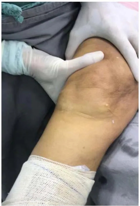
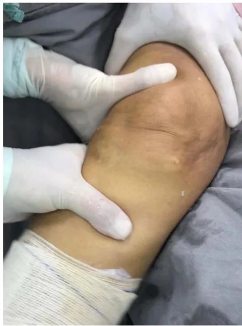
A valgus maneuver was performed at stabilized by the auxiliary in the thigh after intra-articular saline solution was insufflated in the video arthroscopy (Figure 3A and 3B).
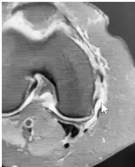
The radiographic femoro-tibial mechanical angles were calculated in AP with monopodal support and verified chondral and meniscal lesions in the pre
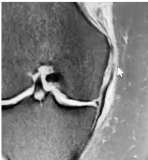
and postoperative radiographic study of Nuclear Magnetic Resonance 30-32 (Figure 4A and 4B).
The Lyscholm grading was used to assess the pre- and postoperative clinical evolution of the operated knees (Chart 1).
| Limping (5 points) Never = 5 Mild or periodically = 3 Strong and continuous ☑0 Support (15 points) No support ☑0 Walking stick or crutches ☑2 Impossible ☑0 Restrainting (15 points) No restraining or restraining feeling ☑15 Has the feeling, but no restraining ☑10 Occasional restraining ☑6 Frequent ☑2 Joint restrained at examination ☑0 Instability (25 points) Never misses a step ☑25 Seldom, during athletic activities or other strong-effort exercises ☑20 Frequently during athletic activities or other strong-effort exercises (or unable to participate) ☑15 Occasionally in daily activities ☑10 Frequently in daily activities ☑5 At each step ☑0 Pain (25 points) No pain ☐ 25 Intermittent or mild during strong-effort exercises ☑ 20 Marked during strong-effort exercises ☑ 15 Marked during or after walking more than 2 Km ☑ 10 Marked during or after walking less than 2 Km ☑ 5 Continuous ☑ 0 Swelling (10 points) No swelling ☑ 10 Upon strong-effort exercises ☑ 6 Upon usual exercises ☑ 2 Continuous ☑5 Climbing stairs (10 points) No problem ☑ 10 Slightly damaged ☑6 One step at a time ☑ 2 Impossible ☑ 0 Squeezing (5 points) No problem ☑ 5 Slightly damaged ☑ 4 Not exceeding 60 degrees ☑ 2 Impossible ☑ 0 Total score: Score table: Excellent: 95 = 130; Good: 84 = 94; Fair: 65 = 83; Poor: < 64 |
III. RESULTS
The procedures were performed in 12 patients (8 females and 4 males) in 16 knees (10 right knees and 6 left) bilateral ones were two females and two males (Table 1).
The femorotibial mechanical angle ranged from of Valgus to of Varus (weighted average of of Varus).
The internal lesions involved grade III medial meniscus and grade I to IV medial chondral lining .
| pat | ag | gen | site | f/tà | mri pré | lysholm pré | lysholm pós |
| mtbr | 55 | fem | rig | vr 2° | m iii ciii | 25 | 93 |
| mtbr | 55 | fem | lef | vr 2° | m iii cii | 25 | 93 |
| jwc | 86 | mal | rig | vl 3° | miii ciii | 18 | 87 |
| jwc | 86 | mal | lef | vl 2° | miii ciii | 18 | 87 |
| rcc | 64 | mal | lef | vr 4° | miii civ | 42 | 99 |
| moa | 71 | fem | tig | vr 0 | miii civ | 19 | 94 |
| rvmc | 65 | fem | rig | vl2° | m iii ciii | 17 | 99 |
| oow | 76 | mal | rig | 0° | m iii cii | 57 | 95 |
| oow | 76 | mal | lef | vr 1° | m iii cii | 57 | 93 |
| dp | 58 | mal | lef | vr 2° | m iii ciii | 25 | 99 |
| ats | 57 | fem | rig | vr 1° | m iii cii | 34 | 92 |
| nca | 68 | fem | rig | vl 2° | m iii ciii | 57 | 89 |
| rci | 63 | fem | rig | vl 1° | m iii ciii | 40 | 95 |
| cmg | 66 | fem | rig | vr3,5° | miii civ | 47 | 98 |
| msb | 72 | fem | rig | 0° | miii cii | 12 | 98 |
| msb | 72 | fem | lef | vr 1° | miii civ | 12 | 98 |
| pat | ag | gen | site | f/tà | mri pré | lysholm pré | lysholm pós |
| mtbr | 55 | fem | rig | vr 2° | m iii ciii | 25 | 93 |
| mtbr | 55 | fem | lef | vr 2° | m iii cii | 25 | 93 |
| jwc | 86 | mal | rig | vl 3° | miii ciii | 18 | 87 |
| jwc | 86 | mal | lef | vl 2° | miii ciii | 18 | 87 |
| rcc | 64 | mal | lef | vr 4° | miii civ | 42 | 99 |
| moa | 71 | fem | tig | vr 0 | miii civ | 19 | 94 |
| rvmc | 65 | fem | rig | vl2° | m iii ciii | 17 | 99 |
| oow | 76 | mal | rig | 0° | m iii cii | 57 | 95 |
| oow | 76 | mal | lef | vr 1° | m iii cii | 57 | 93 |
| dp | 58 | mal | lef | vr 2° | m iii ciii | 25 | 99 |
| ats | 57 | fem | rig | vr 1° | m iii cii | 34 | 92 |
| nca | 68 | fem | rig | vl 2° | m iii ciii | 57 | 89 |
| rci | 63 | fem | rig | vl 1° | m iii ciii | 40 | 95 |
| cmg | 66 | fem | rig | vr3,5° | miii civ | 47 | 98 |
| msb | 72 | fem | rig | 0° | miii cii | 12 | 98 |
| msb | 72 | fem | lef | vr 1° | miii civ | 12 | 98 |
IV. DISCUSSION
The prevalence of knee OA has increased significantly in recent decades and continues to increase, and age, previous knee injuries, obesity (increased body mass index (BMI), joint misalignment and instability resulting in increased mechanical stress are all strong factors. risk for developing knee OA
Physical inactivity is also another important contributor to the increased prevalence of OA, causing greater susceptibility to knee injuries due to less stable and weaker joints . However, weakness of the knee extensor muscles appears to be a weak risk factor, compared to previous knee injuries .
Men are less likely to develop OA than women, making sex one of the risk factors associated with developing OA40. Narrower femurs, thinner patellas, larger quadriceps angles and differences in the size of the tibial condyles make women's knee anatomy different from men's, leading to different kinematics, which influence females to be more likely to develop OA, leading to a higher prevalence of OA in women41,42.
In this study, we selected patients with meniscal injuries associated with knees with varus less than , grade III meniscal injuries and varied chondral injuries.
In our sample, we had an average age of 68.13 years (55 to 86 years) being more prevalent in females, of operated knees, which is in agreement with the literature[43].
The gradation of the tibial femur anatomical angle was of varus (ranging from of valgus to of varus). Respecting the 4th of varus to be considered light varus. Literature agrees that from Moderate Varism (greater than ) the indications for osteotomies and arthroplasties would be of better therapeutic indication.
Meniscal injuries were all grade III, those without chondral or chondral injuries were type I , type II , type III and type IV .
All treated by the same surgeon, with meniscoplasty of the medial meniscus being performed by resection only of the injured area, respecting the maximum amount of intact tissue, chondroplasty by toilet or microfractures when necessary.
Preoperative Lysholm Scorres had a Weighted Average of 31.56 (13 to 57) and all qualified on the score as poor. In the evaluation by the same Score in the Postoperative period, the knees had a Weighted Average of 94.31 (87 to 99) 9 knees had a score as good and 6 as excellent.
Very positive results were obtained for the patients followed.
The purpose of this article serves as a presentation of this technique of ligamentopraxia of the medial collateral complex.
Specific videoarthroscopic meniscoplasty and chondroplasty are always performed for patients with femoro-tibial misalignment with varus to moderate.
V. CONCLUSION
In knees with moderate varus, the technique aims to improve the medial femoro-tibial conflict through the ligamentopraxia of the medial complex (medial collateral ligament and posterior oblique ligament).
Associated with the specific treatment of meniscal and chondral injuries, it improved the pre-topostoperative score from 31.56 to 94.31 points.
As the technique is unprecedented, its efficacy and replicability must be observed with further studies.