I. INTRODUCTION
ROP is one of the leading aetiologies of avoidable blindness in children globally [1]. One out of every five children born prematurely in the world is prone to having ROP [2]. The erroneous thought that ROP was not usually found in the black African child led to many eye care programs neglecting the quest for the disease and its management on the continent [3]. The fact is that ROP is as old as the existence of humanity in all geographical regions of the world, independent of race. Enhancement in retina care coupled with increased survival rates of preterm babies in Africa has deceitfully created impression that the disease is now on the ascendancy on the continent. Although some experts think that the disease is less severe in black than white babies [4,5], this assertion has not been proved scientifically and, therefore can be relegated to the background.
ROP is a vascular disease of the retina which affects premature babies of low birth weight who have received oxygen. Other exacerbating factors include but are not limited to hypercarbia, postnatal hyperglycemia, reduced postnatal weight gain, hypoxemia and neonatal infections [6,7,8]. A baby is said to be premature when born at less than 37 weeks, very premature at less than 32 weeks and extremely premature at or before 28 weeks of gestation [9]. Similarly, birth weight can be low (< 2500g), very low (<1500g) and extremely low (1000g) [9]. Oxygen therapy, essential for the maturation of lungs and survival of a premature baby, may be toxic to the retina leading to ROP [10]. If not diagnosed in time and adequately managed, ROP naturally leads to visual impairment, blindness, social deprivation, psychomotor and cognitive developmental retardation [11].
The purpose of management is to curb detrimental effects from retinal ischemia, neovascularization, tractional bands formation and detachment. Cryotherapy, used for treatment several decades ago, is no more accepted because of the plethora of complications associated with it. Among them are cicatricial disease, ectopic maculae, disc dragging and retinal detachment [12]. The CRYOROP trial also reported on ocular and systemic side effects. Eye-related ones were conjunctival hematoma and laceration, retinal, pre-retinal and vitreous hemorrhage. Systemically, there were bradycardia, arrhythmias and significant apnoea.
Although ILO has currently replaced cryotherapy, the former is equally fraught with complications such as tunnel vision, nystaglia, high myopia and optic disc atrophy[13,14]. Undoubtedly, ILO destroys the retina leaving behind several scars as aftermath. The underlying principal factor which triggers ROP is the release of vascular endothelial growth factors (VEGF) from the avascular retina [15,16]. A modern paradigm shift in ROP management is the use of
IAVEGFIM which, apart from helping in appropriate vascularisation of the retina [17], stops the retinopathy, lacks complications and does not need use of general anesthesia [18].
To date, a few published case series have shown favorable results when IAVEGFIM is utilized in managing undetached ROP[17,18]. To the best of our knowledge, this is the first time such a new treatment is assessed in Africans in a large cohort of patients.
The purpose of the study was to analyze the outcome and complication profile of patients in SubSahara Africa who underwent IAVEGFIM and other modalities of treatment after being diagnosed with ROP.
II. MATERIAL AND METHODS
This article is an analytical retrospective comparative study carried out from January 2022 to review medical records of 50 children(100 eyes) who underwent IAVEGFIM and other treatment modalities from September 2013 to September 2021 after being diagnosed with ROP in the study hospital.
The children in question were followed up for at least five years. One consultant retina surgeon with the help of theatre assistants, performed all the procedures. Institutional ethical approval was acquired for this research. In a broader measure, tenets of the Declaration of Helsinki were used to preserve the human rights of participants whose consents were given by respective parents.
Inclusion criteria- Patients in the study were those who were examined and diagnosed at the retina clinic of 37 Military Hospital in Accra, Ghana. The criterium for IAVEGFIM was clients who had type 1 ROP. Ten eyes went on ILO because the clients could not afford IAVEGFIM. The criteria for surgeries were stage 4 or worse.
Exclusion criteria for IAVEGFIM and ILO were retinal tractional bands, RD, painful red eye, conjunctivitis and blind eye. Out of 68 children whose medical records were reviewed, 18 were excluded from the study because they were either followed up for less than five years, lost to follow up, or did not meet the criteria for IAVEGFIM and other modalities of treatment. Some of the patients had been referred from other Sub-Saharan African countries. In addition to general demographic data, information on procedure indications, post-procedure complications, and latest Best Corrected Visual Acuity (BCVA) were collected and analyzed.
One consultant vitreoretinal surgeon (FKO) performed the IAVEGFIM procedures on all the children in an operation theatre under aseptic and sterile conditions. The skin around the eyelids was cleaned with gauze and povidone-iodine. After the instillation of topical anesthetic drops, povidone-iodine was applied onto the ocular surface, a pediatric speculum was used to open the eyelids, a 30-gauge needle was inserted through pars plana1.5mm away from the limbus and bevacizumab (Avastin, 1.25 mg in 0.025 mL) injected into the vitreous. Shortage of bevacizumab on certain occasions made us use aflibercept and ranibizumab.
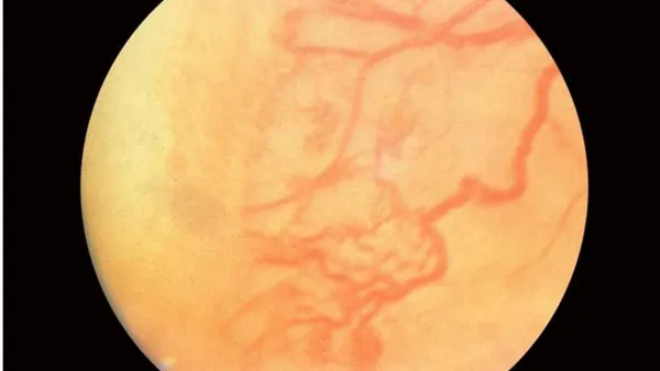
A 15-.second pressure was applied with a cotton-tipped applicator at the site of injection immediately after needle withdrawal. The eye was then covered with gauze and plaster after the instillation of one drop of povidone-iodine onto the ocular surface. Patches were taken away 2 hours after the procedure, and patients were reviewed 24 hours and one week after injection. A treat and extend approach was implemented in all patients with the total number of injections in an eye ranging from 3 to 6 depending on the severity of the disease(fig 1 and 2) on the first examination.
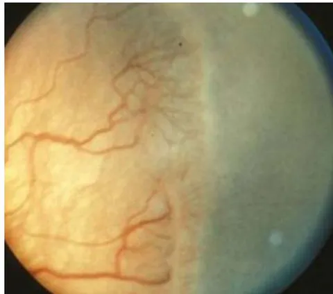
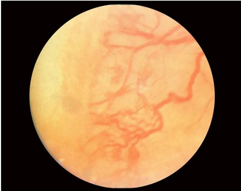
Indication for ILO under local anesthesia was the inability of the parents to afford IAVEGFIM. A wire Vectis was used to indent the anterior retina, rotate and stabilize the globe. ILO was applied starting with 250 milliwatts for 150 milliseconds with repeat mode set at 300 milliseconds to achieve confluent grayish whitish burns at the avascular retina posterior to the ridge in 3600 fashion up to ora Serrata. Three thousand to 4000 spots were delivered in each eye. The baby was reviewed 1, 7 and 28 days after the procedure.
Eyes with tractional RD had lens sparing three port pars plana vitrectomy (1.5mm from limbus), membrane segmentation, membrane delamination, fluid-air exchange and endolaer under general anesthesia, sterile and aseptic conditions. If there was difficulty in membrane dissection between the retina and posterior capsule of the lens, then lensectomy was done. Sclerotomies were closed up with 8-vicryl.
Snellen BCVA was converted into logarithm of minimum angle of resolution (logMAR) units to get a better statistical analysis. Patients whose visual acuities were light perception were assigned an equivalence of 2.70 logMAR units.
Table-1 shows visual outcomes of various procedures or approaches used in the management of ROP. In all, of eyes maintained their excellent visual acuities while the rest had worsened final post-treatment visual acuities compared to pre-treatment measurement.
III. STATISTICAL ANALYSIS
The statistical analysis was done using paired t-test for normally distributed variables. Accordingly, all tests were considered statistically significant if the p-value was 0.05 or less. Chi-square test and paired t-test with SPSS and Graph Pad software were used, respectively.
IV. RESULTS
A total of 100 eyes of 50 children (26 females and 24 males) were identified. The mean postmenstrual age at diagnosis was weeks (range 31-41 weeks) with a minimum follow-up of 5 years (5 - 8).
Children screened, diagnosed and treated early with IAVEGFIM had the best of BCVA. On the other hand, those with complications on presentation had gone blind in the affected eye at the last follow-up visit. Mean pre-treatment visual acuity was 2.70 logMAR units as physiologically, these babies had not started seeing. The mean difference between final post- and pretreatment visual acuity was log MAR units which was statistically significant .
| BCVA quality | Procedure/ Approach | Number of eyes (%) |
| Maintenance | IAVEGFIM | 85 (85%) |
| Worsening | ILO | 10 (10%) |
| Worsening | RDS | 3 (3%) |
| Worsening | Laisser Faire | 2 (2%) |
BCVA - Best Corrected Visual Acuity, IAVEGFIM - Intravitreal AntiVascular Endothelial Growth Factors Injection Monotherapy, ILO - Indirect Laser Ophthalmoscopy, RDS - Retinal Detachment Surgery
Table 2 shows statistically significant vast difference in the means of pre- and post-treatment visual acuities; thus, an improvement in the IAVEGFIMBCVA.
Table 2 T-TEST PAIRS=PRETREATMENT WITH POSTTREATMENT (PAIRED) /CRITERIA=CI(.9500) /MISSING=ANALYSIS. Paired Samples Statistics
| Mean | N | Std. Deviation | Std. Error Mean | ||
| Pair 1 | PRE-TREATMENT VISUAL ACuity | .0010000 | 100 | .00000000 | .00000000 |
| POST-TREATMENT VISUAL ACuity | .900080 | 100 | .2559516 | .0255952 | |
Paired Samples Correlations
| N | Correlation | Sig. | ||
| Pair 1 | PRE-TREATMENT VISUAL ACuity & POST-TREATMENT VISUAL ACuity | 100 | . | . |
Paired Samples Test
| Paired Differences | |||||
| Mean | Std. Deviation | Std. Error Mean | 95% Confidence... Lower | ||
| Pair 1 | PRE-TREATMENT VISUAL ACuity - POST-TREATMENT VISUAL ACuity | -.89908000 | .25595165 | .02559516 | -.94986636 |
Paired Samples Test
| Paired...95% ConfidenceInterval of the...Upper | t | df | Sig. (2-tailed) | ||
| Pair 1 | PRE-TREATMENT VISUALACuity - POST-TREATMENT VISUALACuity | -.84829364 | -35.127 | 99 | .000 |
The major indication for the use of IAVEGFIM was type 1 ROP (n=85 eyes; 85%). Ten eyes (10%), which had ILO because of the inability to afford IAVEGFIM ended up with peripheral visual field loss (n=3;3%), nystagia (n=3;3%), tunnel vision (n=2;2%) and optic nerve atrophy (n=2;2%) in their last follow-up visit. Three eyes (3%) had RDS with poor outcomes. Two eyes from 2 different babies had gone blind on reporting to the study center for the first time.
Eighty-five out of 100 eyes received IAVEGFIM, which did not give rise to any complications (table 3). The most common complications were peripheral visual field loss and nystaglia which resulted from ILO. In 2 eyes, recurrent retinal new vessels following ILO were managed with IAVEGFIM. Surgical intervention in clients who had retinal detachment did not yield a good outcome. Two eyes of 2 patients had gone phthisical on the first examination at the study center.
| SRL | Intervention | Number of Eyes Out of 100 | Complication | Complication N (%) for Each Procedure | Management |
| 1 | IAVEGFIM | 85 | None | 0 (0%) | Observation |
| 2 | ILO | 3 | PVFL | 3 (100%) | Observation |
| 3 | ILO | 3 | Ncytalopia | 3 (100%) | Observation |
| 4 | ILO | 2 | Optic Disc Atrophy | 2 (100%) | Observation |
| 5 | ILO | 2 | RRNV | 2 (100%) | IAVEGFIM |
| 6 | Laisser Faire | 2 | Phthisis | 2 (100%) | Observation |
| 7 | RDS | 2 | PVR | 2 (100%) | Observation |
| 8 | RDS | 1 | Cataract | 1 (100%) | Cataract Surgery |
V. DISCUSSION
ROP is better understood when its pathogenesis is well outlined [15,16]. Vasculogenesis and angiogenesis are physiological processes through which new blood vessels are formed. Whereas the former helps in the formation of the primitive vascular network, the latter aids in remodeling and growing of new capillaries which lack fully developed tunica media. Under ischaemic conditions, new vessels are formed through pathological angiogenesis [19].
Development of retinal blood vessels in humans starts at 16 weeks of gestation in a centrifugal fashion at a rate of [20], with the nasal retina being vascularized at 36 and temporal 40 weeks post menstrual age (PMA). The retina is nourished by choriocapillaris before 16 weeks of PMA.
The pathogenesis and progression of ROP are categorized into phases 1 and 2 [21]. In the first phase, oxygen given to newly born premature babies causes cessation of vascular growth and eventually vasoobliteration on the retina. In phase 2, withdrawal of oxygen and hypoxia lead to ischemia, which triggers the release of various substances, including Vascular Endothelial Growth Factors (VEGF), resulting in pathological angiogenesis and irreversible blindness through retinal detachment if left unattended to [19,21,22,23]. In the study hospital, it has been realized that earlier examinations of premature babies' retinae before age 14 days are not helpful in the detection of
ROP because its clinical signs would not have developed yet. This means that effects of VEGF are manifested on the retina from day 14 and above after birth.
Certain published studies have established that lower oxygen concentration (85 to ) given to premature babies results in less probability in the occurrence of ROP while it leads to increased mortality [24]. Similarly, researchers in another study also discovered that lower concentrations (85-89%) brought about high mortalities and less ROP, while higher concentrations (91-95%) were associated with severe forms of ROP and less mortality [25, 26,27]. Canadian oxygenation trial, however, did not detect any difference in mortality between the higher and lower SpO2 groups[28]. Although in the study hospital, SpO2 of 98-100% is used with the aim of keeping babies alive, there has not been an increase in severity of ROP.
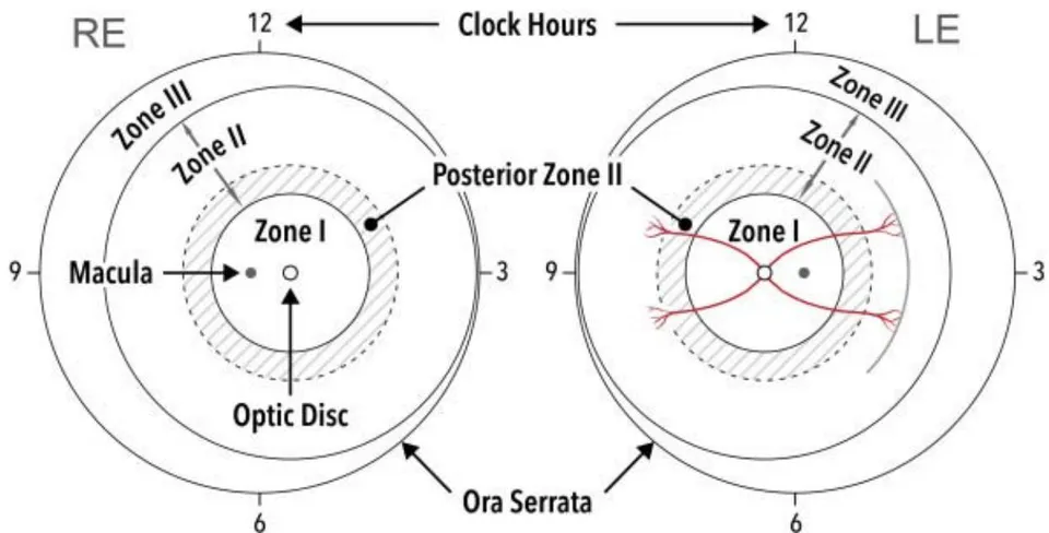
The location of the most posterior retinal vascularization determines the zone of ROP (fig1). The center of the circles is the optic disc. Zone I is the most posterior region and is determined by a circle whose radius is twice the distance between the optic disc center and the center of the fovea. Zone II extends nasally from the outer limit of zone I to the nasal or a Serrata with the same distance temporal, superior, and inferiorly. Posterior zone II is two disc diameters peripheral to zone I border, an area which when perturbed by ROP, usually goes through a guarded prognosis. Zone III is the remaining crescent of the peripheral retina, which extends beyond zone II [29]. In the study hospital, (65), (20) and (15) of eyes were in zones III, II and I, respectively. The extent of the disease is as displayed in clock hours in fig 1.
Stages depict the severity of the disease. In 1, a demarcation line is found between the normally vascularized and peripheral avascular retina. In 2, the demarcation line becomes a ridge. Isolated superficial tufts of neovascular tissue on the retina, commonly called popcorn, can be seen posterior to the ridge but do not constitute stage 3 [30]. Stage 3 is characterized by extraretinal neovascularization with the capacity to progress to 4 and 5, in which there is partial and total retinal detachment, respectively [31]. In 4a, the fovea is spared but involved in 4b. In 5a, b and c, the optic disc is visible, not visible due to retrolental fibroplasia and worsened with anterior segment abnormalities, respectively.
In severe ROP, there is dilation and tortuosity of posterior pole vessels, termed plus disease [32]. In pre-plus, the vascular state is similar to plus disease but insufficient to be called as such [31]. When the condition becomes severe and rapidly progressive within zone I or posterior zone II with the plus disease, it is known as aggressive-posterior ROP or rush disease [33].
In the CRYO-ROP study, threshold disease was defined as stage 3 disease or presence of plus in 5 contiguous clock hours or eight non-contiguous clock hours in zone I or II. It is recommended that babies with threshold disease be treated within 72 hours. Current indications for ILO are based on Early Treatment of Retinopathy of Prematurity (ETROP) study results which state that there are two groups of pre-threshold disease [34]. The first is high-risk or type 1 ROP, defined as any of the following: (1) zone 1 ROP, any stage, with plus disease; (2) zone 1 ROP, stage 3, without plus disease; or (3) zone II, stage 2 or 3, with plus disease. The second is low-risk or type 2 ROP defined as: (1) stage 1 or 2, not accompanied by plus disease in zone I; or (2) stage 3, without plus disease in zone II.ETROP study outlined that early treatment brings about major decline in complications associated with the high-risk pre-threshold disease. In 37 Military hospital, (85) of eyes managed successfully with IAVEGFIM had type 1 ROP.
VEGF plays a crucial role in the angiogenesis of immature retina and the pathogenesis of ROP, as has been elucidated above [35]. Considering the detrimental effects of VEGF in the pathogenesis of the disease [19, 21, 22, 23], it is only scientifically prudent to halt them at the appropriate time by using anti-VEGF in the form of IAVEGFIM. It is on the basis of this theory that the RAINBOW trial was established with the aim of using anti-VEGF to manage severe ROP [36]. IAVEGFIM does not only stop the growth of pathological retinal vessels but also promotes the growth of normal retinal vessels leaving a healthy and intact retina, which ILO would have destroyed [37]. Henaine-Berra A et al published a study in which 47 eyes with ROP had regression of neovascularization and normal retinal vascularization after being treated with IAVEGFIM [38]. Similarly, Wu et al reported a regression rate after having used IAVEGFIM in 41 eyes with stage 3 ROP [39]. In the BEAT ROP study, 286 eyes with zone I-posterior II, stage 3+ disease were randomized to IAVEGFIM with bevacizumab, versus conventional laser. The former showed a significant decline in recurrence rate compared to the latter (6% vs 26%) [37]. Another group of researchers detected that 18 eyes with severe ROP refractory to ILO were managed successfully with IAVEGFIM. [40]. In another study in which 165 eyes with zone I-II, stage 3 disease were managed with IAVEGFIM, there was a regression in , need for additional ILO in and progression to stage 4 disease in [41]. Harder BC and his colleagues used IAVEGFIM in of 57 eyes with type 1 ROP and there was regression of the disease in all the eyes except 2 [42]. Yetik H et al also elucidated that in 122 clients with type 1 ROP managed with IAVEGFIM, there was a regression rate [43]. This result was corroborated by Huang Q et al who applied the same treatment to 283 eyes and had a regression rate [44]. Wallace DK also used the same method of treatment in 58 babies with type I ROP and acquired a rate of regression [45]. In all the evidence-based treatments above, the agent used was bevacizumab. In an attempt to know the efficacy of other anti-VEGFs in the management of the same disease, Chen SN and his colleagues treated 72 eyes that had zone I-II, stage ROP with IAVEGFIM using bevacizumab or ranibizumab. They had regression of the disease without any significant difference in both arms of therapy [46]. In the study hospital, all 85 eyes had had regression of ROP at the last follow-up visit, five years after treatment with IAVEGFIM. The choice of bevacizumab, ranibizumab or aflibercept did not make any difference in the outcome.
Although good in managing ROP, IAVEGFIM may be associated with eye and systemic complications. Endophthalmitis, vitreous hemorrhage, retinal detachment, cataract, choroidal ischemia and rupture, as well as delayed recurrence of ROP have been mentioned by some authors who refused to expound on the level of expertise of those surgeons [47,48,49,50]. Zhou Y et al published a study in which anti-VEGF was found in serum after its use in the eye resulting in a reduction in systemic VEGF [51]. The same study established that while the plasma half-life of ranibizumab was 1, that of bevacizumab was seven days. Since VEGF is vital in the growth of all organs and systems of children, the most significant scientific worry is the rate at which IAVEGFIM may cause systemic growth retardation in children who have received it. Although rare, some children have had complications such as nephropathy [52], upper respiratory tract infections[53], hepatic dysfunction[54], inappropriate lung maturation [55] and respiratory failure [56] from the use of IAVEGFIM on account of ROP. These systemic complications are prevalent in premature children even without ROP, an assertion that scientifically goes against all the systemic side effects published by some authors with the use of IAVEGFIM in babies. In the study hospital, the patients did not get any of the above-mentioned complications, a fact which must be corroborated by another study looking into the protective role melanin plays in adverse effects from IAVEGFIM.
Some ocular diseases are associated with prematurity itself and not ROP treatment. Immaturity of the central nervous system in premature babies may lead to visual, motor and cognitive functional impairment [57,58]. Others are myopia [59], strabismus [60], amblyopia [61] and reduction in contrast sensitivity [62].
Although anatomical success may be achieved in lens sparing vitrectomy for stages 4 and 5 [63,64,65], there are usually permanent visual acuity problems challenging to solve. The patients who had surgery on account of stages 4 and 5 at the center of the study did not get good outcomes when reviewed during the last follow-up visit.
VI. LIMITATION
Limitations of this research include its retrospective nature, one-center focus, different follow-up periods and one retina specialist performing all procedures.
VII. CONCLUSION
The increased survival rate of preterm babies coupled with the relatively increasing number of retina consultants on the continent has revealed that ROP is as common in Africa as it is in other parts of the world. This fact which has been hidden for decades. Our study has proved that there is not a specific oxygen concentration needed for premature babies. Since every premature baby is different, just enough oxygen concentration for survival is safe for the retinae. All preterm babies must be screened at the end of the second week of life for ROP since earlier examinations will not give any clue about the disease because clinical signs would not have developed. Once diagnosed, its management must start within 72 hours with IAVEGFIM when it is type 1 to prevent it from worsening to stages 4 and 5, which require surgery. This study has elucidated that surgical outcomes, even if anatomically good initially, will usually deteriorate into severe visual impairment and eventually blindness after several years. The lesson from this study is that when IAVEGFIM is used applying the treat and extend method, it surpasses the supremacy of all other available treatment armamentaria. We achieved success because we kept changing from one anti-VEGF to another in an eye. ILO or combination therapy ends up destroying the retina.
We noted that without IAVEGFIM, prematurity itself has its own systemic and defective ocular signs, which continue developing as the baby grows. Our study has proved that if properly administered, IAVEGFIM is very safe in the management of ROP. Therefore any ocular or systemic defects found after IAVEGFIM is ascribable to prematurity itself and not IAVEGFIM.
Contribution
FKO initiated the project, implemented and completed the data collection, YBD contributed to statistical analysis. EPA, VKV, PS and RS contributed to revision of the paper.
Financial or Other Competing Interests None