With the purpose of verifying the existence of irregularities in the tracheal rings of horses and correlating it with the presence of equine cervical vertebral malformation, 45 horses were used, including males and females of different breeds, distributed in two groups, the first being horses with lesions cervical lesion of congenital origin (GI 10 animals) and the second group composed of horses with nom-congenital cervical lesions (GII 35 animals), animals from different parts of Brazil, treated in private practice over a period of 2019-2023 (4 years). It was observed that both horses without congenital lesions and those with congenital lesions had tracheal rings of different shapes that did not compromise air flow in the animals studied, and also that in all animals with congenital lesions in the cervical vertebrae, they had tracheal rings in the dorsal portion of the tracheal rings a “ridge.” This was an initial study and we believe that further investigation should be carried out.
## I. INTRODUCTION
The trachea of a medium-sized adult horse is made up of a cartilaginous tube (including cartilaginous rings) 70 to $80~\mathrm{cm}$ long with approximately 48 to 60 rings, with the capacity for dorsal expansion thanks to its muscles (tracheal muscle), conducting air from the larynx to the bronchioles. This anatomical structure bifurcates into two bronchi (Carina region), right and left, which in turn branch more and more within the lungs until the unit called pulmonary alveolus or alveolar sac (Vasconcellos, 2019; Budras Et Al, 2011, Clayton Et Al, 2007).
Obstructive tracheal diseases or malformations are observed in neonates with non-productive cough, without other hematological and biochemical changes, as well as in middle-aged horses (tracheal collapse) with low athletic performance (SimmonS et al, 1988).
Because it is also classified as a congenital disease, equine cervical vertebral compressive malformation (ECVCM or ECVM) is a developmental defect of the neck vertebrae (cervical vertebrae) that causes narrowing of the spaces in the cervical spinal canal. It results from injuries that lead to compression of the spinal cord and damage to the spinal cord tracts. One of the most common non-infectious causes of spinal ataxia in horses, CMVC is commonly called "wobbler syndrome", as affected horses are often unstable or uncoordinated, and may be called other names such as cervical vertebral malformation, cervical vertebral instability, cervical spondylotic myelopathy and cervical spinal stenosis (Pezanite Et Al, 2019; Bedenice Et Al, 2022; Vasconcellos, 2021).
There are also spinal compressions resulting from cervical dislocations, compressions due to fractures, lateral deviations due to osteochondritis dissecans (OCD), trauma to soft tissues (muscular and cervical ligament ruptures) but these pathologies are of traumatic origin and acquired (MannaA, et al, 2023, Vasconcellos, 2021).
## II. MATERIALS AND METHODS
The study was carried out over a period of 4 years (2019-2023), jockey clubs, equestrian stables, stud farms, boarding houses for horses in Brazilian territory, where all horses treated had reduced muscle strength in one or more limbs, muscular atrophies focal, hyposensitivity of the skin and limbs, ataxic (28 males and 17 females, aged between 3 and 12 years, 5 Thoroughbred horses, 30 Quarter Horses, 10 Mangalarga Marchador horses, a group with neurological problems based in the region cervical). In this study group (45 animals), 10 horses had neurological problems with non-traumatic etiology (congenital-GI) and 35 horses had acquired neurological problems (traumas, OCD, dislocations, fractures-GII), but both groups with exclusively localized neurological damage in the cervical region.
After clinical neurological examination (Vasconcellos, 2021), horses with neurological problems based in the cervical region were initially x-rayed (left or right latero-lateral position) for screening, with the purpose of separating the acquired cases (35 horses, traumas, OCD, dislocations, fractures) of the congenital cases (10 horses), and after obtaining the images, the cervical tracheal rings were also analyzed in order to verify their morphology, equality and homogeneity. Images from cervical radiographs of animals with acquired problems and those with congenital problems were used, with the aim of comparing the tracheal rings and associating changes in them with the group with congenital problems. In atypical cases of tracheal rings identified by radiography, videoendoscopy images were taken in an attempt to visualize changes in the cartilaginous rings and record them when present.
## III. RESULTS AND DISCUSSION
According to the images in figures 1 and 2, both in the group with congenital problems and in the group without congenital problems, there is a clear irregularity of the tracheal cartilaginous rings, which does not represent any problem of a functional nature, as the important thing is that there is no resistance to the air passed through the trachea both during exhalation and inspiration and that this finding represents only an anatomical confirmation, a fact that is very little reported whether in anatomy textbooks or findings in this scientific essay.
There is indeed a change in the dorsal region of the tracheal rings seen in the radiographs of GI (fig, 3) where there is the presence of a "ridge", seen in all animals in this group, not occurring in the GII group.
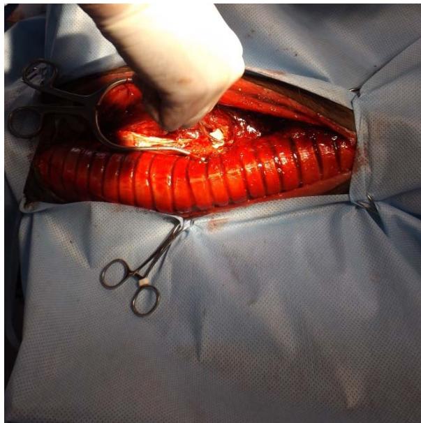
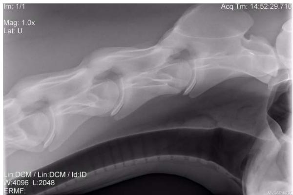 Fig. 1, 2: There is Irregularity of the Tracheal Rings (Blue Arrow)
The image recorded by videoendoscopy (fig. 4) clearly shows the change in the spaces between the rings, visualized and marked, showing a greater space between the rings, clearly showing this irregularity, but only with a visual and superficial assessment, we believe there is no interference in respiratory physiology specifically in this case. The presence of this "ridge" in the dorsal region of the tracheal rings, associated with the cervical malformation in the C6C7 vertebrae (GII), could together with the longus colli muscle, compress the recurrent laryngeal nerve (left or right) causing cases of laryngeal hemiplegia (MAY-DAVIS et al, 2015), which we did not confirm with upper respiratory video-endoscopy, because we did not perform this procedure in all horses in GII..
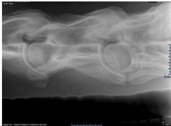 Fig. 3: The Tracheal Rings Seen in the Radiographs of GI where there is the Presence of A " Ridge"(Blue Arrows)
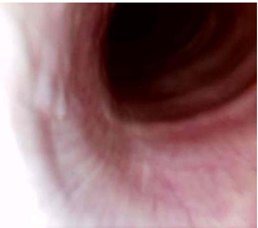 Fig. 4: Videoendoscopy Image Showing Increased Space between the Tracheal Rings of a Horse with Cervical Problems, Non-Congenital Problems (Bule Arrow)
Another important clinical finding in GI and GII were brachial plexus compressions, muscle atrophies in the anterior limbs (right and left), hyposensitivity of the skin (fig. 5) and cervical muscles (fig. 6), (MAY-DAVIS 2015).
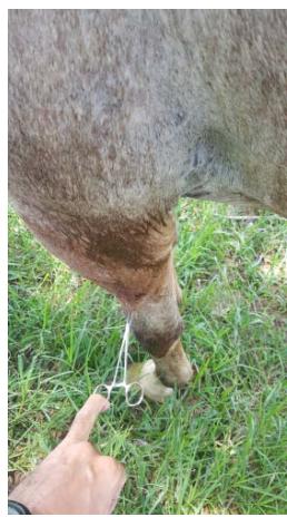 Fig. 5: Image showing complete skin hypoesthesia/ anesthesia in a horse with a non-congenital cervical problem
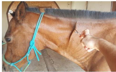 Fig. 6: Cervical region with complete anesthesia resulting, in this case, from a congenital lesion
## IV. CONCLUSIONS
The simple lateral projection radiographs initially showed that the tracheal rings in all horses from both groups are of different sizes and shapes, regardless of whether the animal has or does not have a neurological problem located in the cervical region. Irregularities in the cartilaginous rings ("ridge"), were present in all horses with congenital cervical malformations, that is, we found this lesion in all horses with congenital cervical problems in this study, but we suggest further studies using better investigation techniques.
With these simple findings we can indicate the importance of radiographic examination whether for the purchase of animals that will begin high-performance athletic activity or not, in agreement with DASH (2024), and in the case of animals with findings that indicate congenital problems should be prohibited from entering into a reproductive program.
Generating HTML Viewer...
References
12 Cites in Article
Daniela Bedenice,Amy Johnson (2022). Neurologic conditions in the sport horse.
Klaus Budras,W Sack,Sabine Rock,Aaron Horowitz,Rolf Berg (2011). Anatomy of the Horse.
H Clayton,P Flood,D Resenstein (2007). Anatomia Clínica del Caballo.
R Dash,R Morgan (2024). Should cervical radiographs be included in a pre-purchase examination.
Mazen Mannaa,Ashraf Shamaa,Ahmed Shawky,Islam Hassan,Ashraf Refaey,Ashraf Abu-Seida (2023). A novel surgical technique for treatment of cervical vertebral stenotic myelopathy (wobbler syndrome) in a filly.
Sharon May-Davis,Catherine Walker (2015). Variations and Implications of the Gross Morphology in the Longus colli Muscle in Thoroughbred and Thoroughbred Derivative Horses Presenting With a Congenital Malformation of the Sixth and Seventh Cervical Vertebrae.
Lynn Pezzanite,Jeremiah Easley (2019). Update on Surgical Treatment of Wobblers.
L Siger,J Hawkins,F Andrews,R Henry (1998). tracheal or bronchial stenosis, hypoplasia, collapse, stricture.
T Simmons,M Petersen,J Parker,A Dietze,W ; Rebhun (1988). Tracheal collapse due to chondrodysplasia in a miniature horse foal.
L Vasconcellos (2019). Problemas Respiratórios nos Equinos e seus Métodos de Diagnóstico por Imagens.
Bruno Faustino,Isabel Da Fonseca,Leonor Soares,Joana Alves,Fábio Maio (2022). Research Agenda on Complex Neural Networks Approach to Psychopathology.
D Wong,B Sponseller,E Riedesel,L Couëtil,K Kersh (2008). The use of intraluminal stents for tracheal collapse in two horses: Case management and long‐term treatment.
No ethics committee approval was required for this article type.
Data Availability
Not applicable for this article.
How to Cite This Article
Luiz A. S. Vasconcellos. 2026. \u201cMorphological Irregularity of Tracheal Rings in Horses Associated or not with Cervical Malformation (Initial Study)\u201d. Global Journal of Medical Research - H: Orthopedic & Musculoskeletal System GJMR-H Volume 24 (GJMR Volume 24 Issue H1): .
Explore published articles in an immersive Augmented Reality environment. Our platform converts research papers into interactive 3D books, allowing readers to view and interact with content using AR and VR compatible devices.
Your published article is automatically converted into a realistic 3D book. Flip through pages and read research papers in a more engaging and interactive format.
With the purpose of verifying the existence of irregularities in the tracheal rings of horses and correlating it with the presence of equine cervical vertebral malformation, 45 horses were used, including males and females of different breeds, distributed in two groups, the first being horses with lesions cervical lesion of congenital origin (GI 10 animals) and the second group composed of horses with nom-congenital cervical lesions (GII 35 animals), animals from different parts of Brazil, treated in private practice over a period of 2019-2023 (4 years). It was observed that both horses without congenital lesions and those with congenital lesions had tracheal rings of different shapes that did not compromise air flow in the animals studied, and also that in all animals with congenital lesions in the cervical vertebrae, they had tracheal rings in the dorsal portion of the tracheal rings a “ridge.” This was an initial study and we believe that further investigation should be carried out.
Our website is actively being updated, and changes may occur frequently. Please clear your browser cache if needed. For feedback or error reporting, please email [email protected]
Thank you for connecting with us. We will respond to you shortly.