
Ophtalmologic Cystinosis

§ Hospital Militaire Rabat Maroc

Send Message

To: Author

Ophtalmologic Cystinosis

Article Fingerprint

ReserarchID

PDDTMK35P5

Ophtalmologic Cystinosis Banner

AI TAKEAWAY

Connecting with the Eternal Ground
  • English
  • Afrikaans
  • Albanian
  • Amharic
  • Arabic
  • Armenian
  • Azerbaijani
  • Basque
  • Belarusian
  • Bengali
  • Bosnian
  • Bulgarian
  • Catalan
  • Cebuano
  • Chichewa
  • Chinese (Simplified)
  • Chinese (Traditional)
  • Corsican
  • Croatian
  • Czech
  • Danish
  • Dutch
  • Esperanto
  • Estonian
  • Filipino
  • Finnish
  • French
  • Frisian
  • Galician
  • Georgian
  • German
  • Greek
  • Gujarati
  • Haitian Creole
  • Hausa
  • Hawaiian
  • Hebrew
  • Hindi
  • Hmong
  • Hungarian
  • Icelandic
  • Igbo
  • Indonesian
  • Irish
  • Italian
  • Japanese
  • Javanese
  • Kannada
  • Kazakh
  • Khmer
  • Korean
  • Kurdish (Kurmanji)
  • Kyrgyz
  • Lao
  • Latin
  • Latvian
  • Lithuanian
  • Luxembourgish
  • Macedonian
  • Malagasy
  • Malay
  • Malayalam
  • Maltese
  • Maori
  • Marathi
  • Mongolian
  • Myanmar (Burmese)
  • Nepali
  • Norwegian
  • Pashto
  • Persian
  • Polish
  • Portuguese
  • Punjabi
  • Romanian
  • Russian
  • Samoan
  • Scots Gaelic
  • Serbian
  • Sesotho
  • Shona
  • Sindhi
  • Sinhala
  • Slovak
  • Slovenian
  • Somali
  • Spanish
  • Sundanese
  • Swahili
  • Swedish
  • Tajik
  • Tamil
  • Telugu
  • Thai
  • Turkish
  • Ukrainian
  • Urdu
  • Uzbek
  • Vietnamese
  • Welsh
  • Xhosa
  • Yiddish
  • Yoruba
  • Zulu
Font Type
Font Size
Font Size
Bedground

Ophtalmologic Cystinosis

INTRODUCTION

Cystinosis is a very rare lysosomal, autosomal recessive disease (1/200,000 births) caused by a mutation in the CTNS gene (Chz 17) encoding a protein called cystinosine. The intralysosomal accumulation of cystine induces the formation of insoluble crystals responsible for progressive multiple organ failure. Cystinous nephropathy is manifested by failure to thrive, Fanconi syndrome, damage to the renal glomerulus and manifestations affecting other organs appearing as early as 6 to 12 months of life. The specific treatment for cystinosis is cysteamine.The management is multidisciplinary.

We report the case of a boy, aged 6 years, followed for cystinosis diagnosed at the age of 18 months with polyuropolydipsic syndrome. The patient has been treated with oral cysteamine (Cystagon) since the age of 2 years.

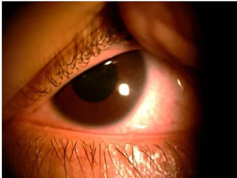
On the ophthalmological level, his corrected visual acuity is evaluated at 10/10 P2 ODG, and the intraocular pressure is at 12 mmHg in both eyes. The main complaint of this patient is photophobia. he is treated with cysteamine eye drops (Cystadrops®) at a dosage of 4 drops per day and additional treatment with artificial tears. With the slit lamp, we find a corneal cystinosis of grade 1.50 according to the classification of Gahl (figure 1): deposits of birefringent spindle-shaped crystals accumulating in the corneal stroma, and progressing from the anterior stroma to the endothelium. The appearance is sparkling and multicolored on biomicroscopic examination.

The severity of the corneal involvement can be assessed by corneal OCT.

The fundus does not show retinopathy. Cystinosis retinopathy is very inconstant. Crystal deposits are sometimes found all over the retina, associated with depigmentation and which can progress to retinal atrophy. It can be explored by retinal angiography. The crystals can also deposit on the conjunctiva, iris, ciliary body, anterior lens capsule, choroid or optic nerve and cause various rarer manifestations, such as glaucoma by closing the angle, papillary edema or visual field changes.

Ophthalmologic monitoring for cystinosis should be annual, with visual acuity assessment, Gahl score estimate supplemented by OCT imaging of the cornea, measurement of intraocular pressure and fundus examination.

This case illustrates a well-followed patient who is coping well with his illness.
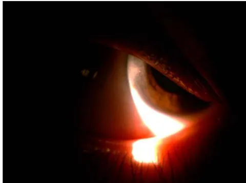
Figure 1: Corneal crystal deposits.

References

3 Cites in Article
  1. Galina Nesterova,William Gahl (2013). Cystinosis: the evolution of a treatable disease.
  2. Rachel Bishop (2017). Ocular Complications of Infantile Nephropathic Cystinosis.
  3. Mohamed Elmonem,Koenraad Veys,Neveen Soliman,Maria Van Dyck,Lambertus Van Den Heuvel,Elena Levtchenko (2016). Cystinosis: a review.

Funding

No external funding was declared for this work.

Conflict of Interest

The authors declare no conflict of interest.

Ethical Approval

No ethics committee approval was required for this article type.

Data Availability

Not applicable for this article.

How to Cite This Article

Fiqhi Aissam. 2026. "Ophtalmologic Cystinosis". Global Journal of Medical Research - B: Pharma, Drug Discovery, Toxicology & Medicine GJMR-B Volume 22 (GJMR Volume 22 Issue B2).

Download Citation

Cystinosis diagnosis and ocular cysts in eye health research.
Journal Specifications

Crossref Journal DOI 10.17406/gjmra

Print ISSN 0975-5888

e-ISSN 2249-4618

Keywords
Classification
GJMR-B Classification DDC Code: 724 LCC Code: NA500
Version of record

v1.2

Issue date
July 9, 2022

Language
English
Experiance in AR

Explore published articles in an immersive Augmented Reality environment. Our platform converts research papers into interactive 3D books, allowing readers to view and interact with content using AR and VR compatible devices.

Read in 3D

Your published article is automatically converted into a realistic 3D book. Flip through pages and read research papers in a more engaging and interactive format.

Article Matrices
Total Views: 421
Total Downloads: 22
All Trends

Request Access

Please fill out the form below to request access to this research paper. Your request will be reviewed by the editorial or author team.
X

This is the heading

Lorem ipsum dolor sit amet, consectetur adipiscing elit. Ut elit tellus, luctus nec ullamcorper mattis, pulvinar dapibus leo.

High-quality academic research articles on global topics and journals.

Ophtalmologic Cystinosis

Fiqhi Aissam
Fiqhi Aissam Hospital Militaire Rabat Maroc