I. INTRODUCTION
Oral condition in pediatric age constitutes a public health problem, reaching a prevalence of (1). Caries is the most frequent ailment, and its origin is multifactorial. However, some reports in the literature show also other significant to matological alterations associated with an underlying systemic disease (2, 3). Some of the most prevalent systemic diseases observed in pediatric patients are Leukemia (4) and Diabetes Mellitus (3). Leukemias are malignant neoplasms of the bone marrow, with a rate of 13 cases per 100,000 inhabitants, with a slight predominance in the male gender (4) Based on the American Cancer Society classification, B-cell Acute Lymphoblastic Leukemia (B-cell ALL) is the most common in this population (4, 5) In United States of America, the incidence of B-cell ALL is about 1.6 per 100,000 population (5) In Mexico, leukemias are among the top 3 causes of mortality in children in the 1 to 14 years range.
In the Mexican State of Chihuahua, leukemias occupy the fifth and second places of mortality in the age groups of one to four years and five to fourteen years, respectively (6). In addition to the clinical picture of systemic manifestations (fever, anemia, bone pain, asthenia, adynamia, ecchymosis, bleeding, adenomegaly, and hepatosplenomegaly) various oral lesions are also frequently observed, mainly: ulcers, xerostomia, gingivitis, and mucositis (4, 7) plus added infections, particularly Candida albicans. Regarding Diabetes Mellitus, between and of people suffer from Type 2 Diabetes Mellitus whereas to are affected by Type 1 Diabetes Mellitus (T1DM), and to corresponds to other variants (8). Of the population of patients with T1DM, the vast majority start at an early age, being currently the most frequent autoimmune disease in childhood (9). Besides the long-term complications (nephropathy, heart disease, diabetic foot, retinopathy, cerebral vascular events, etc.), oral lesions have been seen very frequently; especially those related to periodontal disease: erythema, gingivitis, ulcers, bleeding (10), xerostomia, and caries (11).
This work studied pediatric patients with B-cell ALL or T1DM as an underlying systemic disease; purposes were to find which oral lesions are observed more frequently and find out a relation between these findings with the pathophysiology of the underlying disease.
II. METHODOLOGY
The authorization corresponded to the Bioethics Department of the Autonomous University of Chihuahua (UACH; from Spanish: Universidad Autónoma de Chihuahua) and the Children's Hospital of Specialties of Chihuahua (HIECH; from Spanish: Hospital Infantil de Especialidades de Chihuahua).
a) Data collection
Parents or guardian's participant signed the informed consent letter to take part in the research project. Likewise, each participant was notified about the study and signed the informed letter of consent, according to the criteria set out in the Official Mexican Standard NOM-012-SSA3-2012 and the Regulations of the General Health Law on Health Research.
b) Study groups
This study included fifteen patients, with whom we formed three groups:
- T1DM patients
- B-cell ALL
- Healthy children
Each group consisted of five children, and they were paired by age and sex with children in the other groups (Table S1).
Patients' data were obtained from the file and by direct interrogation to parents, guardians, and patients. We perform a clinical examination with emphasis on the oral cavity. Finally, were analyzed blood count reports.
A pediatric oncologist and pediatric endocrinologist reported diagnosis and managed treatments. All individuals met the inclusion and exclusion criteria (Appendix S1):
- T1DM. All of them with insulin treatment.
- B-cell ALL. They were in the first week of the induction to remission phase of chemotherapy, based on Saint Jude 16 protocol (12, 13); briefly, children received: prednisone, daunorubicin, vincristine, L-asparagine, etoposide, and triple intrathecal therapy during this stage.
- Healthy children. They were obtained from patients who enter for light medical procedures (for example, minor trauma treatments).
c) Study of the clinical case and oral cavity exploration
We interviewed patients' parents or guardians by a questionnaire about the history and clinical evolution, both systemic and buccal. Then, we performed an oral examination which focused on the search for the following alterations:
d) Presence of xerostomia
Initially, it was performed a clinical buccal inspection for signs of xerostomia (opaque mucosa membranes and thick saliva); it was also evaluated the mucosa-membrane hydration level as explained below:
The lower lip was turned from the inside out, the labial mucosa was carefully dried with gauze by a piece of paper over the mucosa; it was searched for saliva droplets formation by minor glands' holes.
For the quantification of the size of saliva production, we set up as parameters:
Low salivary production: more than four drops of saliva in sixty seconds.
Normal salivary production: more than four drops of saliva in sixty seconds.
For the inspection of mucosa hydration, we inspected if the tongue adheres after depression which indicates a positive sign of dehydration.
Therefore, we defined xerostomia as the existence of two positive signs found by the procedures mentioned above.
e) Oral exam of soft tissues
It was performed a detailed examination; different injuries found corresponded to the following ones:
- Traumatic ulcers (14): they have a very irregular loss edge and a frequent unique random distribution.
- Herpetic ulcers (14): they have a scalloped border with an erythematous area. They are usually multiple
and painful; located at the oral corners, lips, and gums.
- Gingivitis (15): they are swollen and erythematous gum, sometimes painful.
- Candida plaques (14): they are white cotton-like and friable regions on gums, mucosa, or tongue; they are easily removed leaving an erythematous and bleeding area.
- Mucositis (16): it is an inflammation of the mucosa membranes with oral erythema, ulcers, and pain.
Gingival index (GI) (17, 18) It was based on the method described by Silness and Loe (Table 1). It is intended to record the degrees of deposit of dental-bacterial plaque
(DBP) without the need to dye it; it is calculated with the formula:
Where is the gingival index of each revised tooth, is the number of explored surfaces, is the presence of DBP on each studied tooth surface. We assessed DBP buildup using a dental scoop and an intraoral mirror, and we codified the findings according to Silness and Loe scale (18) (Table 1). The explored surfaces were vestibular, palatine/lingual, mesial, and distal.
| Degree | Description |
| 0 | DBP absent |
| 1 | DBP only detectable when passing the dentin spoon |
| 2 | Moderate and visible DBP |
| 3 | Abundant DBP; covers beyond the gingival third of the tooth surface |
CPOD and ceod indices. They are used to show the experience of caries in a patient, whether currently or in the past; the only difference between the two is that CPOD index evaluates the permanent dentition and the ceod the deciduous dentition. In order to understand these indices, their components must be broken down: 'C' and 'c' show the decayed teeth in the permanent and deciduous dentition respectively, 'P' refers to the permanent dentition, 'e' writes down the deciduous dentition, or the teeth lost either by caries or by extraction, 'O' denotes the clogged teeth, and 'D' means dentition. These indices are obtained from the individual sum of the values of each element (tooth): decayed, lost, and filled (CPO or ceo), which is divided by the total number of existing teeth (Table 2)(19).
| Interval | Gravity |
| 0.0 – 1.1 | Exceptionally low |
| 1.2 – 2.6 | Low |
| 2.7 – 4.4 | Moderate |
| 4.5 – 6.5 | High |
| >6.6 | Extremely high |
f) Hemocytometry study
Consisted of a blood of carrying out the blood count. Concerning the B-cell ALL group, blood count (BC) study of each patient was conducted on those samples taken in the interval of 1 to 10 days after the first chemotherapy; BC was also checked when this treatment was not started yet. In T1DM cases, BC were taken at the time of the last medical and dental check-up; diabetes treatment had already started. BC in healthy cases were taken during the medical or dental consultation. BC was interpreted by expert physicians based on the normal pediatric values, according to the age range established in the international parameters to Mexican patients (Appendix S2)(20).
g) Statistical analysis
We obtained simple and relative frequencies from nominal and categorical variables. Respect continuous variables, age ranges, means, and standard deviations we used the test for proportions to evaluate significant differences between groups, based on the SPSS-IBM 25.0 program for calculations.
III. RESULTS
This study included fifteen patients, with five children each (see methodology and Table S1).
a) Clinical Findings
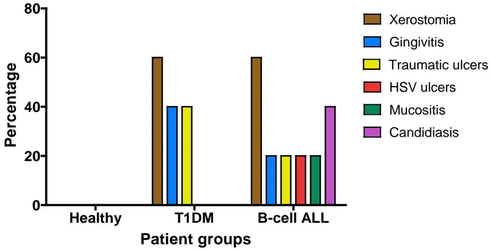
Through the detailed intraoral examination, we found that of both diabetic (Figure 3) and leukemic
(Figure 6) patients exhibited notorious xerostomia; this sign was negative in the healthy group (Figure 1, Table 3).
In the examination of soft tissues, we found that of patients with T1DM or B-cell ALL presented some type of oral alteration; of the healthy group were free of pathology (Table 4).
| Healthy | Percentage* | T1DM | Percentage | B-cell ALL | Percentage | |
| Presence | 0 | 0% | 3 | 60% | 3 | 60% |
| Absence | 5 | 100% | 2 | 40% | 2 | 40% |
| Healthy | Percentage* | T1DM | Percentage | B-cell ALL | Percentage | |
| Presence | 0 | 0% | 4 | 80% | 4 | 80% |
| Absence | 5 | 100% | 1 | 20% | 1 | 20% |
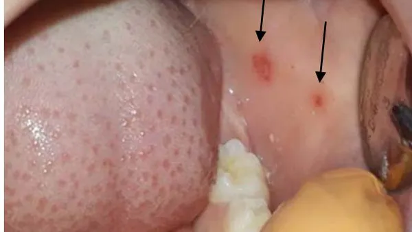
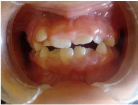
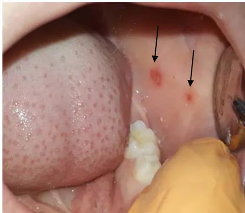
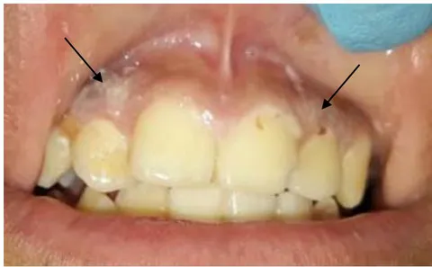
The lesions found in patients with T1DM (Figure 1) were gingivitis (Figure 2) and traumatic ulcers (Figure 3). In patients with B-cell ALL predominated: candidiasis (Figure 4), gingivitis (Figure 5), HSV-likeulcers (Figure 5), traumatic ulcers (Figure 6), mucositis
(Figure 6). Therefore, lesions in the B-cell ALL patients group were more variable than in the T1DM one; moreover, not only a greater variability but also a bigger lesions intensity were observed in the B-cell ALL group.
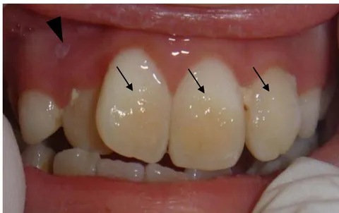
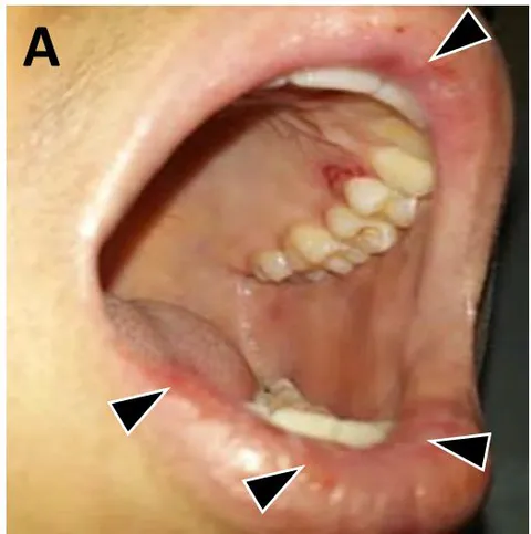
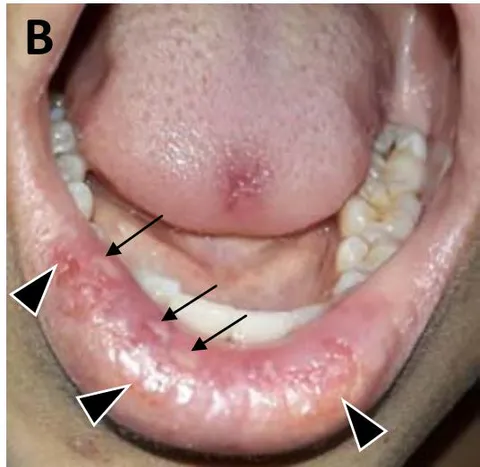
For risk of caries evaluation, we relied on the oral hygiene index standards established by Silness and Loe (50). T1DM children group presented very low values of CPOD-ceod (cariogenic risk) and GI indices (1.0 and 2.2, respectively), healthy patients group had higher risk values (3.2 and 2.7 respectively), and B-cell ALL children set had the highestones (5.2 and4.6 respectively) (Table 5). On the other hand, oral pH values in B-cell ALL and healthy children groups were practically neutral (pH 7.1 and 7.0, respectively); while in the T1DM set the value was acidic (pH 6.1). Nevertheless, these values did not exceed the cariogenic threshold (pH 5.5); they were very close in the three groups. Therefore, there were no significant differences (Table S2).
| Groups | CPOD-ceod* | GI* | GI Interpretation |
| Healthy | 3.2 | 2.7 | Low |
| T1DM | 1.0 | 2.2 | Extremely low |
| B-cell ALL | 5.2 | 4.6 | High |
b) Hemocytometric Study
BC white formula showed that of B-cell ALL patients presented leukopenia secondary to neutropenia and lymphopenia ;
there were notanomalous counts in other morphologically normal leukocytes. There were no leukocyte alterations in healthy and diabetic groups either (Tables 6 and 7).
| Alteration | Healthy | Percentage | T1DM | Percentage | B-cell ALL | Percentage |
| Leukopenia | 0 | 0% | 0 | 0% | 5 | 100% |
| Neutropenia | 0 | 0% | 0 | 0% | 5 | 100% |
| Lymphopenia | 0 | 0% | 0 | 0% | 5 | 100% |
| Eosinophilia | 0 | 0% | 0 | 0% | 0 | 0% |
| Monocytosis | 0 | 0% | 0 | 0% | 0 | 0% |
| Healthy n (%) | T1DM n (%) | B-cell ALL n (%) | p* | |
| Leukopenia | ||||
| Present | 0 (0.0) | 1 (20.0) | 5 (100.0) | 0.003 |
| 4 (80.0) | ||||
| Absence | 5 (100.0) | 0 (12.5) | 0 (0.0) | |
| 5 (100.0) | ||||
| Neutropenia | ||||
| Present | 0 (0.0) | 0 (12.5) | 5 (100.0) | 0.001 |
| 5 (100.0) | ||||
| Absence | 5 (100.0) | 0 (0.0) | 0 (0.0) | |
| 5 (100.0) | ||||
| Lymphopenia | ||||
| Present | 0 (0.0) | 0 (0.0) | 5 (100.0) | 0.001 |
| 5 (100.0) | ||||
| Absence | 5 (100.0) | 0 (0.0) | 0 (0.0) | |
| 5 (100.0) | ||||
| Eosinophilia | ||||
| Present | 0 (0.0) | 0 (0.0) | 1.0 | |
| Absence | 5 (100.0) | 5 (100.0) | ||
| Monocytosis | ||||
| Present | 0 (0.0) | 0 (0.0) | 1.0 | |
| Absence | 5 (100.0) | 5 (100.0) | ||
| Anemia | ||||
| Present | 0 (0.0) | 3 (60.0) | 0.02 | |
| Absence | 5 (100.0) | 2 (40.0) |
BC red formula showed that all B-cell ALL children presented anemia; four patients had macrocytic hyperchromic anemia (80%) consistent with megaloblastic anemia; anisocytosis was reported in 1 case (Table 8).
| Type of Anemia | Healthy | T1DM | B-cell ALL | Diagnosis |
| Hyperchromic Macrocytic | 0 | 0 | 4 (80%) | Megaloblastic anemia |
| Hyperchromic Normocytic | 0 | 0 | 1 (20%) | Anisocytosis |
| Hypochromic Microcytic | 0 | 0 | 0 | Iron Deficiency Anemia |
IV. DISCUSSION
In this study, we performed a global clinical analysis of the oral cavity with the end to identify diverse types of lesions as well as oral cavity hygiene, caries, and tooth losses. We also evaluated the blood tissue state basing us on BC studies. All these parameters were carried out in three groups of pediatric patients: healthy, T1DM, and B-cell ALL (Table S1).
Regarding the clinical analysis, lesions were found in T1DM and B-cell ALL groups; except for poor oral hygiene, the healthy group did not present pathological alterations were found. Respect oral hygiene, T1DM patients had the best CPOD-ceod and GI indices (Table 5) whereas B-cell ALL children revealed the worst ones. These findings can be explained by the fact that diabetic patients eat less sugary foods, and generally receive more oral care than most patients; obviously, the more oral hygiene the fewer complications (21). The most precarious oral hygiene in the B-cell ALL group can be understood as consequence of an important pain presence due to epithelial fragility occasioned by both mucositis -caused by chemotherapy (22)- and leukemia per se. The pain also causes a less teeth brushing frequency, leading to a higher GI (measure of DBP formation) and CPOD-ceod (measure caries and teeth losing) indices (Table 5).
Xerostomia, in general, is more pronounced in Diabetes Mellitus which it was also observed in our T1DM group; it is a consequence of the chronic hypohydration state specially seen in poorly controlled patients (23). This, in the long run, will reduce nonimmunological defense barriers such as:
Physical barriers: saliva will turn more viscous which will decrease an efficient oral mechanical washing (24).
Chemical barriers: salivary hyperviscosity is consequence of the high glucose and electrolyte (calcium and phosphorus) concentrations which occasions a pH decrease and it also affects the enzymatic functions (amylase an alkaline phosphatase) (25).
Biological barriers: physicochemical alterations of microenvironment alter the oral microbiota composition and, therefore, favor growth of pathogenic and opportunistic microorganisms (24).
Immune barriers: the increased oral viscosity and abnormal pH will occasion immune dysfunctions, specifically in the antibodies (26, 27). Diabetic microangiopathy significantly affects the immune function; however, it is not a factor to take into account in our group since this is a long-term complication.
However, there were a lower CPOD-ceod index (lesser caries) in T1DM group than in the healthy set despite of the first one had oral lesions. This can be explained because of the higher tooth washing frequency and lower carbohydrates intake (candies and chocolates especially) by T1D Mchildren.
B-cell ALL group also presented a greater quantity and variety of lesions which were more severe. Like in T1DM, it was detected gingivitis and traumatic ulcers along with exfoliative cheilitis (Figures 5 and 6) and other lacerations: oral candidiasis (Figure 4), herpeticulcers (Figure6), and mucositis (Figure 6). Basing us on the underlying disease's natural history, treatment, and BC results, we suggest that the most relevant lesion-generating factor was the immunosuppression state in this group. So, oral alterations secondary to B-cell ALL pathophysiology can be explained as follows:
The underlying nosological entity itself. The lymphoblastic neoplasm grows in the red bone marrow, so it displaces healthy tissue which is occupied by are non-functional blast cells both in the bone marrow and peripheral blood. Paradoxically, the hemocytometry values of the white formula will be extremely high; however, normal leukocyte counts will be low.
Chemotherapy. Sant Jude 16 protocol (12, 13) (see methodology and Appendix S3), not only kills neoplastic cells but also functional ones which aggravates the state of immunosuppression. In addition treatment is also involved not only in the treatment but also triggering mucositis which turns the mucosa pretty fragile (22). On the other hand, bone marrow damage caused by both leukemia and chemotherapy affect platelet and erythrocyte production causing thrombocytopenia and anemia (megaloblastic anemia) which was proved in the BC (Table 8). This last finding is mainly triggered by methotrexate administration -part of the Saint Jude 16 therapeutic protocol- which is a folic acid antagonist (28).
Oral pH revealed a neutral value in B-cell ALL and healthy patient groups (pH 7.1 and 7.0 respectively); pH in T1DMset was acidic (pH 6.1) (Table S2). However, this record did not reach a critical value to be considered as a cariogenic factor (29). Therefore, CPOD-ceod and Glindices suggest the cariogenic risk is related to other factors such as poor oral hygiene; especially in B-cell ALL children, probably related to the frequent oral pain.
It is also pertinent to clarify that, at the moment, our sample size is small, so we will continue increasing the number of samples of each group to increase the statistical power.
V. CONCLUSIONS
Both B-cell ALL and T1DM are one of the more important systemic diseases in pediatric age which also have a significant impact on the oral cavity. Prominently, the key factor related to B-cell ALL is immunocompromise, provoked by both the neoplastic disease and chemotherapy, whereas in T1DM the main disbalance is ametabolic dehydration and hyperglycemic state. The lowest presence of caries (lower CPOD-ceod index) in the T1DM group is linked to the better oral hygiene associated in the treatment. Finally, our results suggest that systemic diseases alter the buccal cavity homeostatic state, affecting the normal-microbiota development, favoring pathogenic or opportunistic microorganisms' grow and attack(30) and an increase of systemic-infections risk and septicemia (31); so, we suggest that underlying diseases like B-cell ALL and T1DM, and their treatments effects, have a relevant impact in the oral cavity homeostasis as well as over microbiota composition; all this together leads to the formation of oral lesions. Therefore, we propose that it is especially important to carry out metagenomic studies (gene-taxonomy with last generation sequencing) of the oral microbiota to identify specific changes that could specifically identify those pathogenic species that cause oral lesions formation.
ACKNOWLEDGMENTS
We thank the Children's Hospital of Specialties of Chihuahua through Dr. Héctor José Villanueva-Clift (Head of Teaching)," Dr. Sergio Elizalde-Morton (Director), Dr. Lilia Teresa Avenida-Portillo; and Dr. Luz Elena Sanín-Aguirre, from Autonomous University of Chihuahua, for their priceless support in this research.
There are no interest conflicts or disagreements between authors.
Víctor Adolfo Ríos-Barrera and Alejandra Saldivar-De la Torre did most of this research and have the same merits and importance.
SUPPLEMENTARY APPENDICES
Supplementary Appendix S1
a) Inclusion criteria
Group of patients with T1DM: confirmed diagnostic of DM1, insulin treatment already set up. Recent complete blood count (BH) (less than 2 weeks). Age range: 5 to 15 years.
Group of patients with B-cell ALL: Confirmed diagnosis of B-cell or pre B-cell, which will be described together as B-cell ALL. Patients in the induction to remission phase of chemotherapy. Recent full BH (less than 2 weeks). Age range: 5 to15 years.
Control group of healthy children: Age and gender comparable with the groups of B-cell ALL and T1DM. Recent full BH (less than 2 weeks). Hospitalized for trauma-related treatments (fractures, dislocations). Age range: 5 to 15 years.
b) Exclusion criteria
Group of patients with T1DM: Patients outside the age range. Not having written informed consent. Patients with oral appliances. Infectious diseases in the last 4 weeks. Antibiotic therapy 2 weeks prior to sampling.
Group of patients with B-cell ALL: Patients outside the age range. Patients in the consolidation or maintenance phase in their chemotherapy treatment. Not having written informed consent. Patients with oral appliances. Patients with Burkitt lymphoma. Infectious diseases in the last 4 weeks. Antibiotic therapy 2 weeks prior to sampling.
Control group of healthy children: Presence of systemic diseases. Presence of some concomitant syndromes. Not having written informed consent. Patients with oral appliances. Infectious diseases in the last 4 weeks. Antibiotic therapy 2 weeks prior to sampling.
Supplementary Appendix S2
Blood Count Normal pediatric values. According to the patients' age, and established in the international weighting tables adapted to Mexican patients (20).
| Table S3: Red blood cells | |||||
| Age | 1-13 d | 14-60 d | 3 m-10 y | 11-15y | Adults |
| Erythrocytes* (millions/mm3) | 5.1±1.0 | 4.7 ± 0.9 | 4.5± 0.7 | 4.8 | 5.4 ± 0.9 M 4.8 ± 0.6 W |
| Hemoglobin* (g/dL) | 19.5 ± 5.0 | 14.0 ± 3.3 | 12.2 ± 2.3 | 13.4 | 16.0 ± 2.0 M 14.0 ± 2.0 W |
| Hematocrit* (percentage) | 54.0 ± 10.0 | 42.0 ± 7.0 | 36.0 ± 5.0 | 39.0 | 47.0 ± 5.0 M 42.0 ± 5.0 W |
| MCV (fL) | 98-106 | 90 | 80 | 82 | 90 ± 7 M 90 ± 7 W |
| MCH (pg) | 33-38 | 30 | 27 | 28 | 29 ± 2 M 29 ± 2 W |
| CHMC (g/dL) | 34-36 | 33 | 34 | 34 | 34 ± 2 M 34 ± 2 W |
| MCD (μm) | 8.6 | 8.1 | 7.7 | 7.5 ± 0.3 M 7.5 ± 0.3 W | |
Abbreviations: MCV (mean corpuscular volume), MCH (medium corpuscular hemoglobin), MCHC (mean corpuscular hemoglobin concentration), MCD (mean corpuscular diameter); d (days), m (months), y (years), M (men), W (women).
| Table S4: White blood cells | |||||
| Age | 1 y | 4 y | 6 y | 10 y | Adult (>21 y) |
| Total* Leucocytes | 11,400(6.0-17.5 K)100% | 9,100(5.5-15.5 K)100% | 8,500(5.0-14.5 K)100% | 8,100(4.5-13.5 K)100% | 7,400(4.5-11.5 K)100% |
| Neutrophiles | |||||
| Total* | 3,500(1.5-8.5 K)31% | 3,800(1.5-8.5 K)42% | 4,300(1.5-8.0 K)51% | 4,400(1.8-8.0 K)54% | 4,400(1.8-7.7 K)59% |
| Bands* | 350 | 270(0-1.0 K) | 250(0-1.0 K) | 240(0-1.0 K) | 220(0-0.7 K) |
| Segmented* | 3.1% | 3.0% | 3.0% | 3.0% | 3.0% |
| 3,200 | 3,500(1.5-7.5 K) | 4,000(1.5-7.0 K) | 4,200(1.8-7.0 K) | 4,200(1.8-7.0 K) | |
| 28% | 39% | 48% | 51% | 56% | |
| Lymphocytes* | 7,000(4.0-10.5 K)61% | 4,500(2.0-8.0 K)50% | 3,500(1.5-7.0 K)42% | 3,100(1.5-6.5 K)38% | 2,500(1.0-4.8 K)34% |
| Monocytes* | 550(0.05-1.1 K)4.8% | 450(0-0.8 K)5.0% | 400(0-0.8)4.7% | 350(0-0.8 K)4.3% | 300(0-0.8)4.0% |
| Eosinophiles* | 300(0.5-0.7 K)2.6% | 250(0.02-0.65 K)2.8% | 230(0-0.65 K)2.7% | 200(0-0.6 K)2.4% | 200(0-0.45 K)2.7% |
| Basophiles* | 50(0-0.2 K)0.4% | 50(0-.2 K)0.6% | 50(0-.2 K)0.6% | 40(0-.2 K)0.5% | 40(0-0.2 K)0.5% |