Orthodontics Intrication To Maxillo-Facial Surgery Preparation of Cases with Obtrusive Syndrome of Apnea

Send Message

To: Author

Orthodontics Intrication To Maxillo-Facial Surgery Preparation of Cases with Obtrusive Syndrome of Apnea

Article Fingerprint

ReserarchID

JF5Y8

Orthodontics Intrication To Maxillo-Facial Surgery Preparation of Cases with Obtrusive Syndrome of Apnea Banner

AI TAKEAWAY

Connecting with the Eternal Ground
  • English
  • Afrikaans
  • Albanian
  • Amharic
  • Arabic
  • Armenian
  • Azerbaijani
  • Basque
  • Belarusian
  • Bengali
  • Bosnian
  • Bulgarian
  • Catalan
  • Cebuano
  • Chichewa
  • Chinese (Simplified)
  • Chinese (Traditional)
  • Corsican
  • Croatian
  • Czech
  • Danish
  • Dutch
  • Esperanto
  • Estonian
  • Filipino
  • Finnish
  • French
  • Frisian
  • Galician
  • Georgian
  • German
  • Greek
  • Gujarati
  • Haitian Creole
  • Hausa
  • Hawaiian
  • Hebrew
  • Hindi
  • Hmong
  • Hungarian
  • Icelandic
  • Igbo
  • Indonesian
  • Irish
  • Italian
  • Japanese
  • Javanese
  • Kannada
  • Kazakh
  • Khmer
  • Korean
  • Kurdish (Kurmanji)
  • Kyrgyz
  • Lao
  • Latin
  • Latvian
  • Lithuanian
  • Luxembourgish
  • Macedonian
  • Malagasy
  • Malay
  • Malayalam
  • Maltese
  • Maori
  • Marathi
  • Mongolian
  • Myanmar (Burmese)
  • Nepali
  • Norwegian
  • Pashto
  • Persian
  • Polish
  • Portuguese
  • Punjabi
  • Romanian
  • Russian
  • Samoan
  • Scots Gaelic
  • Serbian
  • Sesotho
  • Shona
  • Sindhi
  • Sinhala
  • Slovak
  • Slovenian
  • Somali
  • Spanish
  • Sundanese
  • Swahili
  • Swedish
  • Tajik
  • Tamil
  • Telugu
  • Thai
  • Turkish
  • Ukrainian
  • Urdu
  • Uzbek
  • Vietnamese
  • Welsh
  • Xhosa
  • Yiddish
  • Yoruba
  • Zulu
Font Type
Font Size
Font Size
Bedground

I. INTRODUCTION

Obstructive sleep apnea and its associated risk factors can now be more easily identified by specialists in the oro-facial zone, not only through clinical diagnosis but also with the help of new imaging tools such as CBCT or scanners. Orthodontists, who intervene at the intersection of the aero-digestive spheres, have long had an excellent diagnostic tool called Cephalometrics, which they systematically use on a daily basis. For this reason, they are best equipped to detect early morphological signs of possible apneas or indications that an individual may be prone to obstructive sleep apnea and/or hypopnea, and to recommend more specific examinations and consultations, including a Polysomnography.
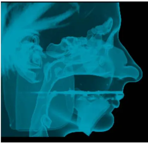
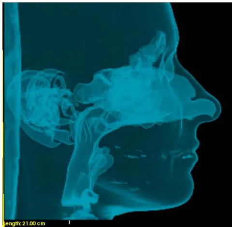
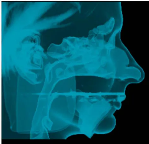
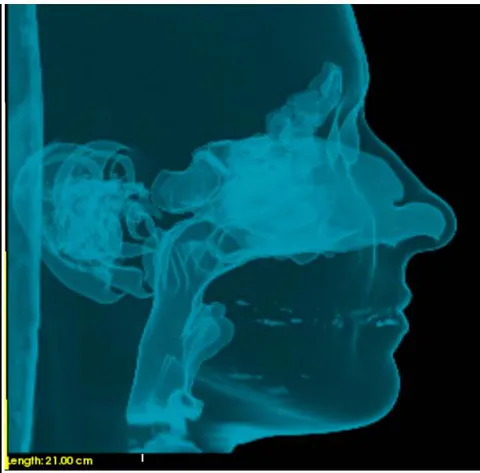
Fig. 1: KC cephalometric X-rays and analysis before and after Surgery showing Narrowness of the retro-basi-lingual area before surgery

This pathology is traditionally addressed with propulsive orthosis, which enlarges the retro-basi-lingual area but can also cause immediate discomfort to the TMJ and teeth, as well as chronic problems when used over the long term (such as slow mesialization of the entire mandibular denture and potential edge-to-edge occlusion for patients starting in CI1). Positive Pressure appliances (PPC) provide an immediate solution to the problem but are burdensome to support and can be embarrassing to wear in marital life.

For this reason, an increasing number of patients, mainly adults, are resorting to a more drastic and definitive solution that resolves this pathology in 90 % of cases. This solution is called "bi-maxillary advancement surgery."

Based on many years of activity in the field of Maxillo-Facial Surgery related to Orthodontics, I have come to understand that major dysmorphosis are often complex and involve a combination of skeletal dysmorphosis and compensatory dental-alveolar changes. They are nothing more than a means of preserving the vital functional role of the masticatory apparatus. However, visualizing possible orthodontic and skeletal improvements that occur in 3D on two moving elements that depend on two delicate joints, the TMJ's, can be quite challenging.

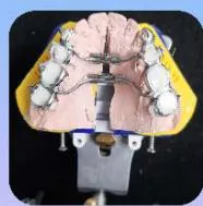
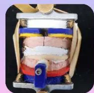
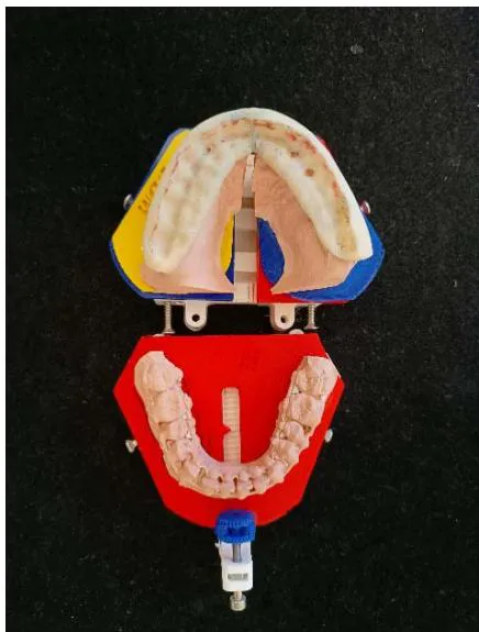
In this context, the subject at hand is the management of the surgical puzzle assembly and its postoperative stability, particularly when significant expansion of the maxilla has been achieved. Focusing on this difficulty, I will present a protocol that allows for a functional and ideal occlusion in the postoperative period using a very precise interface with micrometric adjustments called ORTHOGNATOR®, which perfectly complements the Galetti articulator.
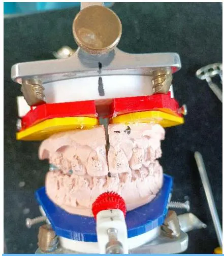
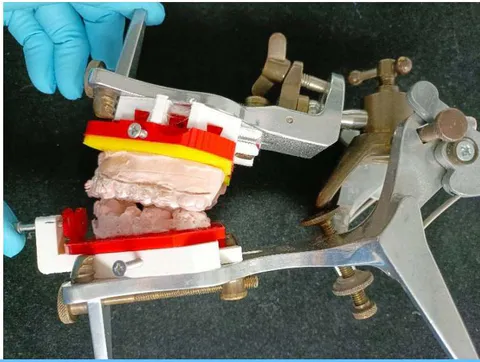
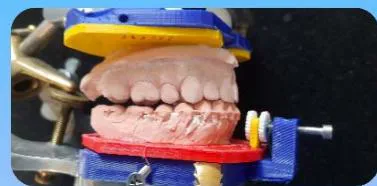
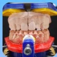
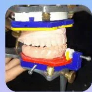
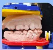
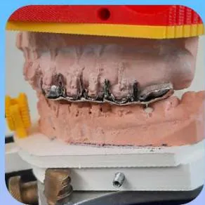
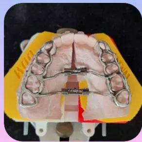
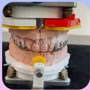
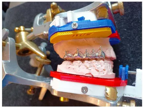
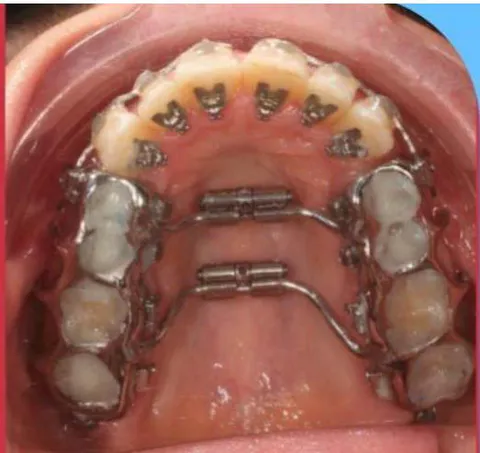
Fig. 2: Orthognator (the Multi colored appliance) and Maxillary dual-screw-expander: An interface to drastically improve the Galletti Articulator

A rigid protocol is necessary because Orthognathic Surgery should only be undertaken with a final resolution in mind that includes all the obligations imposed by our knowledge concerning periodontal longevity 7,8, occlusal stability, and facial harmony, to which smile and beauty contributes. These respond to a number of constraints that we will call our therapeutic envelope.
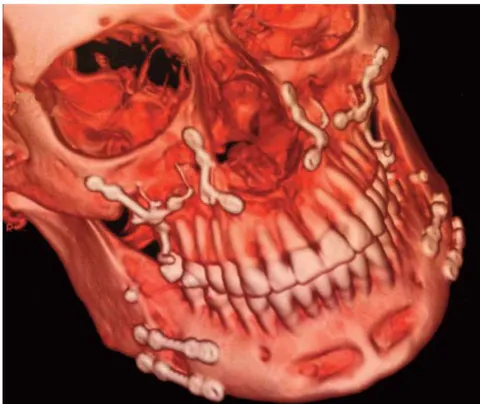
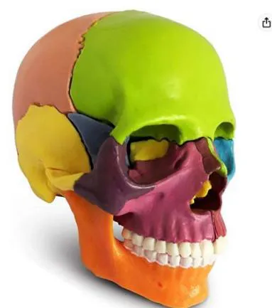
Fig. 3: The assembly of the puzzle, and on the right maxilla an additional osteosynthesis plate as an anchoring supplement. Surgery Jean-François Tulasne showing not only bone syntheses by plates but also intermaxillary grafting by bone transplants

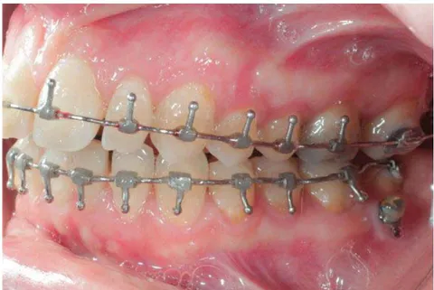
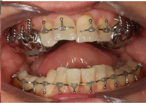
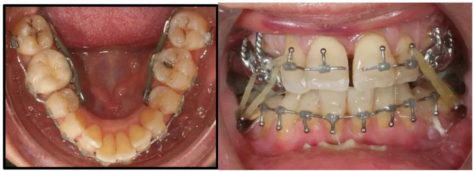
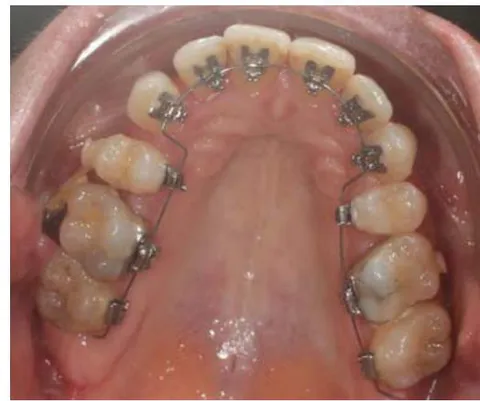
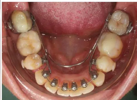
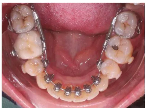
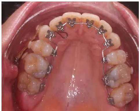
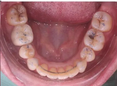
In addition, treatments by lingual approach (often chosen by adults) presents, as everyone knows, particular difficulties. Some of these are related not only to the irregularity of lingual surfaces compared to vestibular surfaces, to access to the working area but also to the height of bondable enamel spaces. These are understood, at least in the maxilla, between the occlusion of the supporting cusps and the gingival cervical limit. When orthognathic surgery has to be linked to this type of orthodontic appliance (Lingual), the problem arises of the relationships of the appliance to the surgical per-operative anchorage (Surgical arches Fig5), and/or to the expansion infrastructures fig 2 and 4 and to the surgical guide (fig4).

The whole system representing superimposed layers Fig 4, which is a vector of possible errors. Moreover, this surgery often involves an intra-arcade modification: rapid palatal expansion aided surgically by a differential expansion gradient or even a three or four-pieces maxillary surgery linked or not to a mandibular displacement. The coherence of the whole system with regard to the final insertion of the surgical guide in the phase preceding bone synthesis is based on well-established protocols and suffers no "prosthetic" imprecision because each error will have a significant impact on post-operative orthodontic outcomes.

The ORTHOGNATOR® permits simulation of simultaneous transversal and rotative movements of the maxillary fragments, full rotative movement of each jaw for midline alignment as well as micrometric sagittal adjustment of the position of the mandible for overcorrection: each full turn of the dented wheel equal a 1 4 of a mm. With the four vertical screws in the posterior area it permits also small adjustments of vertical and transversal tilts.
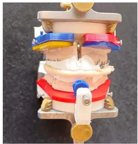
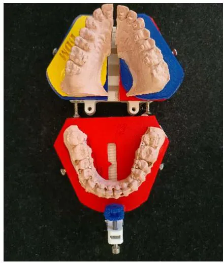
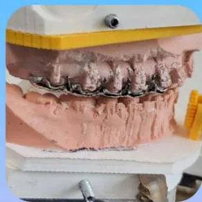
Fig. 4: Predictive assembly, simulation of a Cl3 case and then manufacture of a surgical guide consistent with maxillary expansion and sagittal over-correction, then balanced so that its per-operative insertion is indisputable

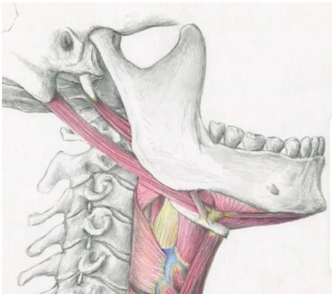
Once these technical difficulties are solved, it is important to also achieve goals with respect to aesthetic finishing. In the case of Sleep Apnea Surgery, a significant advancement of both maxillae is necessary in order to pull the Hyoid bone forward, thus increasing the retro-basi-lingual area. Our priority lies in understanding the implications on occlusion and the alignment of soft tissues. As a matter of fact, Eric Solyom, with whom I work routinely, consistently applies a counter-clockwise rotation of the maxilla whenever feasible. Placing the maxilla first is also a way to ensure a good exposure of the anterior teeth at rest, while speaking, and when smiling.

Fig. 5: TP Stainless Steel 19x26 surgical arcs with glued captive hooks for better hygiene away from embrasures, and Paracrestal screws placed as anchorage in the lower left and right quadrant of the mandible for control of the projection of the lower mandibular incisors during Lingual orthodontic preparation
Fig. 6: Ideal Smile should show sufficient discovery at rest

The analogy with prosthetics is very interesting because as dentists we know that every 1mm in excess in the posterior area creates a bite opening of 3mm in the anterior part of the mouth.

This is exactly what Dr. Solyom's technique does: it creates a huge prematurity that will oblige the mandible to rotate around in the second phase of surgery. Thus something which in turn demands an extra sagittal lengthening of the horizontal branch of the mandible, 15 to 20   mm .

This technique has proven to be extremely efficient in the treatment of SAOS, as many patients report (most of them feel the first benefits in the) awaking room). We also experienced excellent aesthetic result, amazingly in CI3 hyperdivergent cases because the rotation of the mandible highly overcomes its important lengthening.
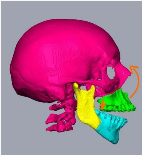
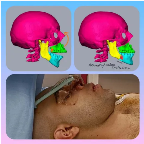
Fig. 8: Counter clockwise rotation of the maxilla induces secondary counter clockwise rotation of the mandible
Fig. 9: Counter rotation of the maxilla by inducing secondary counter rotation of the mandible gives better volume to the chin avoiding genioplasty Surgery which is somehow destructive to the Genioglossus thus counter-productive as far as the cure to Apnea. Dr. Eric Solyom
Fig. 10: The Genioglossus is the transmission "belt" to the advancement of the hyoid bone thus to the enlargement of the Retro-Basi-Lingual area or low Pharyngal area

II. DISCUSSION

Counter clockwise rotation of the mandible has the secondary effect of enlarging the retro-basi-lingual aero-digestive crossway. This helps prevent airway natural tendency to collapse during unconsciousness, as the muscles of the airways tend to relax and the negative pressure from inspiration can exacerbate this collapse.

Traditionally, counter clockwise rotation of the mandible was avoided in mandibular bi-sagittal osteotomy due to concerns about creating an open bite. However, the use of Titanium Osteosynthesis plates with proper compensation, such as temporary posterior disocclusion provided by the surgical guide established on the ORTHOGNATOR©, has demonstrated the ability to overcome this previously feared side effect.

Despite its potential to reduce the effect of surgical mandible retraction on the retro-basi-Lingual area of prognathic cases, there are several morphological contraindications to this procedure. One such contraindication is a prominent but wide symphysis, for which chin reduction is not a favorable aesthetic option. This becomes more problematic in normo-divergent and hypo-divergent Class III cases as it accentuates the concavity of the profile, resulting in an older appearance.

Excessive torque or proclination of the upper incisors can also be a contra-indication or a serious complication when using the counter-clockwise rotation technique for the maxilla, as it can further increase the apparent torque or proclination of the upper incisors. Space is needed to reduce this apparent torque in the maxilla, and while distalization with bony anchorage is a viable option, it can be a time-consuming procedure. Another approach, if necessary and feasible, is to expand the anterior portion of the maxilla to flatten it and reduce the flaring of the incisors.

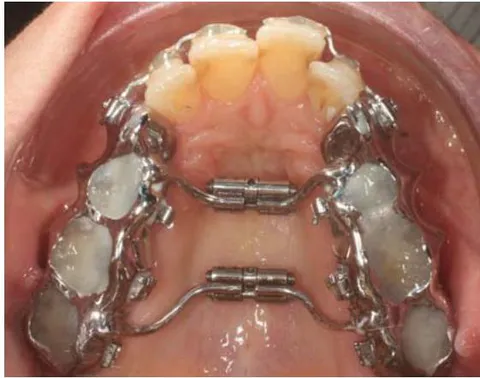
The rapid expansion of the anterior part of the maxilla poses a delicate issue due to concerns about losing the height of the median papilla. This is a recurrent problem reported by many colleagues, but in my practice, it is not an issue thanks to the cast piggyback Chrome-Cobalt dual-screw-expander©, which offers a solution to address this concern.
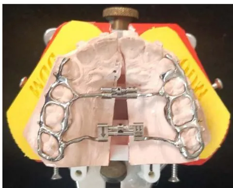
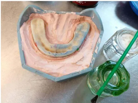
Fig. 7: Ideal Smile should show sufficient discovery in a sketch smile
Fig. 11: A: Cast Chrome Cobalt Dual-screw-rapid expander© permits very precise opening gradients of the palatal vault without damage to the medial papilla. B: Prospective or final surgical guide equilibrated by half arches are subsequently recomposed on a plaster key A

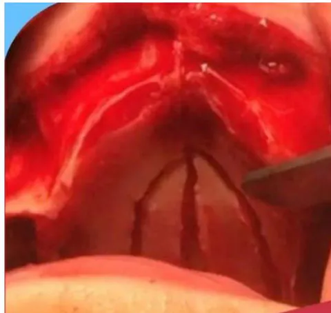
B Fig. 12: A: Nasal fossae and open sinus views of Maxillary 4 pieces osteotomies in closed bird wings giving flexibility to the palatal vault thus avoiding tearing of the palatal mucosa. B Oral palatal view of the Piggy back Chrome-Cobalt dual-screw- expander

i. The Dual-Screw-Expander was devised in my office in 1987
Panel label: A.

Panel label: B.

Panel label: C.

Panel label: D.

Panel label: E.

Panel label: F.

Panel label: G.

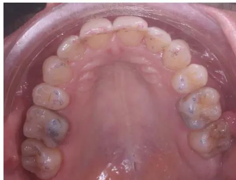
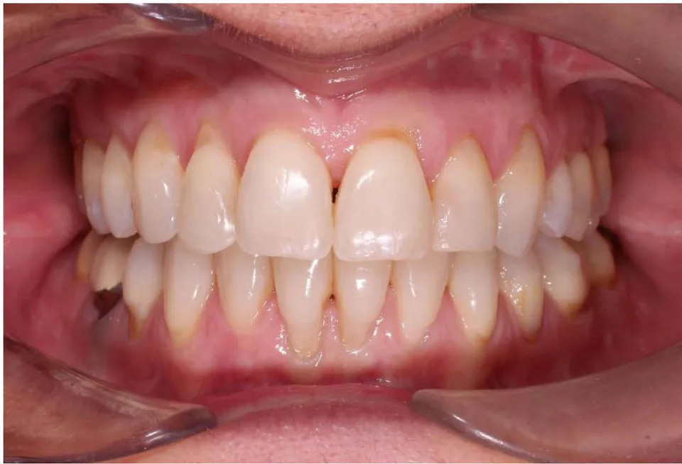
H Fig. 13: Integrity of the median papilla is preserved with the expansion controlling device. (A to B) Oclusal finishing in lingual Technique (C to E) Centering of the midlines on the face (F to G) On a young person it is important for the long term to show a little amount of gum line (G)

Finally the real contra-indication or the serious complication to such surgery happens when patients needing surgery for correction of ASOS also present a serious anterior vertical deficiency[^6] In these types of cases only extensive-intermaxillary grafts could solve the problem.

III. CONCLUSIONS

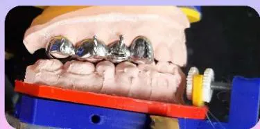
In order to achieve precision and stability in cases of expansion linked to the cure of Sleep Apnea, surgical orthodontic prosthesis is crucial in this complex surgery. It effectively addresses the challenges posed by the powerful connective tissue and elastic fibers of the palatal mucous mantle, which often lead to transverse relapse. Additionally, this procedure allows for bimaxillary surgery to be performed in a single operative time, providing reliability and operative comfort that Maxillo-facial-surgeons have acknowledged, regardless of whether it involves a simple or differential palatal expansion.

In the context of SAOS (Sleep Apnea and Obstructive Syndrome), surgery often becomes a priority due to health concerns. The combination of a Cast Chrome Cobalt dual screw Rapid expander© and the "Orthognator©" tool offers a precise approach for implementing a "surgery first" protocol and managing the aesthetic zone while preserving the delicate gingival tissue, which is challenging to regenerate. Moreover, this protocol enables an assessment of the anterior vertical dimension, which directly impacts the smile.

Compared to the difficult mastery of the virtual aspect and program control in the Workflow technique, as well as the challenges of transferring the results to a solid articulator, the Orthognator® provides a more tangible approach. It allows the practitioner to maintain direct control over the intricate "Puzzle" assembly, including over corrections and relapse simulations that are challenging to convey to a lab technician. By keeping the practitioner closely connected to the surgical reality and its outcomes, the Orthognator® facilitates a concrete approach.

Significant improvements can be observed, such as the unfolding of the labio-mental fold and nasogenien grooves resulting from maxillary advancement. Additionally, there is improved lip support and eversion of the vermilion zone due to better alignment between the incisal border of the upper anterior teeth and the lower lip. When palatal expansion is required in many cases, the use of a bi-expander and of the Orthognator® to model the desired outcome ensures precision, stability, and surgical comfort, leading to a significantly improved prognosis, despite the challenges similar to those encountered with a large prosthesis.
References

31 Cites in Article
  1. J-L Pruvost (1991). Apport des ciments verres ionomères en orthodontie et en occlusodontie.
  2. J-L Pruvost (1994). Utilisation des ancrages bimaxillaires coulés en orthognathie linguale.
  3. J-L Pruvost (2013). Les Ancrages dento-alvéolaires et squelettiques profonds. 1ere Partie Clinic.
  4. J-L Pruvost,J-F Andreani (2013). Les ancrages dentoalvéolaires et squelettiques profonds-2° partie CLINIC.
  5. Lefildentaire Traitements orthodontiques chirurgicaux des insuffisances verticales anterieures en technique lingual "Surgical orthodontic treatments for anterior vertical deficiencies using lingual technique.
  6. Eric Solyom (2023). Étude des voies aeriennes dans la pratique de l’orthodontie et de la chirurgie orthognatique.
  7. P André,Saadoun …… ; 8. Herman Sailer Professor 9,Wa (1969). Pressure-flow relationships in collapsible tubes.
  8. Liann Salman,Rachel Shulman,Jordana Cohen (2020). Obstructive Sleep Apnea, Hypertension, and Cardiovascular Risk: Epidemiology, Pathophysiology, and Management.
  9. R,Doug Mcevoy (2016). CPAP for Prevention of Cardiovascular Events in Obstructive Sleep Apnea.
  10. (2007). Continuous Positive Airway Pressure.
  11. F Jahoo (2016). Le traitement des apnées obstructives du sommeil par les orthèses d'avancée mandibulaires et ses effets secondaires.
  12. C John,S Gandhi,A Sakharia,T James (2018). Maxillomandibular advancement is a successful treatment for obstructive sleep apnoea: a systematic review and meta-analysis.
  13. T Gellée,T Schouman,P Rousseau,R Lockhart (2023). Chirurgie pré-implantaire.
  14. Charles Salvadelli,& Elodie Ehrmann (2023). Apport du dispositif MODJAW dans la planification numérique de la Chirurgiie Orthognathique.
  15. B Neelapu,O Kharbanda,H Sardana,R Balachandran,V Sardana,P Kapoor (2017). Craniofacial and upper airway morphology in adult obstructive sleep apnea patients: a systematic review and meta-analysis of cephalometric studies.
  16. S Hong,S Poomkonsarn,G Millesi,Syc Liu (2020). Upper airway stimulation as an alternative to maxillomandibular advancement for obstructive sleep apnoea in a patient with dentofacial deformity: case report with literature review.
  17. A Zakhar,C Wirth,E Farrow,C Tison,J Ferri,G Raoul (2014). Chirurgie du syndrome d’apnées-hypopnées obstructives du sommeil. Évaluation fonctionnelle.
  18. Makoto Kikuchi (2005). Orthodontic treatment in children to prevent sleep-disordered breathing in adulthood.
  19. Y Chen,L Hong,C-L Wang,S-J Zhang,C Cao,F Wei (2012). Effect of large incisor retraction on upper airway morphology in adult bimaxillary protrusion patients.
  20. D Germec-Cakan,T Taner,S Akan (2011). Uvulo-glossopharyngeal dimensions in non-extraction, extraction with minimum anchorage, and extraction with maximum anchorage.
  21. R Conradt,W Hochban,U Brandenburg,J Heitmann,J Peter (1997). Long-term follow-up after surgical treatment of obstructive sleep apnoea by maxillomandibular advancement.
  22. M Camacho,S Liu,V Certal,R Capasso,N Powell,R Riley (2015). Large maxillomandibular advancements for obstructive sleep apnea: An operative technique evolved over 30 years.
  23. Macario Camacho,Michael Noller,Michael Del Do,Justin Wei,Christopher Gouveia,Soroush Zaghi,Scott Boyd,Christian Guilleminault (2019). Long‐term Results for Maxillomandibular Advancement to Treat Obstructive Sleep Apnea: A Meta‐analysis.
  24. C Jon-Erik,* Holty,Christian Guilleminault (2010). Maxillomandibular advancement for the treatment LEFILDENTAIRE May 2023 p: 48-(àof obstructive sleep apnea: A systematic review and metaanalysis.
  25. Claudio Liguori,Nicola Mercuri,Francesca Izzi,Andrea Romigi,Alberto Cordella,Giuseppe Sancesario,Fabio Placidi (2017). Obstructive Sleep Apnea is Associated With Early but Possibly Modifiable Alzheimer’s Disease Biomarkers Changes.
  26. C Salvodelli Maître De Conf,Ehrmann Elodie,Pr Apport du dispositif MOJDAW dans la planification numérique de chirurgie orthodontique.
  27. W Bacon,J Turlot,J Krieger,J-L Stierle (1990). Cephalometric evaluation of pharyngeal obstructive factors in patients with sleep apneas syndrome.
  28. J-F Tulasne (1990). Vis et plaques vissées en chirurgie des maxillaires Réalités.
  29. Q Wang,P Jia,N Anderson,L Wang,J Lin (2012). Changes of pharyngeal airway size and hyoid bone position following orthodontic treatment of Class I bimaxillary protrusion.
  30. M Zinser,S Zachow,H Sailer (2012). Bimaxillary ‘rotation advancement’ procedures in patients with obstructive sleep apnea: a 3-dimensional airway analysis of morphological changes.
  31. (2013). Light Year.

Funding

No external funding was declared for this work.

Conflict of Interest

The authors declare no conflict of interest.

Ethical Approval

No ethics committee approval was required for this article type.

Data Availability

Not applicable for this article.

How to Cite This Article

Jean-Luc Pruvost. 2026. "Orthodontics Intrication To Maxillo-Facial Surgery Preparation of Cases with Obtrusive Syndrome of Apnea". Global Journal of Medical Research - J: Dentistry & Otolaryngology GJMR-J Volume 23 (GJMR Volume 23 Issue J3).

Download Citation

Orbital Syndrome of Apnea.
Journal Specifications

Crossref Journal DOI 10.17406/gjmra

Print ISSN 0975-5888

e-ISSN 2249-4618

Keywords
Classification
GJMR-J Classification NLM: WU 500
Version of record

v1.2

Issue date
November 18, 2023

Language
English
Experiance in AR

Explore published articles in an immersive Augmented Reality environment. Our platform converts research papers into interactive 3D books, allowing readers to view and interact with content using AR and VR compatible devices.

Read in 3D

Your published article is automatically converted into a realistic 3D book. Flip through pages and read research papers in a more engaging and interactive format.

Article Matrices
Total Views: 667
Total Downloads: 49
All Trends

Request Access

Please fill out the form below to request access to this research paper. Your request will be reviewed by the editorial or author team.
X

This is the heading

Lorem ipsum dolor sit amet, consectetur adipiscing elit. Ut elit tellus, luctus nec ullamcorper mattis, pulvinar dapibus leo.

High-quality academic research articles on global topics and journals.

Orthodontics Intrication To Maxillo-Facial Surgery Preparation of Cases with Obtrusive Syndrome of Apnea

Jean-Luc Pruvost
Jean-Luc Pruvost