The research is based on a comparative analysis of the results after treatment of patients with chronic venous insufficiency (CVI) occurred in the lower extremities, patients were admitted to the hospital of the Tashkent Medical Academy and Akfa Medline hospital during the period 2016-2021. Patients were divided into 2 groups, which includes patients who treated with new treatment method and second group were control for comparison efficiency of the treatment. Conclusions were drawn about the shortcomings of the tactics of managing patients. During the treatment of patients of the main group, android programs were used to measure the area of trophic ulcers, an improved method of sanitation of trophic ulcers and a therapeutic algorithm for managing patients depending on the phase of the process, which led to an increase in the rate of ulcer granulation, and a reduction in the length of stay of patients in the hospital.
## I. INTRODUCTION
Chronic venous insufficiency (CVI) of the lower extremities due to varicose or postthrombophlebitic disease is one of the most common vascular pathologies in the world [1, 3].
The prevalence of CVI among women is several times higher than among men. According to the epidemiological data, various forms of this disease occur in $26\% - 38\%$ of women and $10 - 20\%$ of men of working age [1, 9, 13].
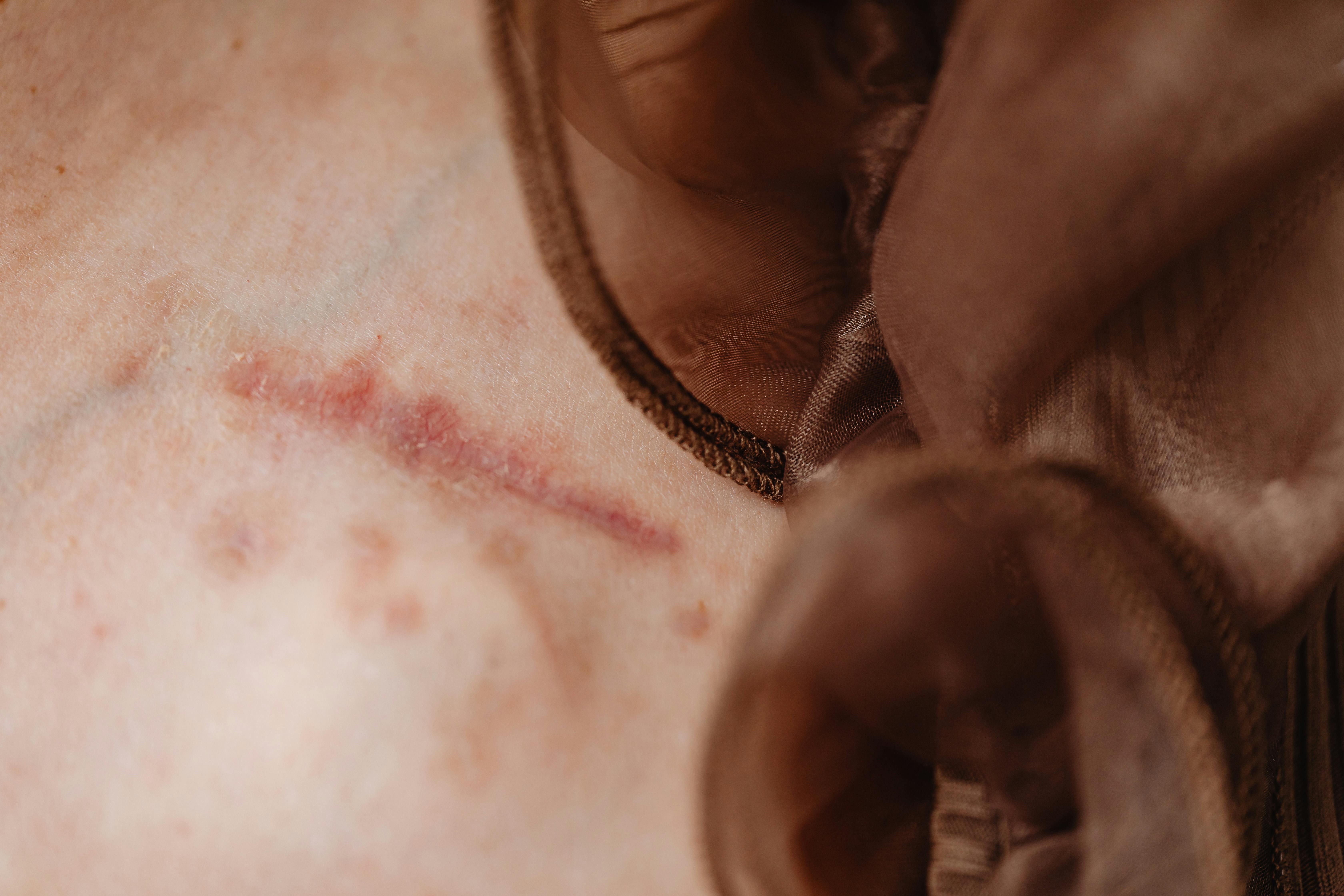
Trophic ulcers are the most common complication of chronic venous insufficiency; it remains an urgent problem in modern medicine [2, 4, 12]. In $15\%$ there are trophic changes in the skin of the lower extremities, $50\%$ of which are complicated by the formation of leg ulcers. Patients with long-term nonhealing purulent wounds often stop working early and become disabled [5, 8].
Only a complex effect allows to achieve healing of a peptic ulcer with a long-term remission of the disease. All this indicates the need to search for new and improve existing therapeutic measures, modern surgical techniques and methods of conservative therapy in this area of surgery [7, 10].
Traditional open intervention or endovascular technologies are usually considered as the method of choice for surgical treatment. According to research data, the use of radiofrequency ablation (RFA), endovenous laser coagulation (EVLC) and echosclerotherapy is preferable. foam-form, the results of which are comparable to those of traditional surgical methods and which allow outpatient treatment, with the patient's ability to work and a satisfactory cosmetic effect [11, 13].
One of the new methods of treatment of wound defects, including in patients with trophic ulcers of venous origin, is vacuum therapy. Vacuum therapy (Vacuum-assisted closure, VAC) is one of the therapies used to improve wound healing [3]. The use of vacuum therapy in the complex treatment of trophic ulcers of venous etiology improves the results of treatment of patients, which is reflected in the acceleration of the course of the wound process, the ability to perform the plastic stage at an earlier date and in the reduction of inpatient treatment [9].
However, despite the large arsenal of surgical interventions, the results of treatment of this category of patients remain insufficiently satisfactory, there are no data on effective methods of local treatment of trophic ulcers and a unified algorithm for managing patients.
In connection with the above, we undertook a study aimed at improving the treatment of patients with chronic venous insufficiency complicated by trophic ulcers.
Purpose of the study. Improving the results of treatment of chronic venous insufficiency complicated by a trophic ulcer by substantiating the complex use of EVLT with vacuum therapy.
## II. MATERIALS AND METHODS
The study is based on a comparative analysis of the results of treatment of patients with chronic venous insufficiency (CVI) of the lower extremities, who were hospitalized in the departments of vascular surgery and purulent surgery of the Tashkent Medical Academy and in the department of surgery of the private clinic AKFAMedline for the period 2016-2021 Depending on the methods of treatment of CVI of the lower extremities, the patients were divided into 2 groups: control and main. The control group consisted of 58 $(47.5\%)$ patients, among whom were patients in the C6 and C6 r stages, who were treated in the period 2016-2018. In this group, patients underwent conservative and traditional methods of treatment. In parallel, drug treatment was prescribed, consisting of phlebotonics, NSAIDs, antihistamines and antibacterial drugs.
The main group consisted of 64 (52.5%) patients with CVI of the lower extremities, which also included patients in the C6 and C6 r stages, who were diagnosed and treated in the period from 2019 to 2021. The main group of patients underwent EVLC + vacuum + autodermoplasty.
The average age of the patients was 51.20.4 years, women prevailed in the gender ratio.
To assess the severity of the disease, we used a clinical scale for assessing the severity of venous diseases.
Examination of patients included diagnostic methods generally accepted for a surgical hospital; of the special research methods, color duplex scanning of the veins of the lower extremities was used, and, according to indications, X-ray contrast phlebography and bacteriological culture from the area of the trophic ulcer were performed. Given the age groups of patients, if necessary, the list of examinations could include echocardiography, examination of narrow specialists with the appointment of appropriate treatment if comorbidities were detected.
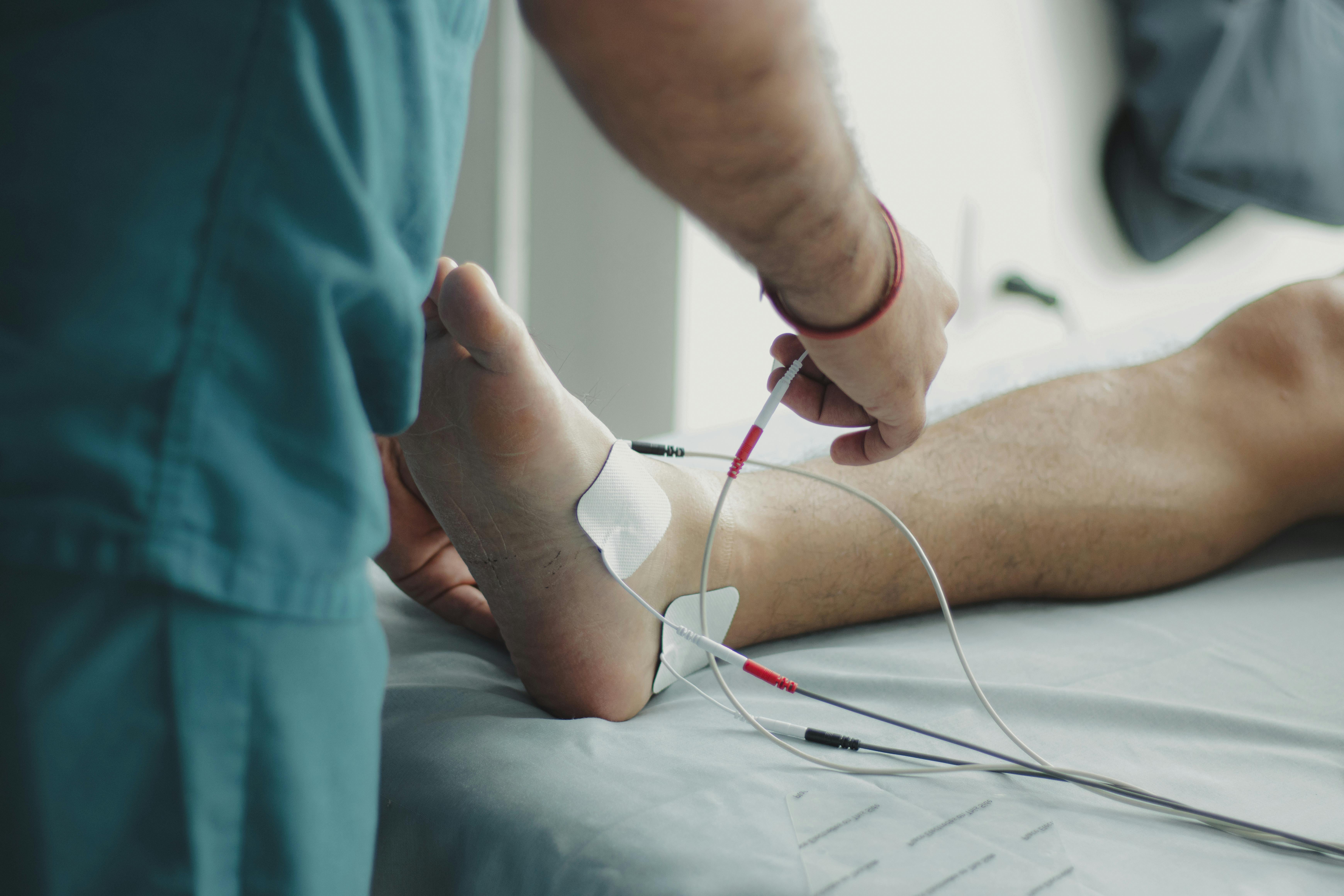
We have developed the program "Program for assessing the area of the trophic ulcer""Wound sizedetector.exe", which allows the non-contact method to determine the size of the trophic ulcer,observe their decrease in dynamics and conduct detailed monitoring with documentary evidence in the database. The interface of the program is convenient, and ease of use makes it possible to recommend it for use, starting with primary health care.
Local treatment of trophic ulcers in the main group consisted of treatment with $0.4\%$ electrolytic aqueous solution (EAS) of sodium hypochlorite in the 1st phase and $0.2\%$ EAS of sodium hypochlorite in the $2^{\text{nd}}$ phase, followed by the use of vacuum therapy in the 1st phase.
## III. RESULTS AND DISCUSSION
According to the results of the color duplex scanning, depending on the detected reflux, patients with vertical reflux prevailed, compared with a combination of vertical with horizontal and horizontal reflux, as can be seen from Table 1.
Table 1: Distribution of patients according to blood reflux on color duplex scanning of the veins of the lower extremities, n=122
<table><tr><td>Reflux</td><td>Main, n=64</td><td>Control, n=58</td><td>TOTAL</td></tr><tr><td>Vertical</td><td>44</td><td>40</td><td>84</td></tr><tr><td>Vertical+horizontal</td><td>17</td><td>16</td><td>33</td></tr><tr><td>Horizontal</td><td>3</td><td>2</td><td>5</td></tr><tr><td>TOTAL</td><td>64</td><td>58</td><td>122</td></tr></table>
For the purpose of additional research, all patients underwent bacteriological culture from a trophic ulcer, as a result of which a variety of gram-positive and gram-negative flora were sown, which gave reason to consider the trophic ulcer identical to the infected one in its microflora.
In the control group, we used local treatment (sanitation with $0.3\%$ hydrogen peroxide, proteolytic enzymes (trypsin, chymotrypsin), hydrophilic ointments (levomekol, levosin, dioxysol). Surgical treatment was carried out depending on the type of reflux: in 40 cases (68, $9\%$ ) vertical reflux, 16 cases $(27.5\%)$ vertical + horizontal reflux and 2 cases $(3.6\%)$ horizontal refluxes, performed traditional types of surgical intervention: phlebectomy according to the Babcock technique (38 cases), phlebectomy according to the Babcock technique+ Linton (3 cases), phlebectomy according to the Babcock technique + Cocket (15 cases) and Endolinton (2 cases).
The assessment of the ongoing treatment was carried out at 1, 5, 10, 15, 20, 30 after its start. The evaluation criteria were the indicators of the VCSS scale. The pain syndrome did not disappear completely in any patient.
Against the background of the ongoing treatment, the duration of the inflammation phase decreased to $5.1\%$, the proliferation phase to $6.7\%$ and the healing phase to $9.8\%$.
The entire course of treatment in the control group was 8-21 bed-days, and the epithelialization period was 7-18 days.
In the control group of patients after the treatment in 10 cases there was a relapse of the trophic ulcer, in the rest of the patients the trophic ulcer was completely cured.
The proposed method for the local treatment of trophic ulcers, treatment with $0.4\%$ EAS sodium hypochlorite in the 1st phase and $0.2\%$ EAS sodium hypochlorite in the 2nd phase, followed by the use of vacuum therapy in the 1st phase, made it possible to achieve the expected results. Assessment of the most common subjective symptom, pain, on a scale showed that from the beginning of treatment, when the initial level was 7.2, on the $15^{\text{th}}$ day of treatment it decreased to 1.1. According to bacteriological culture, the initial level was 7.6, and on the $15^{\text{th}}$ day after the application of local treatment it decreased to 1.0 (CFU/ml).
Taking into account the results of our studies, we proposed an algorithm for managing patients with trophic ulcers, depending on the phase of the process, which we used in the main group of patients.
Conservative therapy in the main group was the same as in the control group, it consisted primarily in compression, which is considered the basis for the treatment of CVI with trophic ulcers, elevated position of the limb, antibiotic therapy, the appointment of phlebotonics, improvement of microcirculatory disorders, as well as the treatment of comorbidities.
Local treatment was carried out according to the method developed by us.
Regardless of reflux, 44 (68.8%) patients with vertical reflux, 17 (26.6%) with vertical + horizontal reflux, and 3 (4.7%) with horizontal reflux underwent EVLC + vacuum therapy.
To assess the effectiveness of the treatment, we carried out a comparative assessment of the results obtained with the results of the control group according to the VCSS classification parameters, during which the following differences were identified:
1. Decrease in the area of the wound surface by $38.7\%$ versus $18.2\%$ in the control group.
2. Minor pains were noted in $73.6\%$ of patients, the remaining $26.4\%$ had no pain symptoms. While in the control group, the absence of pain symptoms was observed only in $10.7\%$ of patients, and the remaining $89.3\%$ of patients complained of moderate and mild pain.
3. In the main group, mild edema and hyperemia were observed in $69.4\%$ of patients, and in $30.6\%$ of cases, edema and hyperemia were absent, compared to the control group, in $85.3\%$ of patients, edema was moderate and insignificant, in the remaining $14.7\%$ of patients There was no edema or hyperemia.
4. Signs of granulation in the main group appeared on the $5^{\text{th}}$ day from the onset of the disease, while in the control group it was on the $10^{\text{th}}$ day.
5. Against the background of the ongoing treatment, it can be noted that the duration of the inflammation phase decreased to $3.1\%$, the proliferation phase to $3.6\%$ and the healing phase to $6.2\%$.
## IV. CONCLUSION
1. Treatment of patients with trophic ulcers against the background of chronic venous insufficiency should be complex and depend on the phase of the process: the most appropriate use of vacuum therapy in the 1st phase, after which we recommend using the method of sanitation of trophic ulcers developed by us in the $2^{\text{nd}}$ phase.
2. The android program developed by us "Wound Sizedetector.exe". Allows non-contact method to measure the area of trophic ulcers and monitor
- during treatment with documentation of the data obtained.
3. The use of our treatment tactics made it possible to improve the results of treatment of patients with trophic ulcers, which resulted in a decrease and disappearance of pain, a decrease in the area of trophic ulcers, a reduction in the duration of the inflammation phase and, accordingly, a reduction in the length of stay of patients in the hospital.
Generating HTML Viewer...
References
10 Cites in Article
F Adylkhanov,A Fursov (2017). Varicose disease of the lower extremities -analysis of the effectiveness of surgical treatment at the present stage.
A Babazhanov,K Tukhtaev Zh,A Toirov,G Akhmedov,U Khudainazarov (2017). Comparison of the effectiveness of endovenous laser coagulation and traditional combined phlebectomy.
L Bokeria,M Mikhalichenko (2014). Complications and pitfalls of endovenous laser therapy for varicose veins of lower extremities.
L Bokeria,M Mikhailichenko,V Kovalenko (2015). Optimization of surgical treatment of patients with varicose veins of the lower extremities.
O Dubrovshchik,G Marmysh,I Dovnar,M Mileshko (2016). MODERN APPROACHES TO THE TREATMENT OF GASTRODUODENAL BLEEDING REQUIRING EMERGENCY SURGERY IN A SPECIALIZED CITY CENTER.
A Kirshin,S Styazhkina,E Goryaeva,L Khannanova (2020). Combined treatment of chronic venous insufficiency in the stage of severe trophic disorders.
Zhuravyev Ou,Kurbaniyazov Zb,Sayinaev Fk (2016). CHRONIC VENOUS INSUFFICIENCY AND TROPHIC ULCERS OF THE LOWER EXTREMITIES.
Alun Davies (2019). The Seriousness of Chronic Venous Disease: A Review of Real-World Evidence.
Eric Depopas,Matthew Brown (2018). Varicose Veins and Lower Extremity Venous Insufficiency.
Caroline Novak,Namrata Khimani,Alan Kaye,R Jason Yong,Richard Urman (2019). Current Therapeutic Interventions in Lower Extremity Venous Insufficiency: a Comprehensive Review.
Explore published articles in an immersive Augmented Reality environment. Our platform converts research papers into interactive 3D books, allowing readers to view and interact with content using AR and VR compatible devices.
Your published article is automatically converted into a realistic 3D book. Flip through pages and read research papers in a more engaging and interactive format.
Our website is actively being updated, and changes may occur frequently. Please clear your browser cache if needed. For feedback or error reporting, please email [email protected]
Thank you for connecting with us. We will respond to you shortly.