Rare Case of Young Patient with Intraventricular Angiomatous Meningioma

Rare Case of Young Patient with Intraventricular Angiomatous Meningioma

Article Fingerprint

ReserarchID

58WV8

Rare Case of Young Patient with Intraventricular Angiomatous Meningioma Banner

Key Research Insights

Synthesized scholarly intelligence & interactive research assistant
  • English
  • Afrikaans
  • Albanian
  • Amharic
  • Arabic
  • Armenian
  • Azerbaijani
  • Basque
  • Belarusian
  • Bengali
  • Bosnian
  • Bulgarian
  • Catalan
  • Cebuano
  • Chichewa
  • Chinese (Simplified)
  • Chinese (Traditional)
  • Corsican
  • Croatian
  • Czech
  • Danish
  • Dutch
  • Esperanto
  • Estonian
  • Filipino
  • Finnish
  • French
  • Frisian
  • Galician
  • Georgian
  • German
  • Greek
  • Gujarati
  • Haitian Creole
  • Hausa
  • Hawaiian
  • Hebrew
  • Hindi
  • Hmong
  • Hungarian
  • Icelandic
  • Igbo
  • Indonesian
  • Irish
  • Italian
  • Japanese
  • Javanese
  • Kannada
  • Kazakh
  • Khmer
  • Korean
  • Kurdish (Kurmanji)
  • Kyrgyz
  • Lao
  • Latin
  • Latvian
  • Lithuanian
  • Luxembourgish
  • Macedonian
  • Malagasy
  • Malay
  • Malayalam
  • Maltese
  • Maori
  • Marathi
  • Mongolian
  • Myanmar (Burmese)
  • Nepali
  • Norwegian
  • Pashto
  • Persian
  • Polish
  • Portuguese
  • Punjabi
  • Romanian
  • Russian
  • Samoan
  • Scots Gaelic
  • Serbian
  • Sesotho
  • Shona
  • Sindhi
  • Sinhala
  • Slovak
  • Slovenian
  • Somali
  • Spanish
  • Sundanese
  • Swahili
  • Swedish
  • Tajik
  • Tamil
  • Telugu
  • Thai
  • Turkish
  • Ukrainian
  • Urdu
  • Uzbek
  • Vietnamese
  • Welsh
  • Xhosa
  • Yiddish
  • Yoruba
  • Zulu
Reading Preferences
Font Size
Line Spacing
Background
This converted HTML version may contain rendering inconsistencies. Please refer to the PDF for the authoritative version, or click here to provide feedback.

Abstract

Pediatric meningiomas are rare and account for only 2.2% of CNS tumors. In this age group, they are more frequently located in atypical sites, such as, mainly, the ventricular system, with a frequency of 8.8 to 13.6%. Adding this to the fact that the angiomatous subtype constitutes only 2.1% of all meningiomas, the rarity of the case reported here i s corroborated. We report a 17-year-old female patient diagnosed with intraventricular angiomatous meningioma; she underwent surgical resection of the tumor in the body and frontal horn of the right lateral ventricle, and there was no neurological sequela. With a follow-up of 4 years, there was no recurrence and the patient had clinical stability. Intraventricular tumors usually have slow growth and reach considerable size until they cause symptoms and then are diagnosed. In addition, the tumor’s deep location and proximity to eloquent areas make such tumors an neurosurgical challenge. The angiomatous subtype, due to the presence of hyper vascularization (consisting of more than 50% of vascular components), may, in some cases, hinder surgical resection as well as be erroneously diagnosed. However, surgical treatment aimed at total resection of the lesion remains the conduct of choice in the case reported here, especially in patients in the first two decades of life, in which the use of radiation is avoided. Specifically when it comes to the surgery, we chose a transcallosal approach that allows a good transoperative visualization of the lesion when located in the body and frontal horn of the lateral ventricle.

I. INTRODUCTION

Meningiomas have a progressively higher incidence with increasing age, with a mean age of presentation of 65 years. They are, therefore, the most frequently reported tumors of the central nervous system (CNS) in adulthood. 1 However, cases in children and adolescents are rare, representing 2.2 to 2.6% of CNS tumors. 1 , 2 In such age group, are more often located in unusual sites, such as in the ventricular system. 3

Such intraventricular meningiomas have the particularity of being slow growing and reaching considerable size until they become symptomatic. 4 In addition, the deep location and relationship with underlying eloquent areas make tumor resection a neurosurgical challenge. 5 , 6 In view of this, and that the angiomatous subtype– defined as presenting more than 50% of vascular components on microscopic analysis analysis – constitutes only 2.1% of all meningiomas, rarity is credited to the case reported here. 7 We emphasize that this is possibly the first report of a patient in the first two decades of life with angiomatous meningioma in an intraventricular site.

In this report, we aim to expose our neurosurgical experience in a case with rare variants and perform a literature review on the main aspects that we deem necessary to support our approach.

II. CASE REPORT

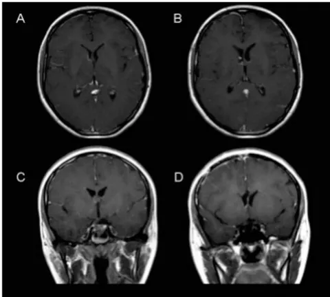
A 17-year-old female patient, previously healthy, presented retro-orbital headache for 3 months, followed by blurred vision and double vision. Physical examination revealed just a convergent strabismus due to paresis of the right lateral rectus muscle. The cranial magnetic resonance (MRI) showed an expansive lesion in the frontal horn of the right ventricle, just in front of the foramen of Monro, with dimensions of 2.0 x 1.3 x 1.8 cm (AP x L x h) in the larger diameter cuts.

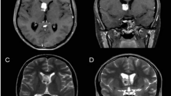
The lesion presented moderate hyperintensity with small hypointense foci on T2-weighted images. On T1, it was isointense, and after contrast, it showed intense and homogeneous uptake, except in the same hypointense foci on T2. There were no signs of dilation of the supratentorial ventricular system (Fig. 1 A - D). Such radiological characteristics suggested the diagnosis of intraventricular meningioma, with areas of calcification. It was considered an occasional finding, since the topography was not compatible with the presenting symptoms.

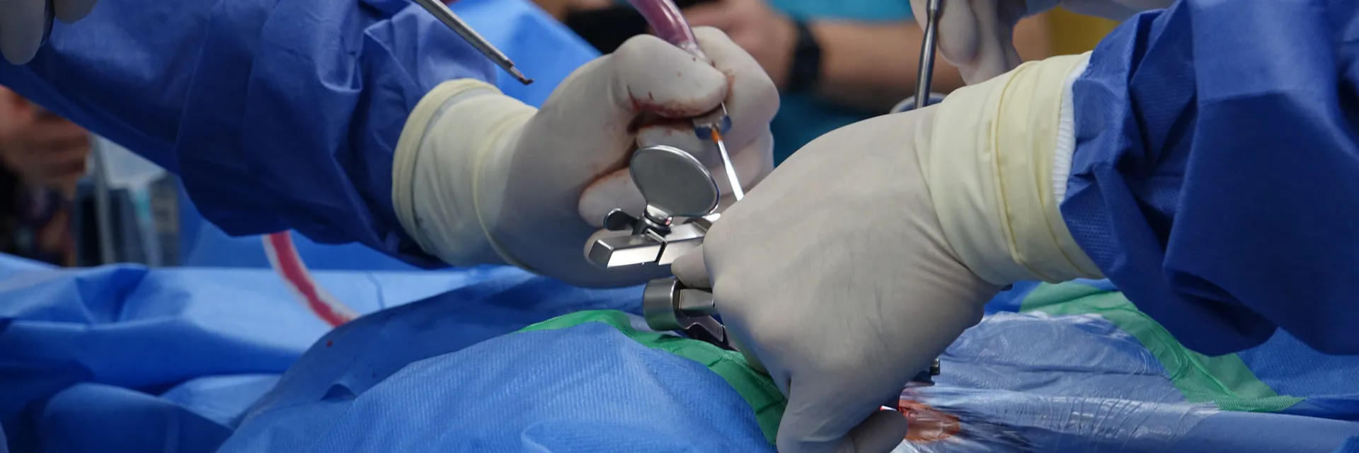
The surgical treatment was performed in the same hospitalization, due to the risk of acute hydrocephalus. The approach was made via the transcallosal route, following the steps: dorsal decubitus and head in a neutral position; bicoronal incision; right frontal paramedian craniotomy, with lateral extension of 5.5 cm from the midline and 5.5 cm from the coronal suture forward; opening the dura mater in a "C" shape, with the base facing the midline; under microscopy, dissection of the inter-hemispheric fissure and retraction of the frontal lobe with placement of the fixed spatula (on a Leyla support) on the medial surface; identification of the cingulate gyrus and pericalosal arteries; callosotomy with 1,2 cm starting from the transition between the knee and the corpus callosum; identified a grayish vegetating lesion inside the right frontal horn, which had a soft consistency and were vascularized; resection of the lesion through coagulation and aspiration and by fragments, leaving a small residue adhered to the ependyma of the caudothalamic groove. Postoperatively uneventful and absence of new deficits, with diplopia and strabismus remaining.

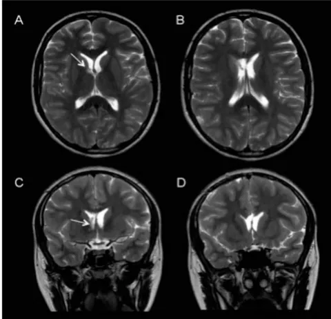
Histopathological analysis and immunohistochemical painel showed extensive vascularization and low mitotic index (Ki67 less than 2%). In addition, that results were visualized: epithelial membrane antigen (EMA), positive; cytokeratin (CK), negative; progesterone receptor, negative; and glial fibrillary acidic protein (GFAP), positive (Fig.2 A-B). Thus, the diagnosis of angiomatous meningioma (WHO grade I) was confirmed. Postoperative MRI showed a small residual focus near the thalamostriate groove (Fig. 3 A-D and Fig. 4 A-D). After 6 months, the patient underwent strabismus correction with an ophthalmologist at another institution. Currently, with approximately 4 years of follow-up, she is asymptomatic and without evidence of recurrence of the residual lesion.

III. Discussion

Meningiomas are tumors that predominate in the fifth and sixth decades of life, with a mean age of presentation of 65 years. 1 In general, they represent 36.4% of primary CNS tumors and approximately 24-30% in adults. 1 , 8 Nevertheless, in the pediatric population, the prevalence among CNS tumors varies between 0.4 and 4.6%.2, 3 , 9 βˆ’ 13 The equivalence between genders also contrasts with what occurs in the adult population, which has a female to male ratio of 2:1. 1 , 2 , 14 This difference is believed to be due, mainly in the prepubertal period, to the absence of the effect of sex hormones on corticosteroid receptors in meningioma cells. 10 , 15 βˆ’ 17

In the first two decades of life, there is a higher incidence of grade II (atypical) and grade III (anaplastic) meningiomas, according to the WHO: 9.9 and 8.9%, respectively. 2 , 18 They are genetically and phenotypically more aggressive with a high frequency of brain invasion. 9 , 14 Among the most frequent grade I meningiomas, the angiomatous subtype occurs in 2.8% of cases, 2 and in 2.1% of all meningiomas at any age. 19 Such subtype is defined when the vascular component exceeds 50% of the total area of the tumor. 7 , 19 However, the differential diagnosis with hemangioblastoma and hemangiopericytoma is necessary, with immunohistochemistry and morphology having essential roles in the diagnostic confirmation: low MIB-1/Ki67 index and positivity for progesterone receptor, EMA, vimetin, cytokeratin, and desmoplaquin. 7 , 19 βˆ’ 23

The characteristics of angiomatous meningiomas, due to their rarity, are covered in few studies. 7 , 19 , 24 , 25 They may present moderate to severe cerebral edema with a frequency of 74 to 88.9%, 7 , 19 due to hypervascularization, increased blood pressure. capillary permeability and secretion of VEGF (vascular endothelial growth factor). 19 On magnetic resonance imaging, they may show more signs of flow voids, rarely present necrosis and tend to have homogeneous enhancement to paramagnetic contrast. 19 , 23

Meningiomas in pediatric patients present in atypical sites more frequently than in adults: lateral ventricles, skull base, posterior fossa. 2 , 3 , 10 , 15 , 26 Intraventricular location occurs in 11%, compared to 0.3-3% in all ages and 0.5-4.5% in adults. 2 , 10 , 27

Intraventricular meningiomas (IVM) are located in 76% of cases in the lateral ventricles (most common on the left side); 16% in the third ventricle; and 7% in the fourth ventricle. 5 , 27 There are studies suggesting that the lateral ventricles are the preferred site for pediatric IVMs. 26 , 28 These originate from the choroid plexus, growing in the t e l a choroidea. 4 The vascularization of the tumor depends on its location in the ventricle: the nutrient vessels are of small caliber and usually originate from the choroidal arteries. 27

Clinically, pediatric IVMs are usually asymptomatic, until they reach large dimensions in the lateral ventricles, where the risk of hydrocephalus is lower. However, when located in the third or fourth ventricle, obstruction to the flow of cerebrospinal fluid can result in manifestations at early stages of tumor growth. 5 , 27 , 29 , 30 Therefore, symptomsβ€”headache, nausea, vomiting and visual disturbances – are more frequently related to tumor compression and an insidious increase in intracranial pressure. 5 , 27 , 31 Indolent cognitive deficits compromising memory and attention may also occur. 32 , 33 Typical symptoms of an acute increase in intracranial pressure are uncommon. 29 The clinic, therefore, correlates with the location of the tumor within the ventricle, tumor size and the direction of its growth. 27 Finally, we emphasize that the clinical presentation of the reported patient – convergent strabismus due to paresis of the right lateral rectus muscle – was not correlated with the tumor. This was still relatively small in size and its location did not justify the signs and symptoms.

IVMs usually have the classic radiological appearance of other meningiomas, well-defined globular shape, however no dural tail. They are usually iso to hypotensive on T1-weighted images, hyperintense on T2-weighted images and suffer strong contrast enhancement. 16 , 27 Especially in the pediatric population, other more frequent intraventricular tumors can make the differential diagnosis difficult: chorioid plexus tumors, ependymoma, primitive neuroectodermal tumor, teratoma, and astrocytoma. 30 , 34 Chorioid plexus tumors usually affect younger than 10 years, and on MRI present a multilobulated mass with intense contrast enhancement and frond-like appearance. Ependymomas represent approximately one third of

CNS tumors in children under 3 years of age and are characterized by necrosis, hemorrhage, cyst formation, and on MRI, they are hypointense on T1 and hyperintense and heterogeneous on T2. 35

The surgical approach of a benign IVM is a neurosurgical challenge, given its deep location and its proximity to eloquent areas and vessels in the ventricular walls. 5 , 34 The extent of the initial resection is an independent prognostic factor, presenting a significant association with recurrence and malignancy. 2 The patient reported here did not present a recurrence in the 4 years of follow-up, which is a result consistent with the literature. In a 2012 review, with 201 cases from different series, there were only eight recurrences; 27 however, in a meta-analysis with 677 cases of meningiomas in the first two decades, the numbers are more significant: there were 141 recurrences with an average presentation of 3.6 years and with mortality from this event in 46 cases. 2 The recurrence in this age group occurs basically in cases of atypical meningiomas and anaplastic or after partial resection. 36 Post-surgical mortality and morbidity in patients in the post-pubertal phase, as is the case of the patient reported here, are similarity to those observed in cases of meningioma in adults. 2 The use of adjuvant radiotherapy should be avoided in young patients, opting for serial evaluation and reoperation in case of recurrence. 2 , 3 , 8 βˆ’ 11 , 14 , 18 , 37

In the literature, there are several surgical approaches for IVM resection: temporoparietal, transfrontal, middle posterior temporal gyrus, posterior inferior temporal gyrus, parieto-occipital and transcallosal. 5 , 34 The choice is individualized and based on the location of the tumor within the ventricle, in tumor size and its vascular network, always with a objective of preserving the adjacent brain tissue, performing small corticectomy and retracting little as possible. 5 , 6 , 27 , 30 , 33 , 34 The justification for the choice the approach is determined by the option that allows better access to the longest axis of the lesion, to minimize transcortical transgression, by the spectrum of preoperative neurological deficits, proximity to the eloquent structures, in addition to anatomical knowledge of the cortical and white matter. 29 We chose the transcallosal approach because it allows better access to the frontal horn and lateral ventricle body. This approach prevents cortical damage; however, some care is needed with the possible presence of tributary cortical veins tributary of the superior sagittal sinus, which can be anticipated in preoperative examinations, and with the corpus callosum, which must be distinguished from the cingulate gyrus by the change in color. 5 Despite the degree of difficulty, IVM surgery has shown low morbidity and mortality rates in recent decades, and most postoperative complications–visual deficits and praxis–are temporary. 27 , 29 , 34 These low rates are consistent with the case reported here, which postoperative complications or sequelae did not occur.

IV. CONCLUSION

Intraventricular angiomatous meningioma is a rare entity, even more in patients in the first two decades of life. The clinic is nonspecific in most cases, making MRI evaluation necessary for the diagnosis and definition of the surgical approach, and the histopathological analysis is what defines the diagnosis of the angiomatous subgroup. Surgical resection is the treatment of choice. However, the goal of total resection should not be above the goal of preserving the patient's functions and quality of life.
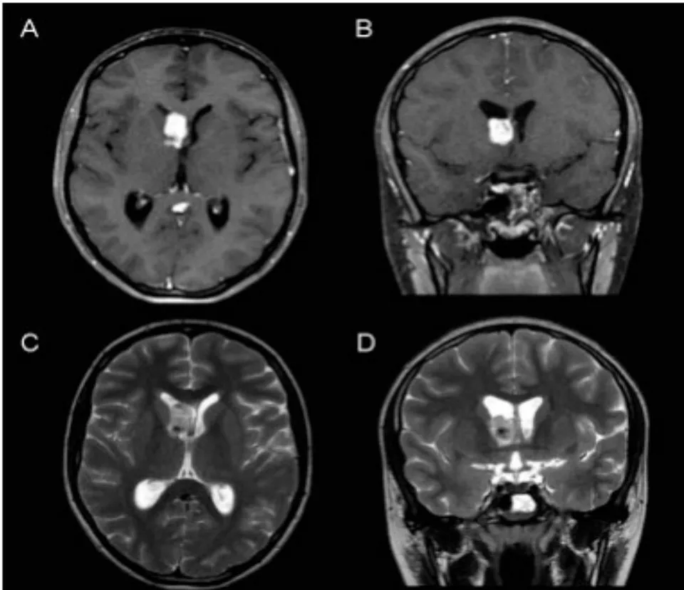
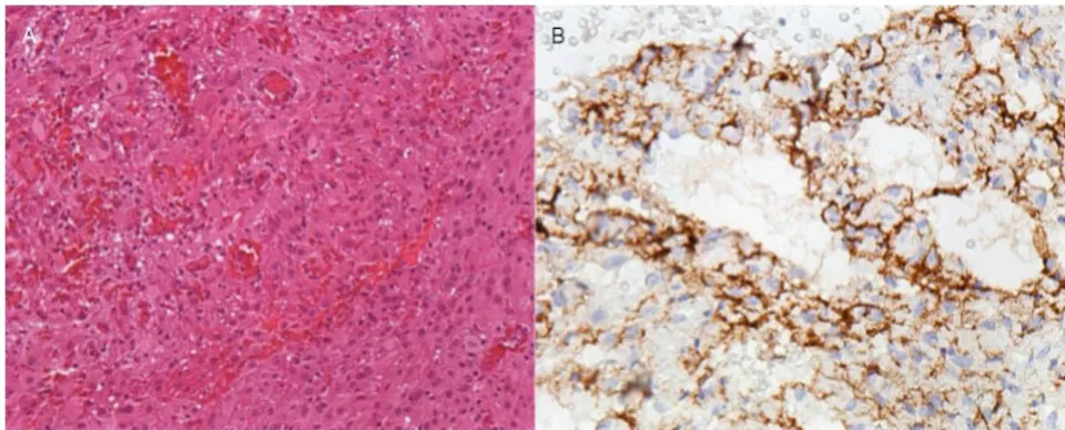
Figure 1: Preoperative MRI: A and B – T1 with gadolinium, axial (A) and coronal (B). Lesion with intense uptake at the level of the right frontal horn, close to the foramen of Monro. C and D – T2 axial(C) e coronal(D). Hyperintense lesion, with a focus of hypointense calcification (also seen in A). Figure 2: A – Hematoxylin and eosin staining, 200 times magnification: image showing histopathological features of meningioma and significant vascular component. B – Immunohistochemistry, 400-fold magnification: tumor cells showing positivity for EMA.
Figure 3: Postoperative MRI: T1 with gadolinium, axial (A and B) and coronal (C and D) planes. Small residual focus near the foramen of Monro, attached to the thalamostriate vein (identified during the transoperative period).
Figure 4: Postoperative MRI: T2 axial (A and B) and coronal (C and D). Small residual focus at the level of the foramen of Monro (arrows).

References

37 Cites in Article
  1. Quinn Ostrom,Haley Gittleman,Jordonna Fulop,Max Liu,Rachel Blanda,Courtney Kromer,Yingli Wolinsky,Carol Kruchko,Jill Barnholtz-Sloan (2015). CBTRUS Statistical Report: Primary Brain and Central Nervous System Tumors Diagnosed in the United States in 2008-2012.
  2. R Kotecha,E Pascoe,E Rushing (2011). Meningiomas in children and adolescents: a metaanalysis of individual patient data.
  3. G Menon,S Nair,J Sudhir,B Rao,A Mathew,B Bahuleyan (2009). Childhood and adolescent meningiomas: a report of 38 cases and review of literature.
  4. Harjinder Bhatoe,Prakash Singh,Vibha Dutta (2006). Intraventricular meningiomas: a clinicopathological study and review of the literature.
  5. Anil Nanda,Shyamal Bir,Tanmoy Maiti,Subhas Konar (2016). Intraventricular Meningioma: Technical Nuances in Surgical Management.
  6. B Lyngdoh,P Giri,S Behari,D Banerji,D Chhabra,V Jain (2007). Intraventricular meningiomas: a surgical challenge.
  7. Martin Hasselblatt,Kay Nolte,Werner Paulus (2004). Angiomatous Meningioma.
  8. L Kwee,M Van Veelen-Vincent,E Michiels,J Kros,R Dammers (2015). The importance of microsurgery in childhood meningioma: a case report.
  9. Yuguang Liu,Feng Li,Shugan Zhu,Meng Liu,Chengyuan Wu (2008). Clinical Features and Treatment of Meningiomas in Children: Report of 12 Cases and Literature Review.
  10. Kapu Ravindranath,M Vasudevan,Anil Pande,Nigel Symss (2013). Management of pediatric intracranial meningiomas: an analysis of 31 cases and review of literature.
  11. M Santos,L Furlanetti,E Valera,M Brassesco,L Tone,R De Oliveira (2012). Pediatric meningiomas: a single-center experience with 15 consecutive cases and review of the literature.
  12. H Cushing,L Eisenhardt (1938). Book Review Meningiomas: Their classification, regional behaviour, life history and surgical end results . Harvey Cushing. 785 pp. Springfield, Illinois: Charles C Thomas, 1938. $15.00..
  13. N Mehta,S Bhagwati,G Parulekar (2009). Meningiomas in children: A study of 18 cases.
  14. A Arivazhagan,B Devi,Sastry Kolluri,R Abraham,S Sampath,B Chandramouli (2008). Pediatric Intracranial Meningiomas – Do They Differ from Their Counterparts in Adults?.
  15. Sushila Jaiswal,Mukul Vij,Anant Mehrotra,Awadhesh Jaiswal,Arun Srivastava,Sanjay Behari (2011). A clinicopathological and neuroradiological study of paediatric meningioma from a single centre.
  16. Ian Whittle,Colin Smith,Parthiban Navoo,Donald Collie (2004). Meningiomas.
  17. Mark Donnell,Glenn Meyer,William Donegan (1979). Estrogen-receptor protein in intracranial meningiomas.
  18. Rishi Kotecha,Reimar Junckerstorff,Sharon Lee,Catherine Cole,Nicholas Gottardo (2011). Pediatric meningioma: current approaches and future direction.
  19. Z Liu,C Wang,Wangh,Y Wang,J Li,Liuy (2013). Clinical characteristics and treatment of angiomatous meningiomas: a report of 27 cases.
  20. S Rao,A Rajkumar,S Kuruvilla (2008). Angiomatous meningioma: a diagnostic dilemma.
  21. Divya Bansal,P Diwaker,P Gogoi,W Nazir,A Tandon (2015). Intraparenchymal Angiomatous Meningioma: A Diagnostic Dilemma.
  22. G Rathod,K Vyas,P Shinde,S Goswami,R Tandan (2014). Angioma-tous meningioma in 49 years old male -A rare case report.
  23. Yu Meng,Wang Chaohu,Liu Yi,Pan Jun,Qi Songtao (2015). Preoperative radiologic characters to predict hemangiopericytoma from angiomatous meningioma.
  24. Juyoung Hwang,Doo-Sik Kong,Ho Seol,Do-Hyun Nam,Jung-Il Lee,Jung Choi (2016). Clinical and Radiological Characteristics of Angiomatous Meningiomas.
  25. Ben Nsir,A Chabaane,M Krifa,H Jeme,H Hattab,N (2016). Intracranial angiomatous meningiomas: A 15year, multicenter study.
  26. Elisabeth Rushing,Cara Olsen,Hernando Mena,Maria-Eugenia Rueda,Youn-Soo Lee,Robert Keating,Roger Packer,Mariarita Santi (2005). Central nervous system meningiomas in the first two decades of life: a clinicopathological analysis of 87 patients.
  27. Kristina Ødegaard,Eirik Helseth,Torstein Meling (2013). Intraventricular meningiomas: a consecutive series of 22 patients and literature review.
  28. H Deen,Bernd Scheithauer,Michael Ebersold (1982). Clinical and pathological study of meningiomas of the first two decades of life.
  29. David Fusco,Robert Spetzler (2015). Surgical Considerations for Intraventricular Meningiomas.
  30. M Liu,Y Wei,Y Liu,S Zhu,X Li (2006). Intraventricular meninigiomas: a report of 25 cases.
  31. Per Rochat,Helle Johannesen,Flemming Gjerris (2004). Long-term follow up of children with meningiomas in Denmark: 1935 to 1984.
  32. Daina Kashiwazaki,Akiko Takaiwa,Shoichi Nagai,Naoki Akioka,Kunikazu Kurosaki,Kyo Noguchi,Naoya Kuwayama,Satoshi Kuroda (2014). Reversal of Cognitive Dysfunction by Total Removal of a Large Lateral Ventricle Meningioma: A Case Report with Neuropsychological Assessments.
  33. Makoto Nakamura,Florian Roser,Otto Bundschuh,Peter Vorkapic,Madjid Samii (2003). Intraventricular meningiomas: a review of 16 cases with reference to the literature.
  34. Danica Grujicic,Luigi Cavallo,Teresa Somma,Rosanda Illic,Mihailo Milicevic,Savo Raicevic,Milica Gazibara,Alessandro Villa,Dragan Savic,Domenico Solari,Paolo Cappabianca (2017). Intraventricular Meningiomas: A Series of 42 Patients at a Single Institution and Literature Review.
  35. Amit Agarwal,Sangam Kanekar (2016). Intraventricular Tumors.
  36. Emanuela Caroli,Michelangelo Russillo,Luigi Ferrante (2006). Intracranial Meningiomas in Children: Report of 27 New Cases and Critical Analysis of 440 Cases Reported in the Literature.
  37. Humphrey Okechi,A Albright (2012). Intraventricular Meningioma: Case Report and Literature Review.

Funding

No external funding was declared for this work.

Conflict of Interest

The authors declare no conflict of interest.

Ethical Approval

No ethics committee approval was required for this article type.

Data Availability

Not applicable for this article.

How to Cite This Article

Gabriel Carvalho Heemann, Vinicius Rosa de Castro, Normando Guedes Pereira Neto, Camila Bocchi, Otavio Garcia Martins, Rafael Silva Paglioli, Ricardo Chmelnistky Wainberg. 2021. "Rare Case of Young Patient with Intraventricular Angiomatous Meningioma". Global Journal of Medical Research - A: Neurology & Nervous System GJMR-A Volume 21 (GJMR Volume 21 Issue A3).

Download Citation

Alt: Young patient with brain tumor diagnosed with intraventricular angiomatosis meningioma.
Journal Specifications

Crossref Journal DOI 10.17406/gjmra

Print ISSN 0975-5888

e-ISSN 2249-4618

Keywords
Classification
GJMR-A Classification NLMC Code: WL 140
Version of record

v1.2

Issue date
July 31, 2021

Language
English
Order Article Reprint
Experiance in AR

Explore published articles in an immersive Augmented Reality environment. Our platform converts research papers into interactive 3D books, allowing readers to view and interact with content using AR and VR compatible devices.

Read in 3D

Your published article is automatically converted into a realistic 3D book. Flip through pages and read research papers in a more engaging and interactive format.

Article Matrices
Total Views: 467
Total Downloads: 41
All Trends

Request Access

Please fill out the form below to request access to this research paper. Your request will be reviewed by the editorial or author team.
X

This is the heading

Lorem ipsum dolor sit amet, consectetur adipiscing elit. Ut elit tellus, luctus nec ullamcorper mattis, pulvinar dapibus leo.

High-quality academic research articles on global topics and journals.

Rare Case of Young Patient with Intraventricular Angiomatous Meningioma

Gabriel Heemann
Gabriel Heemann
Vinicius Castro
Vinicius Castro
Normando Neto
Normando Neto
Camila Bocchi
Camila Bocchi
Otavio Martins
Otavio Martins
Rafael Paglioli
Rafael Paglioli
Ricardo Wainberg
Ricardo Wainberg