

I. INTRODUCTION
Currently population explosion is one of the biggest problems facing by world. It's inevitable consequences are employment, education, housing, health care, economy and environment. At the present growth rates, the population of economically developed countries would double in 120 years. Overpopulation led to serious social and environmental problems such as poverty, overcrowded slums, crime, pollution of air & water and depletion of the protective ozone layer (Vogelsong, 2005) and all around human development especially in developing countries like India (Akbarsha et al, 2001). India is also only the second country to achieve a population of 1.32 billion. Our future well-being depends on increased access to family planning and reproductive health services in developing countries and decreased consumption by people in wealthy countries (Speidel, 2000).
India is first among the countries which adopted an official family planning programmed, as early as 1950. However, fifty years later this has not prevented the population touching the one billion mark (Qian et al, 1995). It is obvious that despite good intentions and concerted efforts we have failed in controlling our population. Since the major responsibilities of pregnancy, birth, and child rearing fell on women, they found methods for controlling fertility and aborting unwanted children, and they have passed down this knowledge as an oral tradition that survives worldwide. It is obvious now that there cannot be an ideal contraceptive (Moudgal and Rao, 1984; Joshi et al, 1977) suitable for everybody.
About 90% of the world's contraceptive users are women. This gender-based usage has occurred due to the emphasis of family planning programs and contraception research (Hazarika and Sarma, 2007). The only male-specific contraceptive methods currently available are withdrawal, condoms, and vasectomy. As concerns regarding side effects and convenience of these existing methods prevent (Beckman et al, 1996; Moore, et al, 1996) their universal acceptance. There is an urgent need for development of male contraceptives drug to prevent unintended pregnancy, of which 80–90 million occur annually (Amory, 2016). The development of additional male methods of fertility control can provide tremendous social and public health benefits. Because methods that require infrequent administration have the lowest typical (user) failure rates (Potter, 1996), considerable research has focused on development of a herbal male contraceptive.
Fertility regulation comprising contraception and management of infertility forms an important (Pankajakshy and Madambath, 2009) component of reproductive health. Though considerable progress has been made in the development of highly effective, acceptable and reversible methods of contraception (Njaret al, 1995) among females. Progress and possibilities on male are still slow and limited, with recent progress towards a better understanding of male reproductive physiology; there is a need to develop new contraceptive modalities for male. Several potential approaches for induction of infertility have been investigated over a long period including hormonal, chemical, and immunological, approaches. However, no suitable method has emerged that is effective and free from side effects (Montanari et al, 1998).
Clearly, there is a need for development of reversible contraceptive from natural resources. The chemical compounds affecting testicular function include steroidal and non-steroidal but application of these compounds has serious reversible antifertility effect (Kulshreshtha and Mathur, 1990; Jensen, 2002). The present study was undertaken therefore, to determine and examined whether plant might have any effect on male reproductive organs, spermatogenesis, and serum hormone levels in rats. Although contraceptives containing estrogen and progesterone are effective and popular, the risks associated to the drugs have triggered the need to develop newer molecules from medicinal plants (Swerdlloff et al, 1998). Hence, there is a need for searching suitable product from indigenous medicinal plants that could be effectively used in the place of pills. More over the phytochemical even today are important resources for medicine (Sundaram and Kumar, 2000).
Plant drugs have been used since time immemorial for their effects upon sex hormones particularly for suppressing fertility, regularizing menstrual cycle, relieving dysmenorrhea, treating enlarged prostate, menopausal symptoms, breast pain and during and after childhood (Williamson et al, 1996). Specific biological effects under the division of fertility regulating category are non-specific contraceptive or antifertility effects, abortifacient, uterine stimulant and uterine relaxants, labour induction and labour inhibition oxytocic and anti-oxytocic, oestrogenic and anti-oestrogenic, progesterogenic and anti-progesterogenic, ovulatory and anti-ovulatory, androgenic and anti-androgenic, spermicidal and anti-spermatogenic effects (Soejarto, et al, 1978). The flavonoids, phytosterol, and terpenoid present in the alcoholic extracts of Piper betle may be responsible for significant antifertility activity (62.2%) (Shah andJhade, 2018).
Antifertility agents may therefore exert their effort at this level either by disrupting (Bullock et al, 1995, WHO, 1990) hormonal function of the hypothalamus or the pituitary, or by interrupting the neural pathway to the hypothalamus that control the liberation of gonadotropins releasing hormones.
The investigation of plant constituents with antifertility properties represents a potential alternative approach to birth control from the existing available methods. If an estrogen from a local source could be shown to be active in humans, it would be of great value as a fertility-regulating agent (United Nations, 1998). The development of new fertility regulating drug from medicinal plants is an attractive proposition, because from times immemorial humans have relied on plants and their products as sources of drugs and therapeutic agents, although in recent times, synthetic drugs are used extensively(Tuxhorn, 2002) in modern medicine. However many modern medicines are developed through the clues obtained from phytochemical.
In view of the importance of plants in the traditional Indian system as a positive health promoter, it was decided to carry out work on the chemical profiling of W. somnifera on the basis of Withaferin-A (Roja et al, 2006, Ganzera et al, 2003, Sengupta Pet al, 2018).
Nonstandardized herbal preparation have not found acceptance in the global market; therefore, there is a need to be chemically standardized on the basis of isolated constituents, preferably bioactive ones. Presently, formulations standardized on the basis of a maximum possible number of biomarkers are accepted readily in the global market. Thus, as part of a long-term evaluation of potential antifertility plant, we have conducted these studies on the effects of Withania somnifera extract and their alkaloids (Withaferin-A) on the fertility of rats. The present investigation elucidated the association of biological activities with specific secondary metabolites known as Withaferin-A present in the Withania somnifera. The aim of present study was to evaluate safety and reversible contraceptive efficacy of alkaloids from this plant to search for an inexpensive, orally effective and reversible male contraceptive. The present study will help in the development of reversible male oral contraceptive from natural resources and to determine whether plant drugs (phytochemical and phytosomes) might have any effect on male reproductive organs, spermatogenesis and serum hormone levels in rats. The availability and use of acceptable male contraceptive methods could reduce the burden traditionally placed almost exclusively on the female partner.
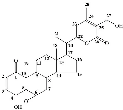
Withaferine A [4β, 27-dihydroxy-1-oxo-5β, 6β, epoxy witha-2, 24-dienolide] (M.P: 245 to 252°C)
II. MATERIAL AND METHODS
a) Collection of Plant Materials and Preparations of ethanol extract
The plant Withania somnifera Dunal (Family: Solanaceae), also known as Ashwagandha, Indian ginseng, winter cherry has been used in Ayurveda, since ancient times to increase longevity and vitality (Mishra et al, 2000). All the parts of the plant Withania somnifera have shown remarkable of pharmacological activities are menstrual troubles, dropsy, rheumatism, sexual and general weakness, asthma and bronchitis, diabetes and inflammation (Al-Hindawi et al, 1992; Andallu and Radhika, 2000; Sree et al, 2008) treatment. The active pharmacological components of Withania somnifera are steroidal lactones of the withanolides and the principal Withanolides in Indian Withania somnifera are Withaferin -A and Withanolide -D (Gupta et al, 1996).
b) Isolation and identification of Withaferin-A
The air-dried root powdered of Withania somnifera (2.4 kg) was extracted with ethanol: water (1:1, 4 L) for 8×3 hours, at 32 °C. The ethanol was evaporated under the vacuum. The eight times extract were combined and finally concentrated to 1/8 of the original volume (590 gm.) under reduced pressure at 50 ± 5°C. The concentrated extract was stirred with chloroform to remove chlorophyll content. The chloroform extract was subjected to four times column chromatography on silica gel (1.2 mt., 250 mesh, 600×4g, 0.580×4L), eluted with a stepwise gradient of increasing order (5 to 10% each step and 2 L solvent eluted in each fraction) ethyl acetate (Khajuria et al, 2004; Baraiya et al, 2005; Mishra et al, 2008) concentration in pet ether was eluted. White crystals (5.4 mg) in ethyl acetate were obtained from ethyl acetate: Petroleum ether (1:3) fraction. The repeated crystallization in acetone from fractions was observed for Thin Layered Chromatography (Merck's silica gel 60F precoated glass plate) in system chloroform: ethyl acetate (1:1).
The comparisons of IR (3445; O-H stretching, 2930, 2880, 1719; -unsaturated- -lactone, 1665; - unsaturated six-membered ketone, 1447, 1370, 1295, 1130, 1025; epoxy), H NMR spectral data (300 MHz, CDCl ) δH: 5.85 (1H, dd, J = 9.8 Hz, H-2), 6.53 (1H, dd, J = 10.2 Hz, H-3), 2.48 (1H, m, H-4), 3.19 (1H, d, J = 3.5 Hz, H-6), 2.25 (2H, dd, J = 3.1 Hz, H-7), 1.90 (1H, m, H-8), 4.47 (1H, dd, J = 12.7, 3.24 Hz, H-22), 2.25 (1H, m, H-23), 2.68 (1H, m, H-23'), 4.38 (3H, s, H-27), 1.98 (3H, s, H-28), 1.25 (3H, s, H-21), 0.97 (3H, s, H-18), 1.22 (3H, s, H-19), C NMR (75 MHz, CDCl ) δC : 200.5 (C-1), 140.1 (C-2), 135.1 (C-3), 64.8 (C-5), 63.2 (C-6), 52.8 (C-7), 48.5(C-10), 25.1 (C-12), 44.0 (C-14), 10.1(C-19), 31.3 (C-20), 18.3(C-21), 78.2 (C-22), 149.3 (C-24), 168.3 (C-26), 58.7 (C-27) data. Melting point (247.50°C) of single spotted compound with previously reported showed the presence of Withaferin-A in availability of other compounds (Withaferin A, Withanone, Withanolide D).
The comparisons of IR (3445; O-H stretching, 2930, 2880, 1719; , -unsaturated- -lactone, 1665; , -unsaturated six-membered ketone, 1447, 1370, 1295, 1130,1025; epoxy).
The H NMR spectrum (δ ppm, CDCl ) displayed a double doublet at 5.85 (J = 9.8 Hz) for one proton present at H-2 position. Proton present at C-3 position form double doublet at 6.53 (J = 10.2 Hz). Multiplet at 2.48 showed many proton nearby at C-4, doublet at 3.19 for C-6, double doublet at 2.25 for C-7 and 4.47 for C-22 showed two type of proton on different environment. Singlet for C-27, C-18, C-19 in the range of 0.97-1.98 showed an isolated proton without neighboring protons. Complex multiple pattern for C-8, C-23, C-27 in the range of 1.90-4.38 showed presence of more proton at neighboring position. On the basis of above discussion compound was identified as Withaferin-A.
c) Animal Model
Colony bred, healthy, fertility proven adult wistar rats (Rattusnorvegicus) 60 days aged. Before using the animals for experiments, their initial body weights was recorded and blood samples was examined. Supervision of qualify Veterinarian was available throughout the study. The animals were housed in well-ventilated animal room and kept in plastic cages under controlled conditions (12 hrs. light: 12hrs dark). The rats was maintain on pallet standard rat feed supplemented with soaked gram and wheat and water was provide.
d) of Alkaloids
Withania somnifera extract has been found to be safe up to 2500mg/kg. Withaferin-A, therapeutic marker of WS3 is reported to have LD50 of 80 mg/kg. This suggests comparatively safe profile of WSE over Withanolide (Sharada et al, 1996).
e) Treatment Protocol
The experiments, namely an antifertility effect and androgenicity, nature of the alkaloid was conducted during the course of study with Withaferin-A. The experiments suspension were prepared daily (10, 20, 40 mg /ml) before administration. The required drug was administered orally at different dose levels for a period of 60 days. Animals were equally distributed into Five treatment groups containing 10 animals in each, as follows:
f) Experiment
Group-A This group was given alone sterile distilled water orally for 60 days. This group was serves as control group.
Group-B Animals of this group were fed with Withaferin-A at the dose level of 10 mg/kg.body wt./day for 60 days, so this group serves as treated group. A suspension of the Withaferin-A was made daily in DMSO for the administration. Freshly prepared drug was administered orally (oral gavages) with a glass syringe fitted with a feeding needle.
Group-C Animals of this group were fed with Withaferin-A at the dose of 20 mg/kg.body wt./day for 60 days.
Group-D Animals of this group were fed with Withaferin-A at the dose of 40 mg/kg.body wt./day for 60 days.
Group-E Animals of this group were receive Withaferin-A 20 mg/kg.bodywt./day for 60 days followed by 30 days recovery period. These groups were served as recovery group.
g) Study parameters
i. Body and Organ Weights
The initial and final body weights of the animals were recorded. Testes, epididymis, seminal vesicles and ventral prostrate were dissected out, freed from adherent tissues and weighed to the nearest milligram on an electronic balance.
ii. Sperm Motility and Density
For sperm motility and density, 50 mg of cauda epididymis was minced in 1 ml of physiological saline. Within 5 minutes after scarification, 1 drop of evenly mixed sample was applied to a glass slide under a cover glass. The percent motility was determined by counting both motile and immotile spermatozoa per unit area. After that caudaepididymal sperm density was made by routine procedure and express as millions/mm suspension (WHO, 1983).
iii. Fertility Test
To check fertility of animal mating was carried out with all the animals 5 days prior to sacrifice (male female ratio 1:2). The mated females were allowed to complete the gestation. The number of pups was recorded and litter size and percent fertility was calculated (WHO, 1983).
iv. Hormone Assay
Blood samples were also collected for serum separation to estimate testosterone, FSH and LH by radioimmunoassay. Serum samples were separated by standard procedures and stored at C for subsequent analysis. Serum levels of testosterone were assayed in duplicate using radioimmunoassay kit (WHO, 2000).
v. Tissue Biochemistry
The testis, epididymis, seminal vesicles and ventral prostrate were dissected out, freed from adherent tissues and weighted at nearest milligram balance. Cholesterol (Mann, 1964), Protein, (Lowry et al, 1951), Sialic acid (Warren, 1957), Ascorbic acid(Roe and Kuether, 1943) and Fructose (Foreman et al, 1973) were estimated in right side of testis and other accessory reproductive organs.
vi. Histological Study
Contra lateral side of the testis, epididymis, seminal vesicle, kidney and liver were fixed in Bouin's fluid, dehydrated in graded ethanol, cleared in xylene and embedded in paraffin wax. Sections were cut at was stained with Harris's hematoxylin and eosin to observe under light microscope. The same sections were used for percentage of normal tubules. Similarly, seminiferous tubules and inter-tubular areas were observed, finally expressed of seminiferous tubules, inter-tubular area and Sertoli cell nuclear area (Abercrombie, 1946) was also determined.
vii. Statistical Analysis
Data are expressed as mean S.E. and analyze for statistical significance by using student's "t" test. The data considered as significant and highly significant at , respectively (Gupta, 1978).
h) Ethical Aspects
III. RESULTS
a) Body and organ weight
The isolated alkaloid Withaferin-A was dissolved in 10% DMSO and administrated orally in male albino rats for period of 60days. Withaferin-A was administrated orally to intact control vehicle treated rats. The weight of Testes, Epididymitis, and Vasa deferens of rats treated with Withaferin-A at the dose level of 10 mg/kg. b. wt., 20 mg/kg. b. wt. group (Group-B and Group-C,) were non-significantly changed while treatment at the dose level of 40 mg/kg. b. wt. (Group-D) weight of testes significantly reduced (P≤0.01) as compared to control intact rats (Group-A). Testicular weight of recovery rats after the treatment (Group-E) however altered non-significantly (P≤ns) (Table 1) Whereas the weight of seminal vesicle, Ventral Prostrate, Kidney, Liver, Heart, an Adrenal gland of Withaferin-A treated rats was non-significantly decreased (P≤ns) in all treated groups (Group-B, Group-C, Group-D and Group-E) as compared to the control intact rats (Group-A) (Table 1), while treatment with Withaferin-A did not alter body weight of the animals when compared with control group animals.
b) Sperm motility and density
Caudaepididymal sperms motility and density were significantly diminished in treatment with Withaferin-A. In recovery group, changes in sperms motility and density were reversibly observed (Table 2).
c) Fertility test
It was observed that the fertility was significantly reduced in Withaferin-A treated rats at different dose levels. The Withaferin-A treatment reduced fertility of rats respectively by, 64%, 51% & 35% in dose depended manner while fertility of rat of recovery revealed that after 30 days withdraw the treatment the percentage of pregnancies were increased up to 72% (Table 2).
d) Hormone Assay
i. Luteinizing and Follicular Stimulating Hormones (LH and FSH)
The levels of Luteinizing and Follicular Stimulating hormones in Withaferin-A treated rats was significantly decreased at the dose of 40 mg/kg b. wt. as compared to control rats, while LH and FSH levels in other groups were non significantly changed after treatment. The withdrawal of the treatment show normal concertation of hormones in the rats. (Figure 1).
ii. Testosterone
Withaferin-A treatment for 60 days at the dose level of 10 and 20 mg/kg. b. wt., dose level the testosterone levels in serum was non-significantly decreased while 40 mg/kg. b. wt., testosterone levels was decreased highly significantly and in recovery group showed non-significantly changes as compared to control intact rats (Figure 2).
e) Tissue Biochemistry
i. Changes in Cholesterol and Glycogen level-Testis, Liver and Heart
The cholesterol content was a marked decreased in Withaferin-A treated rats after the 60 days treatment. It was observed that level of cholesterol in testis and Adrenal gland were non-significantly altered at the dose level of at10 mg/kg. b. wt. while treatment at the level of 20 and 40 mg/kg. b. wt. were significantly decreased cholesterol content in dose depended manner. However in rats of recovery group treatment cholesterol contents were altered up to normal range as compared to cholesterol level in testis and adrenal gland of control intact rats. Observation of cholesterol level in rats treated with Withaferin-A to control intact rats did not altered in liver, heart as compared to control rats (Table 3).
ii. Ascorbic acid and Fructose
Data of ascorbic acid contents of adrenal gland and fructose level of seminal vesicle of rats follows Withaferin-A treatment at different dose level to intact rats show normal changes while rats treated at 40 mg/kg. b. wt. show significantly decreased the both contents (Table 3).
f) Protein and Sialic Acid (Testis, Epididymis (Cauda), Seminal Vesicle, Ventral Prostate, Vas-deferens)
The Protein and Sialic Acid levels of Testis, Epididymis (Cauda), Seminal Vesicle, Ventral Prostate, Vas-deferens in rats follows Withaferin-A treatment at the dose level of 10 and 20 mg/kg. b. wt. was non-significant change while both contain level in treated rats at the dose levels of 40, mg/kg. b. wt. was significantly decreased. However recovery rats show minorchange in both contain level of all reproductive organs as compared to control intact rats (Fig 3a and 3b)
g) Histopathology of testes
Histological studies of control rat's testes showing well develop structure of highly convoluted seminiferous tubules lined by a stratified germinal epithelium, which contained all sprmatogenic cells (Spermatogonia, primary spermatocytes, secondary spermatocytes, spermatid and mature spermatozoa) and Sertoli cell with their distinctive nuclei present in the basal lamina. The spermatogonia can see close to the basal lamina. The seminiferous tubules are well developed and supported by loose connective tissue containing Leydig cells, blood vessels and nerves.
Photomicrograph of testis of treated rats at the dose level of 10 and 20 mg/kg. b. wt. of Withaferin-A show testicular lesions and degenerative changes in germinal epithelium of seminiferous tubules, number of spermatids and spermatozoa was reduced and lumen devoid of mature sperms. The dose level of 40 mg/kg.
b. wt. of Withaferin-A treatment spermatogenesis was completely arrested and atrophied in treated rats. Cellular debris appears in the lumen normal the seminiferous tubules were reduced and inter-tubular in between seminiferous tubules space increase. The testes of the treated animals revealed the arrest of spermatogenesis. Vacuolization was observed in the Sertoli cells, spermatogonia and spermatocytes. Germ cell proliferation beyond the level of the spermatocyte was also affected. The lumen contained sloughed debris and few germ cells. Leydig cell nuclei diameter area and seminiferous tubular diameter were significantly reduced in treated rats. The testes of the recovery animals showed all successive stages of spermatogenesis, and lumen was filled with sperm. Leydig cells were situated in-between the tubules with prominent nuclei (Plate-1).
i. Cauda
Photomicrograph of control rats (Group-A) showing normal histoarchitecture of cauda epididymis, lumen is large, lined with pseudostratified epithelium and columnar cells and full with sperms. Oral administration of Withaferin-A in control intact rats reduced lobular size. Lumens of lobules were appeared narrow and inter tubular stroma was in conspicuous. Epithelium was degenerated as compared to control of rats, however Photomicrograph of recovery rats showed normal histoarchitecture of cauda epididymis (Plate-2).
IV. DISCUSSION
Progress to develop a safe orally effective and reversible male contraceptive is moving at a very slow pace. The development of a safe acceptable reversible contraceptive method for man is important steps to increase option for couples who wish to control their family size (Jenson, 2002). The goal of male contraceptive is focused on the inhibition of spermatogenesis process through suppression of the hormones especially androgens (Nieschlag et al, 2000). An ideal male contraceptive would be acceptable to large segments of the population, and would contribute to stabilize of population growth (Anawalt and Amory, 2001). The observations of the study are sufficient to establish the fact that Withaferin-A reduce fertility in male rats due to contraceptive like action.
The testicular weight was reduced in Withaferin-A treated rats due to the inhibition spermatogenesis of seminiferous tubules particularly spermatid and spermatozoa in the seminiferous tubules (Jones, 1977; Jain et al, 2012). The protein deficiency in reproductive tract of treated rats might be also responsible for decreased testicular weight and arrest of spermatogenesis at spermatocyte or spermatogonial stages (Okamura et al, 2004; Gupta et al, 2012). So decreased protein level of epididymis sperm is possibly responsible for the decreased in weight of epididymis after the treatment of alkaloids (Paulsen, 1978).
Since the weight of testes is known to as index of FSH secretion, it is suggest that both steroidal and non-steroidal agent inhibit Pituitary gonadotropins either acting directly on the pituitary (Morse et al, 1973; Nair, and Bhiwgade 1990) or through the hypothalamus, hypophyseal axis. The decreased in the weight of testes and sex accessories in rats treated with Withaferin-A probably due to suppression of androgen production by Leydig cells in testes (Narayana et al, 2000; and Wang et al, 1999).
The Follicular stimulating hormone responsible for development and function of Sertoli cells by structural proteins and an androgen binding protein, are secreted in the extracellular fluid surrounding the germinal epithelium by Sertoli cells. Androgen binding protein are responsible for the transporting the androgen to the lumen of the epididymis. FSH influences the development of interstitial tissue including LH receptors on Leydig cells. Therefore the reduction in seminiferous tubules and Leydig cell is indicative of reduction in level of FSH and androgen in rats fallows treatment of Withaferin-A. The deleterious effect of Withaferin-A treatment on spermatogenesis of rats suggest impaired Leydig cells functions as evidenced in photomicrographs of testis decreased androgen production arrest spermatogenic process at the primary spermatocytes or spermatid stages (Wu et al, 2004). The decrease in the germ cells number in the germinal epithelial of the testes after the administration of Withaferin-A indicates that the site of inhibition is the testes. The increased inter-tubules space in seminiferous reflects the impairment of Leydig cell also affected their tubules function which may be lead to different androgen production.
It has been known for a long time that sperm concentration is related to male fertility (Craft et al, 1993). Low concentrations are associated with low fertility. The epididymis spermatozoa undergo morphological, physiological and biochemical changes culminating in their functional maturation. Epididymis provide favorable milieu for the storage and survival of spermatozoa. Androgens are essential for the maturation motility and survival of spermatozoa in the epididymis (Kachhawa et al, 2012; Gupta et al, 1974).
The decreased of sperm motility suggests structural defects caused by oral administration of Withaferin-A by changing their membrane permeability (Rao, 1979; Kumar et al, 2012).
These observations of the study suggest that a strong interaction between the alkaloids and plasma membrane of sperm cell. Sialic acid found free or bound to proteins as sialo mucoproteins secreted by the epididymis and its level are considered to be androgen dependent (Warren, 1959; Stanley et al, 1993; Morse et al, 1973). Sialic acid may play role in stabilization of structural integrity of the membrane of spermatozoa, development and maintenance (Rajlakshmi, 1977; Azmeera et al, 2012) of fertility ability of spermatozoa.
The density of testicular and epididymis spermatozoa, was reduced significantly in Withaferin-A treated male rats might be due to a consequence of impaired sperm production (Melis, 1995; Lucinda, et al, 2011) in the testes. The decreased epididymal sperm density in alkaloids treated rats might be due to reduced level of testosterone since the sperm production in testis and maturation in epididymis are under the control of testosterone (Jana et al, 2006).
The result of Withaferin-A treatment in rats marked alteration in sperm counts in dose dependent manner. The androgen deprivation affects sperm density, motility and mature sperms in alkaloids treated rats (Sarvmanglaet al, 1983). The decrease in both testicular and epididymal sperms in rats following treatment suggest inhibition of spermatogenesis process by androgen suppression. Since androgen binding proteins are required to maintain intra-tubular androgen concentration and cytological differentiation in epididymis. The decreased proteins and sialic acid in epididymis suggests that the number of sperms was reduced and suppression of androgen with alkaloids treatment. A decreased testicular and epididymis sperm count in rats followed alkaloids administration suggests inhibition of androgen might affect androgen binding protein by Sertoli cell via action on FSH. FSH and testosterone hormones are required for maintaining normal spermatogenesis in rats. It is shown that testosterone alone could restore qualitatively but not the number of sperms. Optimum level of FSH is required to restore the quantity production sperm (Mudgal et al, 1997; Almenara et al, 2000).
An increase in testicular cholesterol was due to tissue damage increased or decreased the cholesterol has been considered physiological significant. Since cholesterol level involve in inhibition or stimulation of sperm production (Eik-Nes1975). The increased levels of cholesterol in the testes may be considered significant, since it is known to be precursor in androgen biosynthesis in testes and its level is intimately related to fertility and sperm output (Dorfman 1963). Change in level of cholesterol after the Withaferin-A treatment caused degenerative changes in treated rats, might be due to inhibition of steroidgenesis. Adrenal is the main site of steroids synthesis (Saxena and Paul, 1991). Since supplementation of ascorbic acid increased the epididymal sperm concentration and plasma testosterone level and also accelerated degeneration of seminiferous epithelium (Latchomycandane and Mathur, 1999). Therefore decrease ascorbic acid contents in the alkaloids treated rats caused degenerative changes in germinal epithelium of seminiferous tubules resulted decreased number of spermatocyte and sperms in lumen. The fructose level of seminal vesicle alkaloids treated rats was significantly decreased after the treatment at different dose level might be responsible to decreased sperm motility and fertilizing capacity. Therefore androgen supersession effects of treatment may reduce sperm motility and fertility of rats fallows alkaloids treatment (Akbarsha 1995; Rao1988, Mann, 1964). After Withaferin-A treatment of different dose level normal changes were observed in all the hematological indices and serum biochemistry parameters which are show Withaferin-A (WS3) treatment is free from any side effect.
IV. CONCLUSION
This plant-based contraceptive inhibited male fertility, after administration of Withaferin-A at different dose levels. A marked reduction in counts and motility of caudaepididymal sperm in a dose-dependent manner was observed in the treatment group, but after 30 days withdrawal of treatment, all these changes were reversibly observed in the recovery group.
Significantly decrease in fertility (72% negative) was observed in male rats treated at level of dose 40 mg/kg b. wt. of Withaferin-A, and full recovery were also obtained by withdrawal of alkaloids treatment. Since treatment caused reversible antispermatogenic effects and no adverse orside effect was observed on the general health of the treated animal. In view of above scientific evidence and discussion, Withaferin-A may use as antifertility agent whereas further detail study required for the development of an ideal male contraceptive.
ACKNOWLEDGEMENT
University Grand Commission, New Delhi, for the financial assistance and the Head, Department of Zoology, University of Rajasthan, Jaipur and Head, Department of Chemistry, Shri Varshney College, Aligarh for providing laboratory facilities, are gratefully acknowledged.
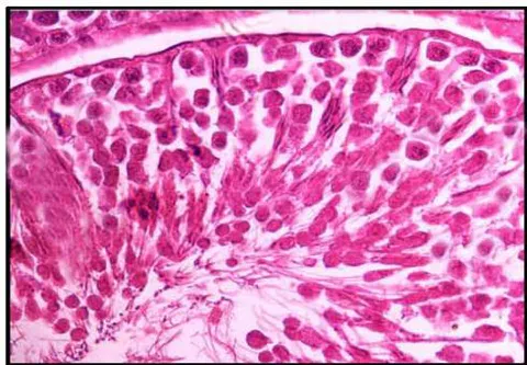
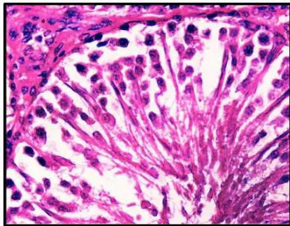
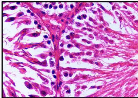
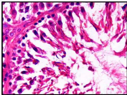
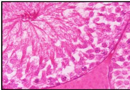
Plate 1: Photomicrograph of testis of rat fig-1 showing normal histoarchitecture of seminal vesicle and spermatozoa clearly visible in lumen, fig-2-4 showing degenerative changes in dose dependence manner whereas fig-5 showing normal histoarchitecture of spermatogenesis.
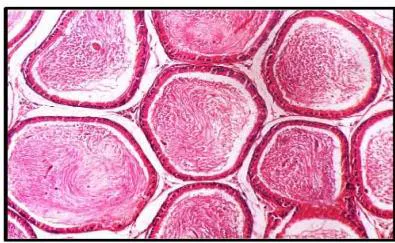
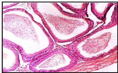
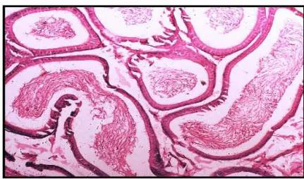
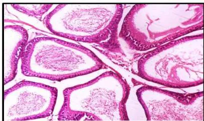
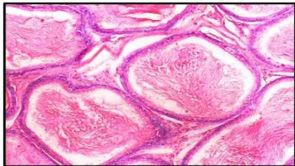
Plate 2: Photomicrograph of Cauda Epididymis of rat fig-1 showing normal histoarchitecture of germinal epithelial and stereocillia and lumen filled with sperm. Fig-2-4 showing degenerative changes in lumen dose dependence manner, whereas fig-5 showing normal histoarchitecture of cauda.
| Treatment | Initial B. wt. (gm.) | Final B. wt. (gm.) | Testes (mg/100 gm b. wt.) | Epididymides (mg/100gm B. wt.) | Vas-defere ns (mg/10 0gm B. wt.) | Seminal vesicle (mg/100gm B. wt.) | Ventral Prostate (mg/100 gm B. wt.) | Kidney (mg/100 gm B. wt.) | Heart (mg/100 gm B. wt.) | Liver (mg/100 gm B. wt.) | Adrenal (mg/100gm B. wt.) |
| Group A Control Intact | 106.50 ± 1.50 | 157.0 ± 2.00 | 1155.12 ± 14.71 | 507.48 ± 16.24 | 163.89 ± 2.11 | 425.00 ± 14.27 | 96.51 ± 1.84 | 580.72 ± 12.84 | 274.67 ± 4.84 | 2454.68 ± 33.91 | 20.48 ± 0.54 |
| Group B 10mg/kg. b. wt. | 105.00 ± 1.67ns | 155.0 ± 2.24ns | 1144.30 ± 13.53ns | 503.21 ± 10.09ns | 162.37 ± 3.21ns | 423.66 ± 14.17ns | 96.49 ± 2.13ns | 612.73 ± 15.82ns | 271.13 ± 4.78ns | 2449.18 ± 24.74ns | 20.23 ± 0.41ns |
| Group C 20mg/kg. b. wt. | 102.00 ± 1.33ns | 159.0 ± 1.94ns | 1137.57 ± 20.20ns | 492.99 ± 10.90ns | 158.54 ± 2.64ns | 421.55 ± 14.30ns | 95.75 ± 2.12ns | 551.76 ± 7.86ns | 268.31 ± 7.76ns | 2415.94 ± 35.64ns | 19.74 ± 0.34ns |
| Group D 40mg/kg. b. wt. | 102.00 ± 1.33ns | 155.0 ± 1.67ns | 1105.35 ± 17.87* | 464.77 ± 10.62* | 153.41 ± 3.33* | 399.05 ± 15.77ns | 94.32 ± 2.06ns | 551.74 ± 12.83ns | 240.63 ± 14.71ns | 2412.85 ± 27.45ns | 19.68 ± 0.43ns |
| Group E Recovery 20mg/kg. b. wt. | 104.00 ± 1.63ns | 156.0 ± 1.94ns | 1142.34 ± 13.27ns | 498.87 ± 10.84ns | 159.98 ± 3.41ns | 423.63 ± 13.99ns | 95.86 ± 2.22ns | 573.26 ± 11.76ns | 270.58 ± 4.58ns | 2446.01 ± 32.72ns | 19.84 ± 0.44ns |
| Treatment | Sperm motility (Cauda) (%) | Sperm density | Fertility (%) | Number of pups delivered | |
| Cauda (million/mm ) | Testes (million/mm ) | ||||
| Group A | 74.57 | 15.15 | 3.42 | 100% (+ve) | 70 |
| Control | ± | ± | ± | ||
| Intact | 0.97 | 0.19 | 0.17 | ||
| Group B | 73.89 | 14.90 | 3.11 | 64% (-36%) | 45 |
| 10mg/kg. b. wt | ± | ± | ± | ||
| 0.75ns | 0.11ns | 0.10ns | |||
| Group C | 73.17 | 14.88 | 2.98 | 51% (-49%) | 36 |
| 20mg/kg. b. wt | ± | ± | ± | ||
| 1.02ns | 0.22ns | 0.05* | |||
| Group D | 68.14 | 14.10 | 1.97 | 35% (-65%) | 24 |
| 40mg/kg. b. wt. | ± | ± | ± | ||
| 0.87** | 0.14** | 0.08* | |||
| Group E | 73.31 | 14.90 | 3.08 | 72%(-28%) | 50 |
| Recovery | ± | ± | ± | ||
| 20mg/kg. b. wt. | 0.71ns | 0.11ns | 1.98ns | ||
| Treatment | Cholesterol (mg/gm) | Glycogen(mg/gm) | Ascorbic acid (mg/gm) | Fructose (mg/gm) | |||||
| Testis | Liver | Heart | Adrenal | Testis | Liver | Heart | Adrenal | Seminal Vesicle | |
| Group A Control Intact | 12.62 ± 0.40 | 14.11 ± 0.62 | 14.80 ± 0.83 | 9.40 ± 0.42 | 3.62 ± 0.17 | 4.55 ± 0.17 | 4.16 ± 0.15 | 3.05 ± 0.25 | 7.56 ± 0.41 |
| Group B 10mg/kg. b.wt. | 11.56 ± 1.19ns | 14.08 ± 0.50ns | 14.49 ± 0.74ns | 8.82 ± 0.48ns | 3.38 ± 0.10ns | 4.20 ± 0.13ns | 3.93 ± 0.22ns | 2.84 ± 0.20ns | 7.49 ± 0.34ns |
| Group C 20mg/kg. b.wt. | 11.44 ± 0.42* | 13.75 ± 0.68ns | 13.88 ± 0.67ns | 8.31 ± 0.53ns | 3.25 ± 0.19ns | 4.22 ± 0.14ns | 3.85 ± 0.07ns | 2.66 ± 0.21ns | 6.11 ± 0.44* |
| Group D 40mg/kg. b.wt. | 10.81 ± 0.85* | 13.58 ± 0.57ns | 13.81 ± 0.72ns | 8.25 ± 0.36* | 2.74 ± 0.12* | 4.07 ± 0.40ns | 3.57 ± 0.35ns | 2.38 ± 0.13* | 5.96 ± 0.39* |
| Group E Recovery 20mg/kg. b.wt. | 11.00 ± 1.13ns | 13.94 ± 0.54ns | 14.50 ± 0.74ns | 8.56 ± 0.44ns | 3.28 ± 0.10ns | 4.12 ± 0.12ns | 3.71 ± 0.05ns | 2.73 ± 0.19ns | 7.45 ± 0.36ns |