


Bulbar Syndrome Revealing Anti-PM/SCL Antibody-Positive Scleromyositis: A Case Report

§ Centre Hospitalier National Universitaire de Fann

Send Message

To: Author

Bulbar Syndrome Revealing Anti-PM/SCL Antibody-Positive Scleromyositis: A Case Report

Article Fingerprint

ReserarchID

MRP321C

Bulbar Syndrome Revealing Anti-PM/SCL Antibody-Positive Scleromyositis: A Case Report Banner

Key Research Insights

Synthesized scholarly intelligence & interactive research assistant
  • English
  • Afrikaans
  • Albanian
  • Amharic
  • Arabic
  • Armenian
  • Azerbaijani
  • Basque
  • Belarusian
  • Bengali
  • Bosnian
  • Bulgarian
  • Catalan
  • Cebuano
  • Chichewa
  • Chinese (Simplified)
  • Chinese (Traditional)
  • Corsican
  • Croatian
  • Czech
  • Danish
  • Dutch
  • Esperanto
  • Estonian
  • Filipino
  • Finnish
  • French
  • Frisian
  • Galician
  • Georgian
  • German
  • Greek
  • Gujarati
  • Haitian Creole
  • Hausa
  • Hawaiian
  • Hebrew
  • Hindi
  • Hmong
  • Hungarian
  • Icelandic
  • Igbo
  • Indonesian
  • Irish
  • Italian
  • Japanese
  • Javanese
  • Kannada
  • Kazakh
  • Khmer
  • Korean
  • Kurdish (Kurmanji)
  • Kyrgyz
  • Lao
  • Latin
  • Latvian
  • Lithuanian
  • Luxembourgish
  • Macedonian
  • Malagasy
  • Malay
  • Malayalam
  • Maltese
  • Maori
  • Marathi
  • Mongolian
  • Myanmar (Burmese)
  • Nepali
  • Norwegian
  • Pashto
  • Persian
  • Polish
  • Portuguese
  • Punjabi
  • Romanian
  • Russian
  • Samoan
  • Scots Gaelic
  • Serbian
  • Sesotho
  • Shona
  • Sindhi
  • Sinhala
  • Slovak
  • Slovenian
  • Somali
  • Spanish
  • Sundanese
  • Swahili
  • Swedish
  • Tajik
  • Tamil
  • Telugu
  • Thai
  • Turkish
  • Ukrainian
  • Urdu
  • Uzbek
  • Vietnamese
  • Welsh
  • Xhosa
  • Yiddish
  • Yoruba
  • Zulu
Reading Preferences
Font Size
Line Spacing
Background
This converted HTML version may contain rendering inconsistencies. Please refer to the PDF for the authoritative version, or click here to provide feedback.

Abstract

Scleromyositis is a new clinical entity, an overlapping syndrome involving two distinct autoimmune diseases, systemic scleroderma and polymyositis or dermatomyositis. We report an observation of a 43-year-old female patient with a history of thyroidectomy, who has been presenting with phonation and swallowing disorders for over 6 months. Clinical examination revealed neuro-myogenic syndrome. Biological findings included elevated creatine phosphokinase at 1497 IU/L and lactate dehydrogenase at 954 IU/L, and the presence of a chronic inflammatory syndrome on serum protein electrophoresis. The electroneuromyography showed sensitive and motor polyneuropathy associated with polymyositis. Immunological tests revealed positive anti-PM/Scl-75 antibodies in the DOT myositis assay. The patient was treated with corticosteroid therapy 1mg/kg/day plus adjuvant plus methotrexate 15mg/week. The evolution was marked by a progressive clinical improvement of the motor deficit and biological improvement with a clear decrease in creatine phosphokinase.

1. Introduction

Scleromyositis is an overlap syndrome combining two distinct entities: systemic sclerosis and either polymyositis or dermatomyositis [2, 5]. It is a rare autoimmune disease characterized by relapses and remissions [4, 9]. It may be life-threatening due to bulbar, respiratory, and/or cardiac involvement [4, 9]. We report a case managed in our medical neuro-intensive care unit involving a patient with scleromyositis and positive anti-PM75 antibodies. The outcome was favorable with corticosteroid therapy and methotrexate.

2. Clinical Observation

This was a 43-year-old woman, in a monogamous marriage, mother of six children, and a housewife. Her medical history included a subtotal thyroidectomy performed in October 2021, followed by treatment with Levothyrox 200 μg/day.

Nine months before admission, she developed phonation disorders characterized by dysphonia associated with swallowing difficulties, which developed gradually and progressively worsened without fluctuation over time. Four months later, she progressively developed motor weakness affecting all four limbs, predominantly involving the proximal muscles.

The clinical picture evolved in a context of inflammatory polyarthralgia involving the peripheral joints (knees, elbows, ankles, and hands), associated with myalgia.

Physical examination on admission revealed a neuromyogenic syndrome involving all four limbs, with proximal-predominant motor weakness, more marked in the upper limbs; thenar and hypothenar muscle wasting; limited mouth opening; a pseudobulbar syndrome; dropped head syndrome; bilateral pitting edema of the lower limbs; reduced chest expansion; and absent idiomuscular reflexes at the quadriceps.

On extra-neurological examination, the patient presented with erythematous lesions on the eyelids and symmetrical erythematous scaly lesions over the metacarpophalangeal and interphalangeal joints.

After five days of hospitalization, the clinical course was marked by the development of acute respiratory distress requiring transfer to the neurological intensive care unit.

Laboratory investigations revealed elevated creatine phosphokinase (CPK) at 1,271 IU/L and lactate dehydrogenase (LDH) at 954 IU/L (Figure 1), evidence of a chronic inflammatory syndrome on serum protein electrophoresis, and elevated C-reactive protein.
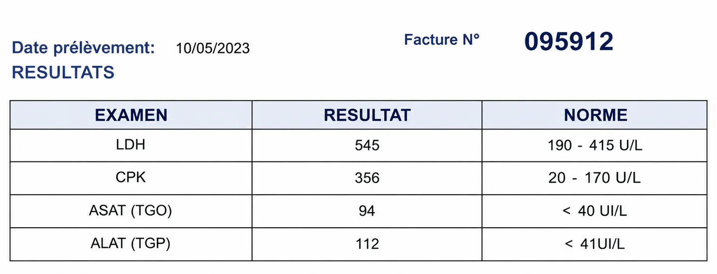
Initial CPK and LDH results
Figure 1. Initial CPK and LDH results

Renal function, thyroid hormone levels, serum calcium, and serum albumin were normal. Arterial blood gases showed mild hypercapnia at 50 mmHg and hypoxemia at 60 mmHg.

The immunological work-up revealed positive anti-PM75 antibodies on the myositis DOT assay (Figure 2).
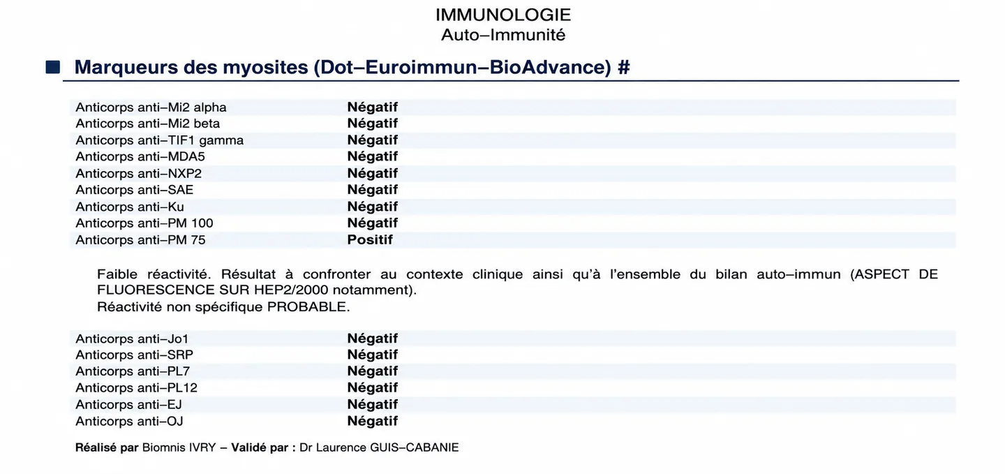
Myositis DOT assay results
Figure 2. Myositis DOT assay results

A cervico-thoraco-abdomino-pelvic CT scan (Figure 3) showed micronodules and nodules of varying sizes in the basal regions of the lungs, associated with a 10-mm right cervical lymph node, suggestive of interstitial lung disease. Echocardiography was normal.
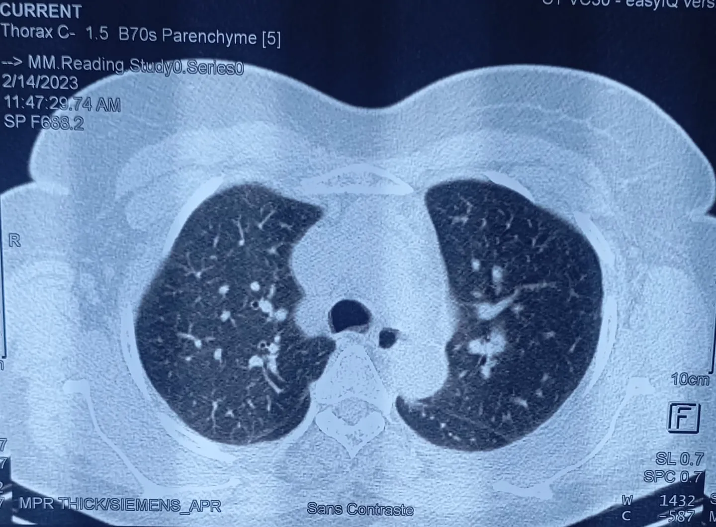
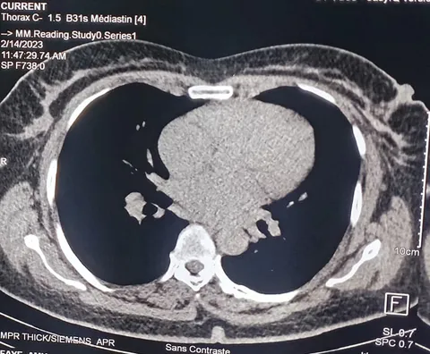
Figure 3. Chest CT angiography showing interstitial lung disease

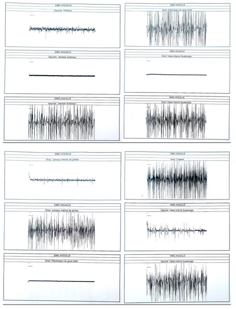
Electroneuromyography (Figures 4 and 5) showed an axonal sensorimotor polyneuropathy associated with myogenic patterns, particularly involving proximal myotomes, suggesting polymyositis.
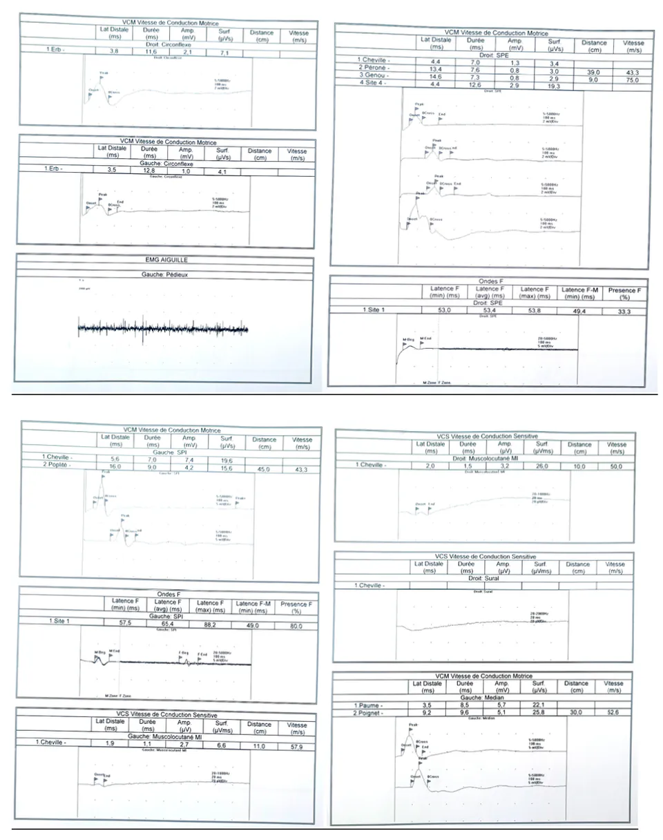
Neurography showing decreased sensory potentials in musculocutaneous nerves and decreased motor potentials in extensor digitorum brevis and anterior tibialis
Figure 4. Neurography showing decreased sensory potentials in musculocutaneous nerves and decreased motor potentials in extensor digitorum brevis and anterior tibialis
Electromyography showing fibrillation potentials in the left extensor digitorum brevis, right medial gastrocnemius, and left quadriceps
Figure 5. Electromyography showing fibrillation potentials in the left extensor digitorum brevis, right medial gastrocnemius, and left quadriceps

The diagnosis of scleromyositis with positive anti-PM75 antibodies was therefore established.

The patient was treated with corticosteroid therapy at 1 mg/kg/day with adjunctive treatment, together with methotrexate at 15 mg/week. The clinical course was characterized by progressive improvement in motor weakness and laboratory parameters, with a decrease in CPK and LDH levels (Figure 6). The modified Rankin Scale score at 6 months was 0.
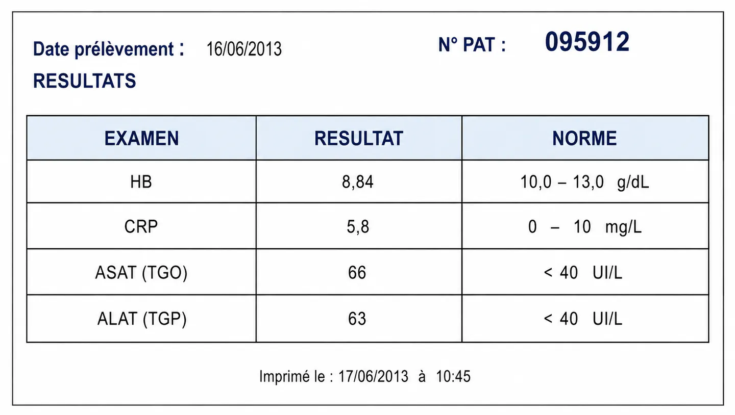
Follow-up CPK and LDH results
Figure 6. Follow-up CPK and LDH results

3. Discussion

Scleromyositis with positive anti-PM75 antibodies is an overlap syndrome between autoimmune myositis and systemic sclerosis. It is a condition that most commonly affects young adults [1, 8].

The clinical manifestations are varied but relatively stereotyped, most frequently involving the muscles, peripheral nervous system, and skin [2, 4]. The clinical features observed in our patient met the 2017 ACR/EULAR diagnostic criteria [10].

In our patient, proximal motor weakness was more severe in the upper limbs. These findings were similar to those reported by Gaudin et al. in 2006, who reported that 10 of their 18 patients had severe proximal muscle weakness [5]. Furthermore, Lorenzo et al. reported that this proximal weakness was more severe in the arm abductors and hip flexors [2].

In addition to peripheral nervous system involvement, extra-neurological manifestations may occur. This was the case in our patient, who presented with both cutaneous and pulmonary involvement [2, 7, 8, 12].

Our patient developed respiratory failure secondary to diaphragmatic involvement. The prevalence of interstitial lung disease associated with systemic sclerosis is 56% (95% CI: 49–63%). Pulmonary involvement is one of the most serious complications and may be life-threatening [11]. Thus, in the EUSTAR cohort, more than 50% of deaths were related to pulmonary involvement [10].

Chest CT may detect interstitial lung disease in up to 90% of patients. These findings were almost identical to those observed in our patient, who presented with respiratory distress and interstitial lung disease on chest CT angiography [5]. Pulmonary CT angiography allowed us to rule out pulmonary embolism, which could have worsened the clinical condition of these patients.

Patients with scleromyositis have serum CPK levels greater than five times the upper limit of normal. This elevation in serum CPK is part of the 2017 ACR/EULAR criteria [10]. These findings were similar to those observed in our patient, whose CPK level was more than nine times the upper limit of normal.

Electromyography is abnormal in approximately 60% of patients with scleromyositis. Spontaneous activity, including positive sharp waves and/or fibrillation potentials, may be observed. Interestingly, a neurogenic pattern may also be found in 25% of patients with scleromyositis [6, 9].

The electroneuromyographic findings in our patient were consistent with those described in the literature, showing myogenic patterns predominantly involving the proximal myotomes, suggestive of polymyositis, associated with a neurogenic pattern [4, 5].

A 2017 meta-analysis reported that 31% of patients with scleromyositis were positive for anti-PM/Scl antibodies [6]. In our patient, anti-PM/Scl antibodies were positive on the myositis DOT assay. However, muscle biopsy could have provided further information regarding muscle ultrastructure [3].

The management of this condition is complex. We initially started treatment with prednisone at 1 mg/kg/day combined with adjunctive therapy, which helped reduce the inflammatory phase and slow respiratory deterioration. Based on the immunological findings, methotrexate 10 mg/week was added, together with folic acid. Methotrexate has also been used in several studies, including that of Marie et al. [11].

The patient also underwent motor rehabilitation sessions. The clinical outcome was favorable, with removal of the nasogastric tube, motor recovery, and a decrease in CPK and LDH levels. However, our patient did not receive intravenous immunoglobulins.

The modified Rankin Scale score at 6 months was 0. This favorable outcome has been described in the literature among patients positive for anti-PM/Scl autoantibodies [5, 7, 11]. Muscle involvement appears to be responsive to immunosuppressive therapy, and patients tend to recover well during the first year of treatment.

4. Conclusion

Scleromyositis is a low-prevalence autoimmune disease that is frequently associated with anti-PM75 antibodies. Some clinical forms may be severe, particularly when the respiratory system is involved. Therefore, rapid diagnosis and multidisciplinary therapeutic management are essential. Corticosteroids and immunosuppressive agents constitute the mainstay of treatment for scleromyositis.

References

12 Cites in Article
  1. 1. Chatterjee,Prayson (2020). Concurrent anti-PM-Scl antibody-associated systemic sclerosis and inclusion body myositis - report of two cases and review of the literature. Semin Arthritis Rheum, 50(3), 498-502.
  2. 2. De Lorenzo,Pinal-Fernandez,Huang,Albayda,Tiniakou,Johnson,Milisenda,Casal-Dominguez,Corse,Danoff,Christopher-Stine,Paik,Mammen (2018). Muscular and extramuscular clinical features of patients with anti-PM/Scl autoantibodies. Neurology, 90(23), e2068-e207676.
  3. 3. Ellezam,Leclair,Troyanov,Meyer,Hudson,Landon-Cardinal (2021). Capillary basement membrane reduplication in myositis patients with mild clinical features of systemic sclerosis supports the concept of « scleromyositis ». Acta Neuropathol (Berl), 142(2), 395-397.
  4. 4. Fernandez,Mendoza,Pinto Garcia,Garcia,Garcia (2023). Scleromyositis: A Novel Entity Beyond Systemic Sclerosis and Autoimmune Myositis. Cureus, 15(8), e44292.
  5. 5. Gaudin,Fortenfant,Adoue,Beyne-Rauzy (2006). Autoanticorps anti-PM-Scl. Étude de prévalence et de signification. Rev Médecine Interne, 27(9), 665-670.
  6. 6. Giannini,Ellezam,Leclair,Lefebvre,Troyanov,Hudson,Senecal,Geny,Landon-Cardinal,Meyer (2022). Scleromyositis: A distinct novel entity within the systemic sclerosis and autoimmune myositis spectrum. Implications for care and pathogenesis. Front Immunol, 13, 974078.
  7. 7. Guillen-Del Castillo,Pilar Simeón-Aznar,Fonollosa-Pla,Alonso-Vila,Reverte-Vinaixa,Muñoz,Pallisa,Selva-O Allaghan,Fernandez-Codina,Vilardell-Tarres (2014). Good outcome of interstitial lung disease in patients with scleroderma associated to anti-PM/Scl antibody. Semin Arthritis Rheum, 44(3), 331-337.
  8. 8. Jablonska,Blaszczyk (1999). Scleroderma overlap syndromes. Adv Exp Med Biol, 455, 85-92.
  9. 9. Lefebvre,Giannini,Ellezam,Leclair,Troyanov,Hoa,Bourre-Tessier,Satoh,Fritzler,Sénécal,Hudson,Meyer,Landon-Cardinal (2021). Histopathological features of systemic sclerosis-associated myopathy: A scoping review. Autoimmun Rev, 20(7), 102851.
  10. 10. Ingrid E. Lundberg,Anna Tjärnlund,Matteo Bottai,Victoria P. Werth,Clarissa Pilkington,Marianne De Visser,Lars Alfredsson,Anthony A. Amato,Richard J. Barohn,Matthew H. Liang,Jasvinder A. Singh,Rohit Aggarwal,Snjolaug Arnardottir,Hector Chinoy,Robert G. Cooper,Katalin Dankó,Mazen M. Dimachkie,Brian M. Feldman,Ignacio Garcia-De La Torre,Patrick Gordon,Taichi Hayashi,James D. Katz,Hitoshi Kohsaka,Peter A. Lachenbruch,Bianca A. Lang,Yuhui Li,Chester V. Oddis,Marzena Olesinska,Ann M. Reed,Lidia Rutkowska-Sak,Helga Sanner,Albert Selva-O’Callaghan,Yeong-Wook Song,Jiri Vencovsky,Steven R. Ytterberg,Frederick W. Miller,Lisa G. Rider (2017). European League Against Rheumatism/American College of Rheumatology classification criteria for adult and juvenile idiopathic inflammatory myopathies and their major subgroups. Ann Rheum Dis, 76(12), 1955-1964.
  11. 11. Marie,Lahaxe,Tiev,Duval-Modeste,Vittecoq,Levesque,Jouen (2010). Myopathies inflammatoires et anticorps anti-PM-Scl : à propos d’une série et revue de la littérature. Rev Médecine Interne, 31(8), 540-544.
  12. 12. Vandergheynst,Ocmant,Sordet,Humbel,Goetz,Roufosse,Cogan,Sibilia (2006). Anti-pm/scl antibodies in connective tissue disease: Clinical and biological assessment of 14 patients. Clin Exp Rheumatol, 24(2), 129-133.

Funding

No external funding was declared for this work.

Conflict of Interest

The authors declare no conflict of interest.

Ethical Approval

No ethics committee approval was required for this article type.

Data Availability

Not applicable for this article.

How to Cite This Article

Serigne Abdou Aziz Fall, Dr. Grace TSEMO, Prof. Ndiaga Matar GAYE, Prof. Adjaratou Dieynabou SOW, Prof. Marième Soda DIOP, Prof. Anna Mbodji BASSE, Prof. Lala Bouna SECK, Prof. Moustapha NDIAYE. 2026. "Bulbar Syndrome Revealing Anti-PM/SCL Antibody-Positive Scleromyositis: A Case Report". Global Journal of Medical Research - A: Neurology & Nervous System GJMR-A Volume 26 (N/A).

Download Citation

Global Journals publishing group case report on anti-PM/SCL antibody-positive scleromyositis.
Journal Specifications

Crossref Journal DOI 10.17406/gjmr

Print ISSN 0975-5888

e-ISSN 2249-4618

Keywords
Classification
MeSH D012595
MeSH D017285
Version of record

v1.2

Language
English
Order Article Reprint
Experiance in AR

Explore published articles in an immersive Augmented Reality environment. Our platform converts research papers into interactive 3D books, allowing readers to view and interact with content using AR and VR compatible devices.

Read in 3D

Your published article is automatically converted into a realistic 3D book. Flip through pages and read research papers in a more engaging and interactive format.

Article Matrices
Total Views: 36
Total Downloads: 0
All Trends

Request Access

Please fill out the form below to request access to this research paper. Your request will be reviewed by the editorial or author team.
X

This is the heading

Lorem ipsum dolor sit amet, consectetur adipiscing elit. Ut elit tellus, luctus nec ullamcorper mattis, pulvinar dapibus leo.

High-quality academic research articles on global topics and journals.

Bulbar Syndrome Revealing Anti-PM/SCL Antibody-Positive Scleromyositis: A Case Report

Serigne Abdou Fall
Serigne Abdou Fall Centre Hospitalier National Universitaire de Fann
Grace TSEMO
Grace TSEMO Centre Hospitalier National Universitaire de Fann
Ndiaga Matar GAYE
Ndiaga Matar GAYE Centre Hospitalier National Universitaire de Fann
Adjaratou Dieynabou SOW
Adjaratou Dieynabou SOW Centre Hospitalier National Universitaire de Fann
Marième Soda DIOP
Marième Soda DIOP Centre Hospitalier National Universitaire de Fann
Anna Mbodji BASSE
Anna Mbodji BASSE Centre Hospitalier National Universitaire de Fann
Lala Bouna SECK
Lala Bouna SECK Centre Hospitalier National Universitaire de Fann
Moustapha NDIAYE
Moustapha NDIAYE Centre Hospitalier National Universitaire de Fann

