

I. INTRODUCTION
Intra-abdominal abscesses are common after surgery, trauma, severe gastrointestinal infection, intestinal perforation, or acute pancreatitis [1, 2]. Thirty percent of episodes are related to inflammatory illnesses of the abdominal organs caused by gastric/duodenal perforation, twenty percent to the liver/biliary system, and one-third to appendicitis [1, 3]. Between the diaphragm and abdominal organs like the liver and spleen, sub-diaphragmatic abscesses occur. Surgical drainage, percutaneous drainage, and endoscopic drainage are all options for abscess drainage.
The mortality rate of a sub-diaphragmatic abscess is significant, and failing to recognize or postpone treatment might be harmful to the patient [4]. The sub-diaphragmatic purulent collection has a significant mortality rate, and if undetected or delayed, it can lead to intra-thoracic problems [5]. Fever with chills and rigors, upper quadrant pain, and leukocytosis are some of the symptoms. The best way to diagnose a sub-phrenic abscess is to use imaging, particularly a CT scan [6].
The patient is at risk of developing chronic episodes of unrelenting fever, sepsis, peritonitis, and death unless treated. Early percutaneous drainage and empiric intravenous antibiotics may be enough to treat the abscess. Surgeons must always be cautious when dealing with post-operative persistent pyrexia that does not respond to antibiotics, and the possibility of a sub-diaphragmatic abscess must always be addressed.
We report a case of sub-diaphragmatic abscess in a post-operative case of perforated duodenal ulcer treated with ultrasound guided percutaneous drainage, with a favorable post-treatment course.
II. CASE REPORT
A 48-year-old male patient came with chief complaints of pain in the abdomen, breathing difficulties, fever, loss of appetite since four days. Patient is a post-operative day 14 case of pre-pyloric perforation and was previously explored and managed by modified graham's patch repair. His pelvic drain was removed on post-operative day 3 and the abdominal drain in Morrison's pouch was removed on post-op day 5 with minimal to nil drain output. The patient was discharged in a healthy state on post-op day 7 with no complaints. On routine post discharge follow up day 3, he had no complaints and his general and per abdomen examination were satisfactory. On post-operative day 11, patient started experiencing pain in right hypochondrium and fever. Pain was dull aching, and progressively increasing in nature with spikes of low-grade fever throughout the day. Gradually he further developed breathing difficulties and loss of appetite. Patient is a known chronic cigarette smoker. Patient did not have any comorbidities.
At admission, on general physical examination, patient was febrile (38.6 degrees centigrade) with tachycardia (130/min), patient had right hypochondriac tenderness with localized guarding with a palpable lower margin of liver, Respiratory system showed decreased breath sounds on the right lower zone of the chest on auscultation. Per-Rectal examination showed no abnormalities. After initial resuscitation with Intravenous fluids, patient was investigated with routine laboratory investigations which revealed a marked inflammatory response, with a white blood cell count of 14,500 cells/
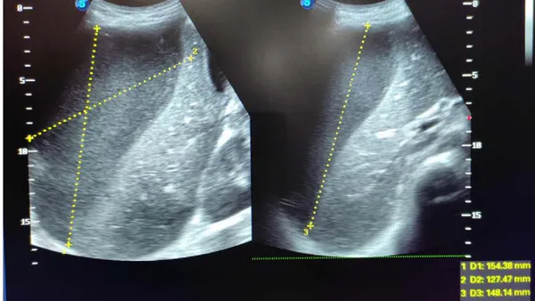
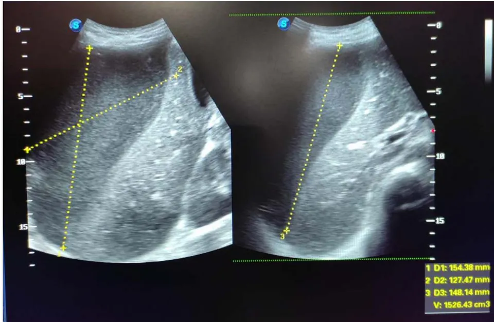
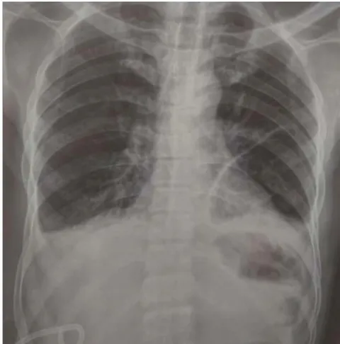
l with a predominant neutrophilic differential count. A chest and abdominal roentgenogram revealed minimal right sided pleural effusion. An Abdomino-pelvic ultrasonography was then performed which revealed a right sided sub-phrenic collection of about 1500 cc pushing liver down with minimal interbowel free fluid, most likely suggestive of infective etiology, right sub-phrenic abscess.
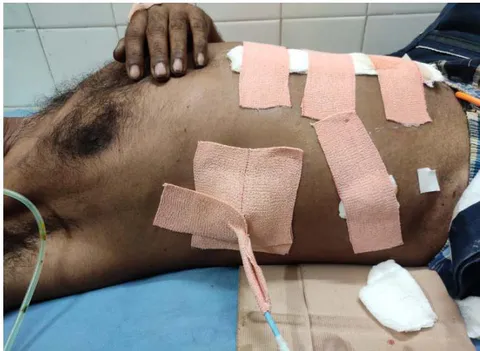
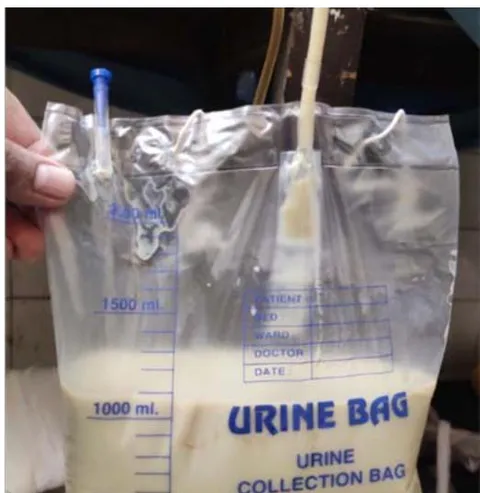
The Patient was started on third-generation cephalosporin antibiotics, metronidazole, and amikacin. An Emergency percutaneous draining pigtail catheter of 16 F was inserted in the suprahepatic right subdiaphragmatic space between and intercostal space and fixed under ultrasound guidance, and frank pus was drained. Microbiological and biochemical samples for testing were collected and pus sample was sent for culture and antibiotic sensitivity. The residual pus got drained over the later 2 days. A repeat chest and abdominal roentgenogram and abdominopelvic ultrasonography were done on day 3 of percutaneous catheterization, to look for any residual collection. With no radiological or clinical evidence of any sub-phrenic collection, the pigtail catheter was removed after 3 days. The Post-procedure course was uneventful, and patient improved clinically. The patient was later discharged and followed up and managed conservatively thereafter.
III. Discussion
The area between the diaphragm and the transverse colon and mesocolon is the sub-diaphragmatic zone. The liver further divides this region into suprahepatic and infrahepatic compartments. These sub-diaphragmatic abscesses can progress to pleurisy with or without effusion, empyema, lung abscess, and bronchial fistula, as well as peritonitis and mortality in rare cases.
The difficulties are exacerbated as the clinical picture is frequently obscured by the causative disease and prior operational intervention. Pain, fever, malaise, cough, or pleural effusion are common symptoms in patients with thoracic or abdominal symptoms. In any patient with unexplained fever, tachycardia, or leucocytosis, the likelihood of a localized intra-abdominal pus accumulation should be considered.
A perforated viscus or a distant extra-abdominal source, probably by hematogenous dissemination, might cause an abscess. Intraoperative contamination and anastomotic leakage are usually quickly disseminated throughout the peritoneal cavity, gathering in the left subphrenic area due to peritoneal fluid flow. According to Oschner and Graves' study, 31 percent of 3372 instances were caused by a ruptured appendix, and 29 percent were caused by a perforated stomach or duodenum [1]. Wetterfors discovered that of his cases were related to previous surgery [7].
According to culture results, the organisms most typically found in a subdiaphragmatic abscess were Staphylococcus, Streptococcus, E. coli, B. Proteus, and pseudomonas.
The relevance of radiological diagnostics in verifying and localizing the Subdiaphragmatic abscess is crucial. Few indications highly suggestive of a subphrenic collection include loss of diaphragmatic mobility and elevation of the diaphragm, pleural effusion, loss of posterior costophrenic angle in lateral view, gas or fluid beneath diaphragm, and enlargement of liver shadow.
The treatment technique for each patient must be determined based on the severity and origin of the subdiaphragmatic abscess. A single sufficient drainage using an extraserous technique may be all that is required in individuals with a single, accessible, well-localized abscess [8]. Sherman et al. (1969) found a 23 percent mortality rate in patients with drainage and an 80 percent mortality rate in those without drainage [9]. If the aspiration is done low down, with the needle pointing up towards the subphrenic spaces, the lung will not be damaged. When the diaphragm is raised, it adheres to the chest wall, allowing aspiration to be conducted safely without concern of injuring the lung or contaminating the pleura. For individuals with numerous abscesses, a transperitoneal technique should be used. An upper paramedian or a subcostal incision can be used to execute this procedure. The key to efficient care of a Subphrenic abscess is early detection and appropriate drainage under the influence of antibiotics.
IV. CONCLUSION
We experienced a case of sub-diaphragmatic abscess associated with a post-operative perforated duodenal ulcer that was cured by percutaneous drainage.
Since the advent of antibiotics, spontaneous subphrenic abscess has become less prevalent, but post-surgical occurrence has become more common.
Declarations
Funding: No Funding sources
Conflict of interest: None declared
Ethical approval: Not required