Introduction
A 21 year old male presented with Right hydrocele with pain in Right hemi scrotum which was diagnosed clinically and radiologically as torsion and subjected to orchidectomy. Histopathology revealed filariasis. The case has been presented for its rarity.
Introduction
Filariasis is a parasitic infection endemic to tropical and subtropical regions. The disease primarily affects the body’s lymphatic system. In India, heavily infected areas with filariae found in Orissa, Uttar Pradesh, Bihar, Andhra Pradesh, Tamil Nadu, Kerala, and Gujarat. Genital filariasis in India more commonly presents as a secondary vaginal hydrocele with an associated epididymo-orchitis. While testicular involvement is rare, the clinical manifestations of filariasis vary from person to person, depending on the course of infection and the worm load. It is very uncommon to find adult worm in the testis.
Case History
A 21-year-old young man presented with a history of pain and swelling in the Right hemiscrotum for 3–4 days with a history of trauma to scrotal area due to a fall 7–8 days back.
On examination, a tense cystic swelling of Rt hemiscrotum, firm in consistency, and a firm cord in consistency were found. USG examination of Rt testis was done and reported as Rt hematocele with Rt testicular torsion.
Treatment and Histopathology
Patient underwent a right orchiectomy. The specimen was sent for histopathological examination in 10% formalin. On examination, the entire right testis was dark brown. Histopathological examination showed a scanty rim of testicular tissue with extensive area of hemorrhage and necrosis, adult filarial worm surrounded by dense inflammatory cell infiltration of neutrophils, lymphoplasmocytes, and eosinophils were seen. No significant pathology was detectable in the epididymis or spermatic cord. A histopathological diagnosis of right-sided filarial orchitis was given.
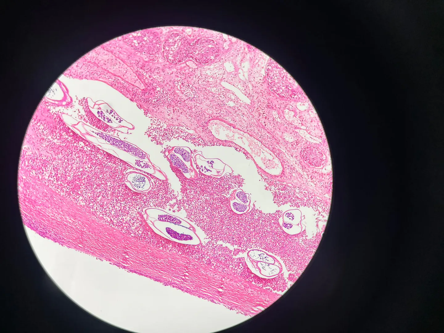
Female adult filarial worm in testicular parenchyma (H&E stain, 100X)
Double-barrel uterus of female adult filarial worm with microfilariae seen against the background of mixed inflammatory infiltrate (H&E stain, 400X)
Discussion
The filarial orchitis simulates grossly with neoplastic and non-neoplastic testicular and paratesticular lesions that include non-specific orchitis, tuberculous epididymo-orchitis, malignant mesothelioma, adenomatoid tumors, mesothelioma cyst and reactive mesothelioma hyperplasia, malakoplakia, sarcoidosis, and inflammatory pseudotumor.
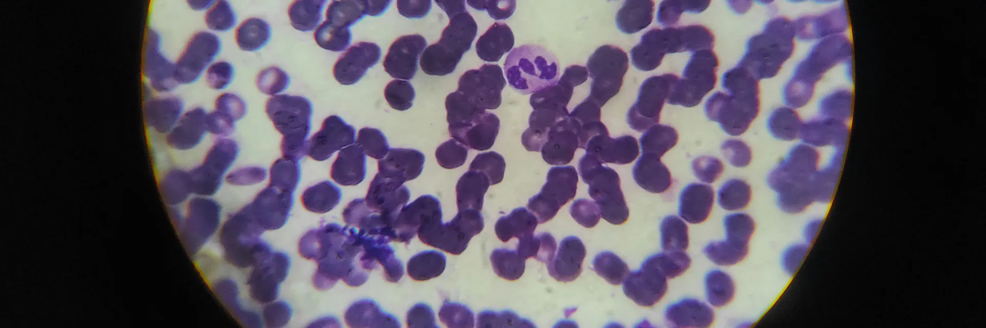
The filarial infection is prevalent in both urban and rural areas. Adult worms are found in the lymphatic vessels and lymph nodes of humans only; there is no animal reservoir. Adult worms in the lymphatic channels produce clinical manifestations of the disease due to lymphatic dysfunction, obstruction, and inflammation. Genital bancroftian filariasis may manifest in several ways including hydrocoele, lymph varix, lymph scrotum, filarial penis or elephantiasis of the genitalia, and chyluria. Hydrocoele accounts for 90% of the morbidity due to genital filariasis. Diagnosis of genital filariasis can be confirmed by direct demonstration of microfilaria in blood or aspirated fluid unequivocally. The tools available for the detection of active infection in an amicrofilaremic patient are circulating filarial antigen (CFA) tests and ultrasound with a high-frequency probe showing filaria dance sign (FDS). This case report highlights an unusual presentation of genital filariasis where intense congestion of the testis and epididymis with microfilaria resulted in acute scrotum masquerading as testicular torsion.
Conclusion
Filarial orchitis may simulate clinically with many neoplastic and non-neoplastic lesions. The various differential diagnoses must be kept in mind when dealing with testicular swellings, especially in endemic areas, to avoid unnecessary orchidectomy.
All the authors contributed equally to the preparation of this manuscript.
Written informed consent could not be obtained in this case as the patient was lost to follow-up. Institutional ethical committee approval was obtained after declaring that the case report does not contain any patient identifiers.