I. INTRODUCTION
Schulte and Heimke initially described protocol for immediate implant placement about 30 years ago. Following which Lazzara in 1989 reintroduced immediate implant placement into fresh extraction sockets.
Immediate implant placement may be defined as implant placement immediately following tooth extraction and as a part of the same surgical procedure, or as implant placement immediately following extraction of a tooth which must be combined in most patients with a bone grafting technique to eliminate peri-implant bone defects.
Immediate implant placement for replacing missing anterior teeth has become undoubtedly a predictable treatment option, at the same time it is challenging both surgically and prosthetically.
Further, a variety of classifications were proposed to facilitate placement of implants in freshly extracted sockets. One of the most common of all is that given by Elian et al 2007. This classification scheme was based on presence or absence of adequate hard and soft tissues and states: Class I (adequate facial and palatal hard and soft tissues), Class II (inadequate facial soft tissue but adequate facial and palatal hard tissue), Class III (Inadequate hard and soft tissue on facial aspect but adequate hard and soft tissue on palatal aspect).
Although appropriate decontamination along with debridement of the surrounding hard and soft tissues of the freshly extracted socket is essential for placement of dental implant, to further prevent spread of infection to peri-implant tissues.
In cases with active infection like pain, draining sinus, purulent discharge, swelling and/or mobility; a 6 day protocol is carried out. Initially the infected tooth is extracted atraumatically with periotomes followed by complete curettage irrigation using bone curette with irrigation connection. Repeated irrigation using 500mg infusion solution of Metronidazole. Following extraction, the root was trimmed to its half-length, cleaned using ultrasonic scaler and reinserted into the extraction socket with its clinical crown bonded facially to adjacent natural teeth. Subsequently post 6 days, the root was removed and the VST protocol is advised. On the other hand, the cases that show no active infection, could be treated directly with VST.
Araujo and Lindhe 2005 advocated use of combined ridge augmentation (contour augmentation with guided bone regeneration) for placement of dental implants in areas of inadequate hard and soft tissues.
Implants placed in presence of inadequate hard and soft tissues may result in gingival recession altering future implant prognosis. Inadequate hard tissues could be replaced with appropriate bone grafts while soft tissues could be enhanced with mucogingival surgeries like connective tissue grafts. The cases with narrow extraction socket orifice, a cortical membrane shield of 0.6mm thickness is advised. Before use it has to be trimmed, hydrated and then introduced from the socket orifice through the tunnel apically till the vestibular access incision where it is stabilized by placing membrane tacks or micro-screws. This prevents possible risk of interdental papilla recession.
As a result, a novel method called VST was developed by Dr Abdelsalem Elaskary in 2019. This clinical protocol could be used to treat a wide variety of fresh extraction sockets with alveolar defects (thin, deficient facial plate with active infection). It follows the protocol of restoring freshly extracted infected socket with simultaneous implant placement. This not only reduces the treatment time but also allows immediate rehabilitation of infected socket with predictable esthetic outcomes which is the need of the hour.
Inclusion Criteria for VST
Single or multiple teeth with hopeless prognosis in maxillary anterior teeth with either type I or type II extraction sockets.
Deficient labial bone plate in width (type I) and height (type II) sockets.
Minimal of 3.0mm basal bone present for immediate implant placement to achieve optimum primary stability.
Exclusion Criteria for VST
Current smokers
Patients with debilitating systemic disease
Patients who have undergone any sort of radiotherapy and chemotherapy in past 2 years.
Pregnant and Lactating females
Vestibular Socket Therapy Instrument Kit Comprises of
Vestibular elevator: Used to carefully elevate periosteum with perforating and leaving behind any residual tissue, available in 4mm or 8mm varieties.
Membrane holding forceps: Angulated membrane holding forceps that allows vertical delivery of membrane through the vestibular access incision, available in 4mm size.
Bone curette with irrigation connection: This bone curette provides dual combination of debriding and disinfecting socket at the same time, available in 3mm size. Simultaneous irrigation while curettage helps to remove all diseased and infected tissues.
Hammerhead periotome: Its shape adapts to the curved sockets and prevents laceration of surrounding tissues, available in 10mm size.
Forklift vestibular retractor: This retractor allows complete visualization of the surgical site and also aids in appropriate placement of bone graft materials.
Soft tissue graft holding forceps: This forceps allows holding of soft tissue grafts against the surgical site and also helps in optimal stabilization of graft at the orifice of the socket while suturing.
7) Scalpel blade holder
8) Cooley's atraumatic 0.8mm straight forceps
9) De Wijs's Periosteal elevator: 5mm in size
10) Kelly's toothed angled scissors: 16mm in length
11) Barraquer Micro-needle holder: straight in shape and 0.8mm in dimensions
Pre-Operative Procedures
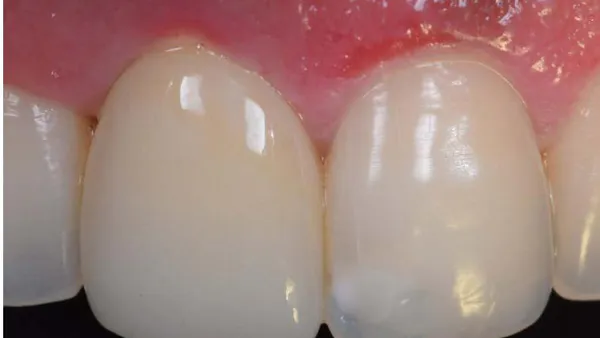
Non-surgical periodontal therapy followed by rinsing with 0.12% Chlorhexidine mouth-rinse for one week. (Figure No: 1)
Pre-operative CBCT (Cone beam computed tomography) scan to determine anatomy of the surgical site. (Figure No: 2)
Computer guided scans that aid in preparation of guide for guided implant placement.
Surgical Protocol for Vestibular Socket Therapy
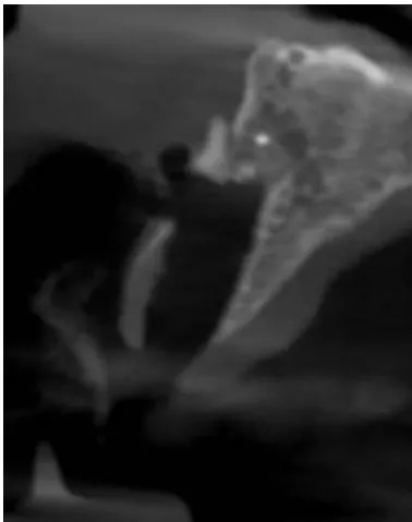
i. Atraumatic tooth extraction (Figure No: 3) was carried out using periotomes (Stoma, Storz am Mark) under local anesthesia (ARTINIBSA 4% 1:100,000, Inibsa Dental S.L.U.) and a post extraction CBCT was taken inorder to evaluate labial bone plate (Figure No: 4).
ii. Following which, the socket was thoroughly curetted and debrided and repeatedly irrigated with 100 mL of anti-anaerobic infusion solution of 500 mg metronidazole (Minapharm Pharmaceuticals).
iii. The socket was curetted and rinsed with saline, and the VST protocol was implemented:
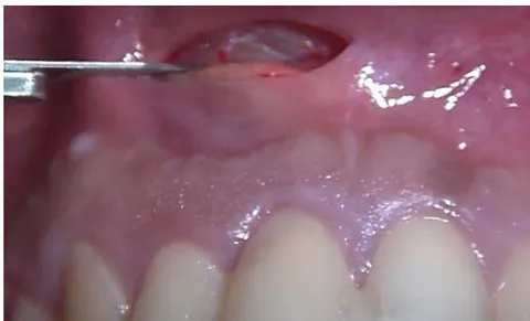
iv. A 1-cm long vestibular access incision (Figure No: 5) was made using a 15c blade (Stoma, Storz am Mark GmbH, Emmingen-Liptingen Germany), 6-8 mm apical to the mucogingival junction of the involved tooth.
v. The socket orifice and the vestibular access incision were connected via a subperiosteal tunnel (Figure

No: 6) that was created using a periotome and a micro-periosteal elevator (Stoma, Storz am Mark GmbH, Emmingen-Liptingen Germany).
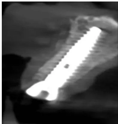
vi. Implants, (tapered pro Biohorizons, Birmingham, Al, USA) (Figure No 7) were used for its aggressive thread design to provide an optimal primary stability, as well as to benefit from the platform switched to enhance the peri implant tissue thickness to its planned location 3 to 4 mm apical to the socket orifice with adequate primary stability, were installed using the 3D printed surgical guide (Surgical Guide Resin, Form 2, Formlabs).
vii. A flexible cortical resorbable membrane (OsteoBiol® Lamina, Tecnoss®, Torino, Italy) of heterologous origin, 0.6 mm in thickness was prepared by hydrating and trimming it.
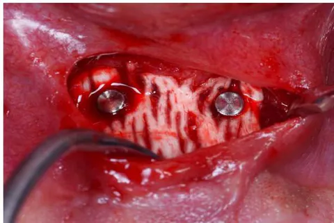
viii. The membrane was then tucked through the vestibular access incision (Figure No: 8), till it reaches 1 mm apical to the socket orifice, and stabilized using two membrane tacs (AutoTac System Kit, Biohorizons Implant Systems, Birmingham, Alabama Inc, USA) to the sound apical bone.
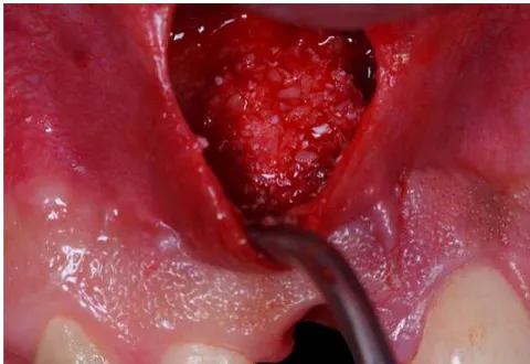
ix. The gap between the implant and the shield/the labial plate was then filled with particulate bone graft [autogenous bone chips harvested form local surgical site mixed with inorganic bovine bone mineral matrix (MinerOssX, Biohorizons, Birmingham, Al, USA)](Figure No: 9).
x. Finally, the vestibular incision was secured with 6/0 nylon sutures (Stoma, Storz am Mark GmbH, Emmingen-Liptingen Germany) (Figure No 10).
xi. A temporary Peek abutment (Figure No: 11) (hexed PolyEtheerEtherKetone Temporary Cylinder, Biohorizons Implant Systems, Birmingham, Alabama Inc, USA) was trimmed to the socket orifice level and the gap was filled with composite resin (FiltekTM Supreme Ultra Flowable Restorative, 3M Corporate Headquarters, MN, USA) to create a sealed chamber that protected the bone graft.
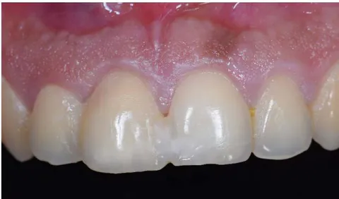
xii. Post-operative follow-up advised 10 days after surgery (Figure No: 12).
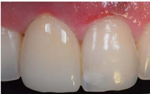
xiii. Final crowns (full anatomical zirconia, bruxzir, Glidewell, CA, USA) were cemented 2 months post-implant placement (Figure No: 13).
xiv. Patient recalled again at 6 months for follow-up visit (Figure No: 14).
FIGURE LEGENDS
1. Intra-Oral Pre-Operative
2. Pre-Operative CBCT
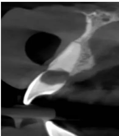
3. Atraumatic Tooth Extraction
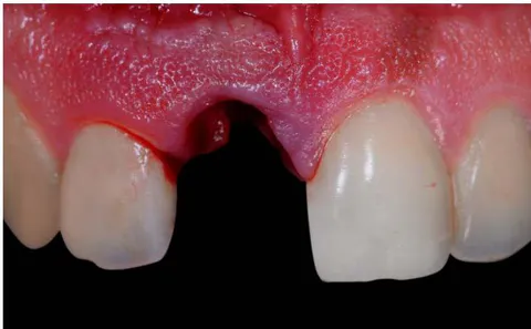
4. Post-Extraction CBCT
5. Vestibular Access Incision
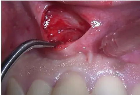

7. Implant Placement Through 3D Printed Surgical Guide
- Insertion of Cortical Membrane from the Vestibular Access Incision Up Till Socket Orifice and Stabilized with Auto-Tacs
9. Placement of Particulate Bone Graft Filling the Bone Defect
10. Suturing
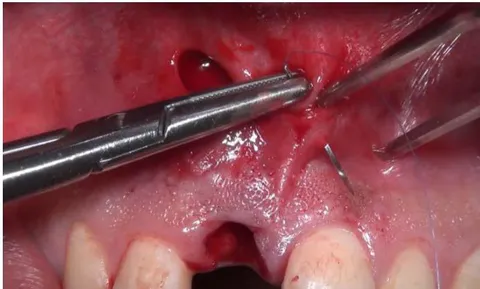
11. Preparation of Temporary Peek Abutment to Cover Socket Orifice with Customized Healing Abutment
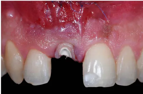
12. Post-Operative One Week
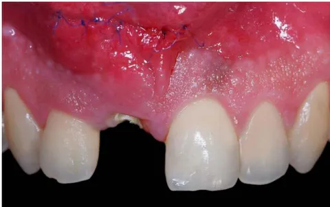
13. Final Prosthesis at 2 Months
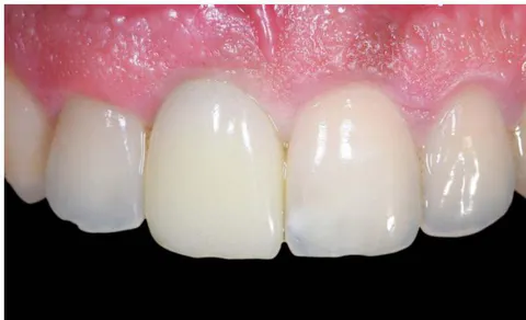
Post-Operative Phase
Antibiotics like Ciprodiazole (Combination of Ciprofloxacin 500mg and Metronidazole 500mg) and Analgesics (any NSAID (non-steroidal anti-inflammatory drug) are prescribed for a 5 day course along with rinsing with Chlorhexidine mouthwash 0.12% twice a day for 1 week.
Advantages of VST
- Used in infected freshly extracted sockets (thin, lost and infected)
2) Treat multiple teeth at a time
3) Single surgical entry
4) Minimal or no facial gingival recession
5) Pre-prosthetic profiling not required
6) Minimal post-operative soft tissue trauma
7) Treatment time reduced to 8 weeks
The main advantage of VST is that it is the only technique available till date that reduces post-extraction ridge resorption of surrounding hard tissues as well as soft tissues.
VST technique is known to show promising results as the flexible labial shield made up of membrane undergoes slow resorption, until then helps maintain appropriate dimensions of extraction socket.
Furthermore, the vestibular access incision aids in stabilization of the labial shield, further ensuring stability of the bone graft materials. Additionally, a sub-epithelial connective tissue graft allows formation of thicker gingival biotype around extraction socket which not only decreases chances of mucosal gingival recession but at the same time also enhances soft tissue profile around the implant.
Studies Carried Out on VST
Elaskary et al 2020 carried out a single-arm clinical study on 12 fresh extraction sockets which were divided into two groups: Group 1: those with intact facial plate of bone and Group 2: those with deficient plate of bone. Sockets divided under both the groups were treated with VST. Pre-operative and post-operative CBCT scans were taken. Pink esthetic score (PES) was recorded 6 months and 13 months following VST protocol. At 3 months, marked co-incidence of facial plate of group 2 sockets was seen with the sockets of group 1 while an increase of mm in the group 2 at 13 months. On the other hand, the PES score at 6 months and 13 months was a total of 14 with 11.33 being for both the groups. The authors concluded that the VST protocol was a minimally invasive treatment and showed predictable results in cases of deficient facial bone plate and hence advocate use of this technique to treat such compromised sockets.
Elaskary et al 2021 conducted one-arm cohort study including 16 implants that aimed to assess radiographic, esthetic and periodontal outcomes 1 year post implant placement in cases of compromised sockets in esthetic zone using VST. In this study, implants were directly placed into type II sockets whereas cases with active infection were treated with a 6 day protocol of anti-microbial therapy. Total parameters assessed were bone height, labial plate thickness at 3 levels at baseline and after 1 year in addition to that PES was determined along with certain periodontal parameters like modified sulcus bleeding index and peri-implant probing depth 1 year post implant placement. The results showed significant increase of bone height and thickness of bone at the middle and crestal thirds with mean PES of 12.63 (1.71), mean modified sulcus bleeding index was 1.19 (0.40) and lastly mean peri-implant probing depth of 1.97 (0.46) mm. Thus, the authors concluded VST protocol along with the 6-day protocol of anti-microbial therapy protocol was successful for treating such compromised sockets and thus aided to minimize total treatment time and surgical interventions providing predictable esthetic outcomes for the patient.
Elaskary et al 2021 assessed regeneration of hard and soft tissues following immediate implant placement in compromised fresh extraction sockets using VST 2 years post-operatively. This study included 27 compromised fresh extraction sockets that were treated with VST followed by immediate implant placement. The defects within the sockets were filled with particulate bone grafts (75% autogenous bone chips harvested from local surgical site infused with 25% de-proteinized bovine bone mineral (DBBM) of equine origin fully enzyme de-antigenised). Thickness of labial bone plate along with bone height were assessed through CBCT scans 2 years post-operatively. A statistically significant difference was observed for increase in total bone height crestally, mid-facially, apically along with bone thickness. Although, the changes in PES and probing depth were not as significant as that compared to thickness of labial plate. Lastly the authors concluded, a combination therapy of immediate implant placement followed by VST helps to manage such compromised sockets and provide prosthetic rehabilitation at an early stage.
II. CONCLUSION
The VST protocol has offered predictable hard and soft tissue regeneration with preserving original anatomy of the extraction socket and allows immediate implant placement in altered and infected socket. The 6-day anti-microbial therapy protocol for cases showing active infection has shown promising results too in limited span of time.
Conflicts of Interests: None