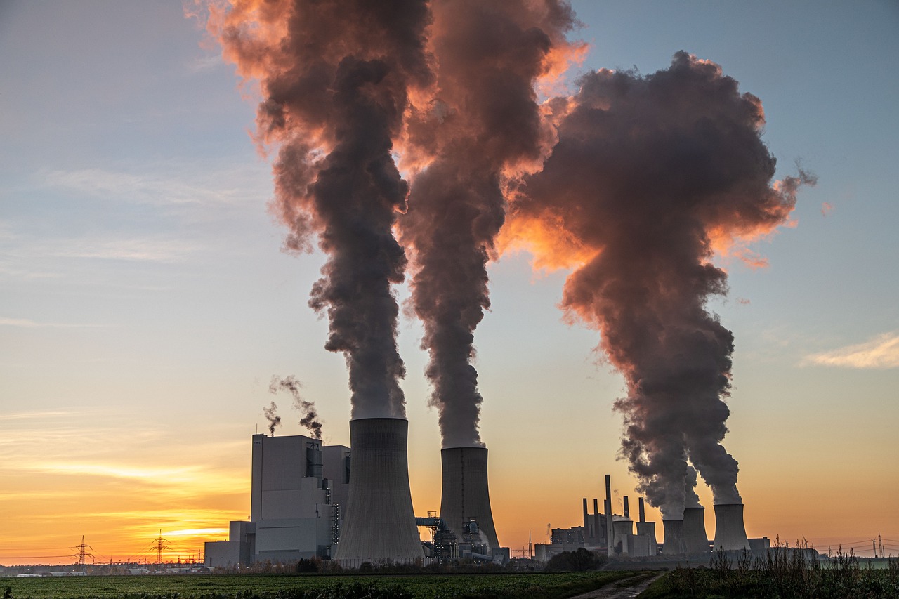
This paper is based on a general analysis of pneumoconiosis and how it is related to work. Breathing in solid particles can cause the lung disease pneumoconiosis. The inhalation of solid particles is what causes it. Exposure to particles such as asbestos, silica, and coal dust is discussed as a central factor in the genesis of the disease, resulting in complex and specific inflammatory and repair processes for each etiologic agent. The methodological design was a comprehensive literature review between the years 2000 and 2023; the texts chosen were in the English language and searched in PubMed and SciELO databases. Coal dust in mining represents a risk, especially for workers in coal transportation and use, as well as for those involved in mineral excavation and mining. Silicosis, a disease linked to miners, resurfaces due to a lack of understanding of modern work practices.
## I. INTRODUCTION
The study explores the etiology, pathophysiology, occupational effects, diagnosis, and treatment of pneumoconiosis. Asbestos, silica, and coal dust exposure become major topics of conversation, with a focus on how these sneaky particles can cause inflammation and repair through a complex and unique mechanism in each of the causes, leading to pneumoconiosis. A meticulous anamnesis, physical examination, imaging investigations, and laboratory tests are crucial for diagnosis and for determining the best course of action based on stopping the progression and exposure. In order to reduce the dangers associated with pneumoconiosis, this analysis emphasizes the urgent need for education, strong regulation, and preventative measures.
## II. METHODOLOGY
The SciELO and PubMed data sources served as the basis for this review of the literature. The search period was July 2023, and the inclusion criteria were full-text, online, English papers published from 2000 to 2023. To analyze the texts more effectively, the health descriptors "Pneumoconiosis," "Work," and "Exposure" were applied.
## III. DISCUSSION
Breathing in solid particles causes a lung condition known as pneumoconiosis.1 Particles such as asbestos, which causes asbestosis; silica, which causes silicosis; and coal dust are considered occupational risk factors for pneumoconiosis. Asbestos, silica, and coal dust exposure can result in pneumoconiosis due to the fibrotic degeneration of lung tissue.1 Mechanisms like apoptosis, iron complexation, oxidative stress, and inflammation aid in thisprocess.1 Studies on animals indicate a link between these chemicals and pneumoconiosis.1 Exposure to coal dust in mining can contain different types of coal, silicates, and asbestos fibers, depending on the specific mineral composition of the mined substance. The only risk that workers who transport bulk materials and use coal at work face is coal dust.2 In addition, people who labor in coal mines and mineral extraction are the groups most harmed by coal dust exposure.2
The inhalation of inhalable crystalline silica particles causes ilicosis, an ancient and potentially fatal pulmonary condition. 2 The historical documentation surrounding silicosis predominantly stems from the experiences of miners. However, the present-day resurgence of silicosis can be attributed to a dearth of awareness regarding contemporary occupational procedures, including but not limited to jeans sandblasting, the production of synthetic stone countertops, construction laborers, individuals employed in the glass industry, as well as workers in the mining, oil, and gas extraction sectors, among various others.2
It is imperative to underscore the gravity of asbestos exposure, a substance that has already been prohibited in numerous nations owing to its inherent health hazards, notably pneumoconiosis. 2 Occupations associated with potential asbestos exposure encompass construction workers, individuals employed in the automotive sector, personnel engaged in the oil and gas industry, workers handling insulation materials, textile industry professionals, and individuals involved in the removal of asbestos-containing materials from aged or contaminated structures.
The inflammatory process causes alveolitis and fibrosis, which are the pathophysiologies of pneumoconiosis.1 Alveolar macrophages phagocytozese silica granules or asbestos fibers after they enter the alveoli.1 Alveolitis begins when macrophages that have been injured or activated emit cytotoxic oxidants, proteases, and inflammatory mediators that attract inflammatory cells to the alveolar wall and to the alveolar epithelial surface.1 Although lymphocytes and neutrophils are also involved, alveolar macrophages are the primary cells that cause alveolitis.1 Inflammatory mediators also increase the production of mucus in the airways.1 After the inflammatory phase, the repair phase begins. During this phase, growth factors cause type II pneumocytes, fibroblasts, fibronectin, and collagen to recruit and multiply, which leads to fibrosis.
The most typical signs of pneumoconiosis include nodular opacities, fibrous masses, or scars in the lung tissues. The diagnosis of pneumoconiosis is made primarily based on questions about the work history, a physical exam, and imaging studies, which are initial instruments to assess the presence of pulmonary changes. 3 Pulmonary function tests can be done to check for irregular breathing patterns, decreased lung capacity, and blocked airways. 3 To determine whether inflammation is present, laboratory testing can also be used. In more complicated and uncommon circumstances, a biopsy may be required. 3 As there is no specific treatment for pneumoconiosis that may reverse the harm brought on by exposure, therapeutic strategies focus on symptom management and delaying the disease's progression. 5 The strategy involves removing exposure, treating symptoms, managing problems, and providing emotional and educational support. 5 The most important of the strategies is the implementation of safety precautions in the workplace, such as dust management, the use of personal protective equipment, a better ventilated environment, and routine worker health monitoring.
## IV. FINAL CONSIDERATION
In conclusion, silica, asbestos, and coal are categorized as the main risks related to this exposure in the work environment, causing fibrotic degeneration of lung tissue. This is an important factor for the diagnostic questionnaire, and for treatment, it is necessary to stop the exposed occupation and provide symptomatic support. To reduce the effects and incidence of pneumoconiosis, it is therefore essential to be aware of its predisposing factors as well as its pathophysiology and prevention. It is also crucial to create safe working environments for employees' health, which calls for cooperation between the union of workers, health professionals, employers, and regulatory authorities.
Generating HTML Viewer...
References
12 Cites in Article
Daniele Mandrioli,Vivi Schlünssen,Balázs Ádám,Robert Cohen,Claudio Colosio,Weihong Chen,Axel Fischer,Lode Godderis,Thomas Göen,Ivan Ivanov,Nancy Leppink,Stefan Mandic-Rajcevic,Federica Masci,Ben Nemery,Frank Pega,Annette Prüss-Üstün,Daria Sgargi,Yuka Ujita,Stevie Van Der Mierden,Muzimkhulu Zungu,Paul Scheepers (2018). WHO/ILO work-related burden of disease and injury: Protocol for systematic reviews of occupational exposure to dusts and/or fibres and of the effect of occupational exposure to dusts and/or fibres on pneumoconiosis.
Ryan Hoy,Daniel Chambers (2020). Silica‐related diseases in the modern world.
D Perlman,L Maier (2019). Occupational Lung Disease.
A Weston (2011). SAGES guideline for clinical application of laparoscopic bariatric surgery.
Chinatsu Nishida,Kazuhiro Yatera (2022). The Impact of Ambient Environmental and Occupational Pollution on Respiratory Diseases.
Chinatsu Nishida,Kazuhiro Yatera (2022). The Impact of Ambient Environmental and Occupational Pollution on Respiratory Diseases.
Ryan Hoy (2021). Artificial stone silicosis.
Veruscka Leso,Luca Fontana,Rosaria Romano,Paola Gervetti,Ivo Iavicoli (2019). Artificial Stone Associated Silicosis: A Systematic Review.
Naoki Fujimura (2000). Pathology and pathophysiology of pneumoconiosis.
Peng Shi,Xiaoyue Xing,Shuhua Xi,Hongmei Jing,Jiamei Yuan,Zhushan Fu,Hanqing Zhao (2020). Trends in global, regional and national incidence of pneumoconiosis caused by different aetiologies: an analysis from the Global Burden of Disease Study 2017.
P Liubchenko (2004). Pnevmokonioz v sovremennom meniaiushchemsia mire [Pneumoconiosis in contemporary changing world.
A Pliukhin,T Bourmistrova,L Postnikova,A Kovalyova (2013). Pneumoconioses in contemporary industry.
Explore published articles in an immersive Augmented Reality environment. Our platform converts research papers into interactive 3D books, allowing readers to view and interact with content using AR and VR compatible devices.
Your published article is automatically converted into a realistic 3D book. Flip through pages and read research papers in a more engaging and interactive format.
Our website is actively being updated, and changes may occur frequently. Please clear your browser cache if needed. For feedback or error reporting, please email [email protected]
Thank you for connecting with us. We will respond to you shortly.