Cleft palate is the most common cause of velopharyngeal dysfunction (VPD), and primary palatoplasty should aim to establish the anatomical and functional conditions for adequate closure of the velopharyngeal mechanism during speech. The most common symptoms of VPD are hypernasality, characterized by excessive nasal resonance in the production of normally non-nasalized sounds due to the inability of the velopharyngeal sphincter to remain closed enough to prevent nasal resonance of orally articulated sounds; nasal air emission, characterized by the inappropriate escape of air through the nose during the production of pressure consonants, which may or may not be audible; and compensatory articulation disorders, considered strategies developed by individuals, replacing orally articulated sounds with those articulated in places posterior or superior to the velopharyngeal sphincter to compensate for the inability to impose pressure on the oral cavity and avoid pressure loss during speech.
## I. INTRODUCTION
Cleft lip and palate (CLP) are congenital facial defects characterized by openings or discontinuities in the structures of the lip and/or palate, with variable location and extent, affecting the nose, alveolar ridge, teeth, hard and soft palate, and any region of the face and skull. It occurs during intrauterine life, specifically in the embryonic period, between the 4th and 12th week of gestational life. Orofacial clefts can be diagnosed during prenatal care, but it is not possible to treat them during the gestational period (Silva Filho, et al., 2005).
Cleft lip and palate are the most frequent facial deformities in the human population, with an accepted global average prevalence between 1 and 2 cases per thousand live births, varying according to ethnicity, gender, and geographic location (Monlleó, et al., 2006). Regarding etiological aspects, studies report that most clefts are associated with multifactorial inheritance, linked to both hereditary and environmental factors (Altmann, et al., 1998). Among the environmental factors influencing the occurrence of isolated CLP, the following stand out: viral infections, stress, maternal folic acid deficiency, alcohol and drug use, the use of medications such as corticosteroids during pregnancy, malnutrition, smoking, and radiation exposure. There are cases where CLP is associated with other anomalies and genetic syndromes, with around 400 syndromes including CLP in their phenotype, according to information recorded in the London
Dysmorphology database (Freitas, et al., 2011). Velopharyngeal insufficiency is the incomplete closure of the sphincter between the oropharynx and nasopharynx, often resulting from anatomical abnormalities of the palate, with cleft palate being the most frequent cause of velopharyngeal dysfunction (VPD). In patients with cleft palate, the structures fail to achieve velopharyngeal closure due to structural alterations, leading to velopharyngeal insufficiency (VPI). In these conditions, part of the sonorized airflow is diverted to the nasal cavity, compromising speech production in various ways. The most common symptoms of VPI are hypernasality, characterized by excessive nasal resonance during the production of sounds due to the inability of the velopharyngeal sphincter to remain sufficiently closed to prevent nasal resonance of orally articulated sounds; nasal air emission, characterized by the inappropriate escape of air through the nose that occurs during speech (Kummer, et al., 2001).
The production of pressure consonants may or may not be audible, and compensatory articulation disorders are considered strategies developed by individuals, substituting orally articulated sounds with those articulated in locations posterior or superior to the velopharyngeal sphincter to compensate for the inability to impose pressure in the oral cavity and avoid pressure loss during speech (Mituuti, et al., 2011).
Primary palatoplasty should aim to establish the anatomical and functional conditions for adequate velopharyngeal closure during speech (Yamashita, et al., 2010). Palatoplasty is the surgical procedure for the reconstruction of the hard and soft palate, aiming to correct the cleft. The cleft should be anatomically and functionally corrected, restoring functions such as speech, chewing, and breathing, while preserving maxillofacial growth potential. However, the primary focus of palatoplasty is speech restoration. To achieve this, the palatal levator muscle must be realigned to a transverse and posterior position in the soft palate. Studies indicate that surgical closure of the palate before 24 months of age provides better speech and hearing development (Trindade, et al., 2005). Despite the recognized effort to normalize muscle orientation in primary palate correction, some patients who undergo primary palatoplasty may still exhibit VPI symptoms, possibly due to the disorientation of muscle fibers and their anteriorized insertion on the posterior edge of the palatine plates. In such cases, secondary palate surgery becomes necessary (Funayama, et al., 2014).
The modified palatoplasty technique aims to reposition the muscles and elongate the soft palate, offering good mobility to the velum and consequently improving velopharyngeal competence.
Considering the above, the present study proposes a prospective clinical cohort study of patients undergoing primary palatoplasty, evaluating velopharyngeal closure and function.
The study is characterized as a prospective clinical cohort study, where the function of velopharyngeal closure during speech was evaluated through nasopharyngoscopy and hypernasality tests in patients undergoing primary palatoplasty using the modified technique.
## II. MATERIALS AND METHODS
The research was conducted at the Association for the Rehabilitation and Social Integration of People with Craniofacial Congenital Malformations of Ceará (Associação Beija Flor), located in Fortaleza, Ceará. For data collection, an individualized evaluation form, attached to this document, was used.
### Sample
A sample of 09 patients operated on at an average age of 2 years, 4 female and 5 male, who underwent primary palatoplasty using the technique, performed by the same operator with a 2-year clinical follow-up at the Beija Flor Association. The evaluation was carried out between March 2023 and January 2024.
## III. DESCRIPTION OF THE MODIFIED
### PALATOPLASTY TECHNIQUE
The modification of the palatoplasty technique can be performed in conjunction with the Bardach technique, first described in 1967, or the Von Langenbeck technique described in the 1800s. Both involve incisions along the edges of the cleft and paramedian relaxing incisions to create mucoperiosteal flaps that may be monopediculated or bipediculated depending on the chosen initial technique. The incision begins along the medial edge of the soft palate where the line between the palate and nasal mucosa is located. Through this incision, the muscles on both sides are exposed. The incision is made to the tip of the uvula, followed by dissection with Metzenbaum scissors in the posterior region of the palate, dividing the tensor and levator veli palatini muscles from the palatoglossus and palatopharyngeus muscles. The next step is to isolate the mucoperiosteal flaps on the oral surface, inserting a Molt elevator into the lateral incision and sliding it between the bone and periosteum until reaching the incision at the medial edge of the cleft, tunneling the palatine artery in its anterior region to the bundle. By moving the Molt elevator up and down, the upper portion of the mucoperiosteal flap is lifted.
After full-thickness mucoperiosteal elevation, at the junction between the hard and soft palate of the cleft, where the muscles insert vertically, the muscles must be divided with the insertion of the Molt elevator in this region. Moving downward and backward, the oral mucosal flap is elevated. This procedure aims to complete the division of the muscles forming the palate. In this division, the tensor and levator veli palatini muscles remain inserted into the nasal mucosa, and the palatoglossus and palatopharyngeus muscles remain inserted into the oral mucosa. The muscles of the soft palate attached to the nasal mucosa are not dissected as in the intravelar veloplasty technique; instead, a horizontal incision is made from medial to lateral into the muscle and nasal mucosa ensemble. This maneuver will cause a posterior displacement and lengthening of the soft palate, promoting proper positioning for the velopharyngeal mechanism (Figure 1).


 Figure 1: Modified Technique for Mucosa Closure and Palate Lengthening - Bilateral Nasal Mucosa Incision and Horizontal Suturing of the Musculature
After hemostasis review, the flap closure should be done in layers and without tension using absorbable sutures (e.g., Vicryl 4.0).
The closure pattern should follow the sequence: 1) suturing the levator and tensor veli palatini muscles attached to the nasal mucosa, positioning them horizontally; 2) suturing the palatoglossus and palatopharyngeus muscles attached to the oral mucosa; and 3) finalizing with "U" sutures in the oral mucosa along the entire cleft length.
## IV. VELOPHARYNGEAL MECHANISM
### EVALUATION
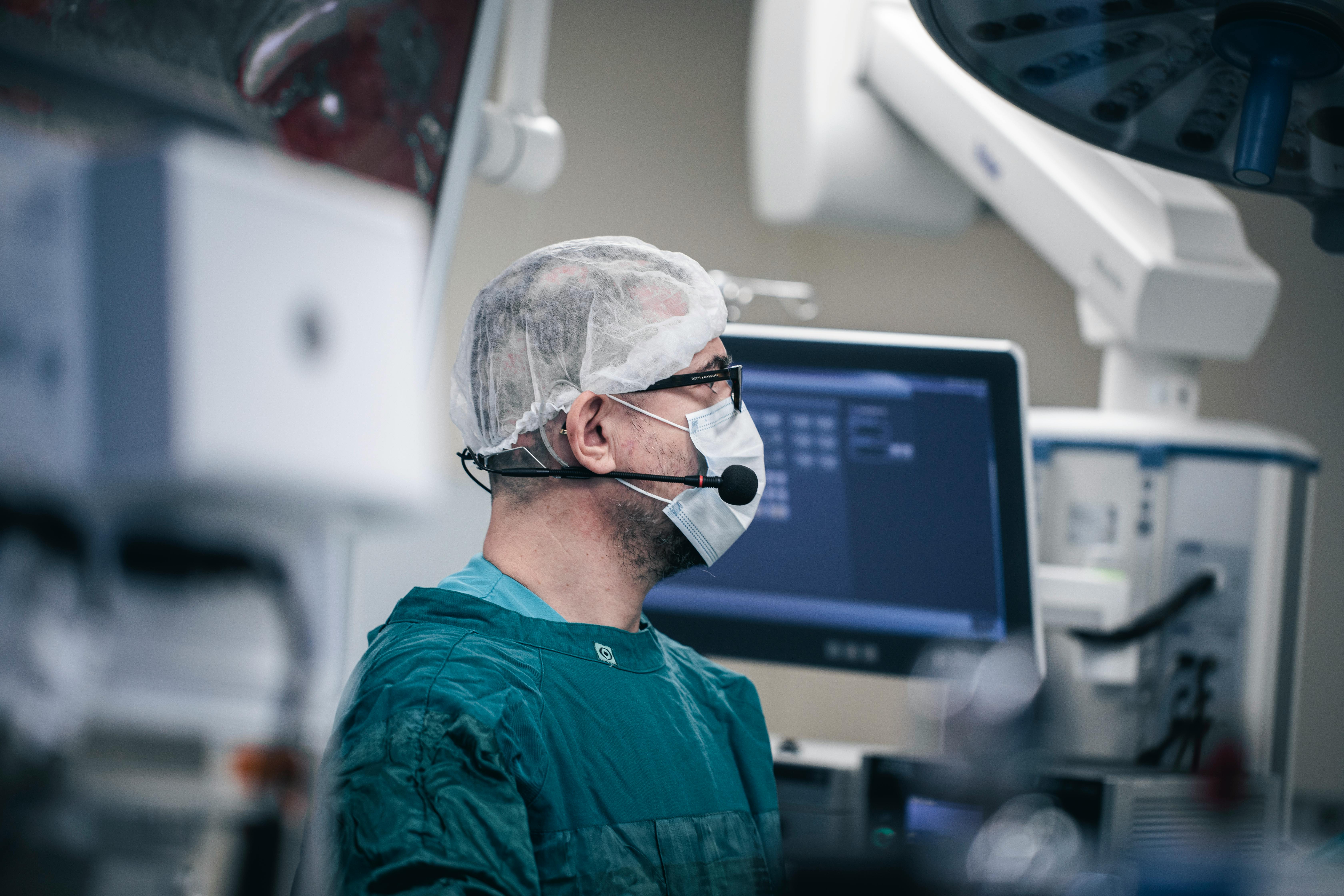
For velopharyngeal mechanism evaluation, the hypernasality test and nasopharyngoscopy were used. Videonasendoscopy is one of the most commonly used procedures in clinical practice, allowing for endoscopic evaluation of velopharyngeal function and better visualization of the nasal and pharyngeal-laryngeal cavities. It is one of the most appropriate tools for observing the closure patterns of the velopharyngeal sphincter (VPS), especially during speech, with characteristics and degrees of movement of the soft palate and pharyngeal walls. During the examination, the videonasendoscope is inserted through the nasal passage after the application of local topical anesthesia to the nasal mucosa, advancing to the VPS region. This direct instrumental evaluation provides valuable information pertinent to the diagnosis, such as the investigation of soft tissue obturator contact during speech, palatal conformation, velopharyngeal motor actions, type of closure, among others, in addition to diagnoses and prognoses for postoperative follow-up (Trindade, et al., 2007).
In our study, the nasopharyngoscopic evaluation was performed using the Machida Flexible Nasopharyngolaryngoscope model ENT-30PC. For the speech sample used in nasopharyngoscopy after surgery, plosive phoneme emissions (PA, TA, CA) and fricative sounds (FA, SA, XA) were evaluated, with movements classified as good, regular, poor, or no closure. The symmetry of movements and velopharyngeal function were also assessed (Genaro, et al., 2004).
For the hypernasality test, the mirror test for nasal air escape evaluation, proposed by Morley (1973), was used. A mirrored surface or a card was positioned just below the nostrils during exhalation to determine if airflow escaped through the nostrils. The Altmann Nasal Mirror (ENMA) consists of a calibrated metal plate accompanied by a Reference Block, which is a replica of the mirror, for transcribing results. This material aims to quantify nasal aeration and measure nasal air escape. The evaluation involved light blowing, with syllabic sequences using the vowel /i/ and sentences. However, if there was a need to assess a specific sound, the speech therapist produced the isolated phoneme for an extended period or used sentences containing the desired sound with greater frequency (Morley, et al., 1973). For this study, evaluation was performed during and blowing, and classified as either absent or present. In cases where nasal air escape was present, it was quantified based on the ENMA gradation as small, medium, or large.
For data collection, a customized evaluation form was generated to improve understanding and clarity of result analyses. The evaluation form is attached.
## V. DATA ANALYSIS
For our study, a convenience sample was used, taking into account the availability of patients to be part of the sample within a specific time frame. In our study, a sample of 09 patients was evaluated, operated on at an average age of 2 years, who underwent primary palatoplasty using the Modified technique with a 2-year clinical follow-up conducted at the Beija Flor Association. The technique was performed by the same operator, and clinical evaluation was conducted from March 2023 to January 2024. (Table 1)
## VI. ETHICAL ASPECTS
The Informed Consent Form (ICF) will be provided to the guardians during the routine consultation, where the researcher will explain the details described in the ICF. After agreement and signature, the research will begin, following the evaluation protocol.
## VII. RESULTS
Table 1: In the Table Below, We Observe and Have the Results of the Research Data from the 9 Operated Patients
<table><tr><td>Evaluation Period</td><td>Type of Cleft</td><td>N (9)</td><td>Number of Surgeries</td><td>Clinical Examination</td><td>Teste de HP</td><td>VPI</td></tr><tr><td>2023/2024</td><td>6 unilateral left-sided transforamen</td><td>3 bilateral transforamen</td><td>2 anos</td><td>Absence of fistulas in 100% of cases</td><td>Absence of nasal escape during speech and blowing in 100% of cases</td><td>Absent in 100% of cases</td></tr></table>
## VIII. DISCUSSION
Cleft palate is one of the most common congenital malformations and occurs due to a failure in the fusion of the frontonasal and maxillary processes during intrauterine life, resulting in a cleft between the premaxilla and the lateral slopes of the maxilla. The cause is multifactorial, linked to both genetic and environmental factors such as alcohol consumption, tobacco use, drugs, infections, or nutritional deficiencies. It may be associated with syndromes such as Pierre Robin syndrome, Van Der Woude syndrome, and popliteal pterygium syndrome (Amorim JG, et al., 2014).
The levator muscle in the palate, which normally develops, forms a strip across the posterior half of the soft palate. When the normal closure of the palatal raphes does not occur, the levator palatini muscle extends longitudinally along the cleft margin and inserts into the posterior edge of the hard palate, resulting in ineffective contraction and the inability to close the palate, leading to speech alterations and abnormalities in the function of the Eustachian tube (Neligan PC, et al., 2015).
Velopharyngeal function is essential for correct speech production. The sphincteric action of the velopharyngeal structures, soft palate, and pharyngeal walls is responsible for directing expiratory airflow and acoustic vibrations to the oral cavity during the production of oral sounds and to the nasal cavity during the production of nasal sounds (Chait, et al., 2002).
The most frequent cause of VPI is cleft palate, in which the insertion of the palate muscles, particularly the levator veli palatini, is directed anteriorly, remaining in a sagittal position and inserting into the posterior edge of the hard palate. As a result, there is no formation of the muscle sling necessary for velopharyngeal closure, leading to altered resulting vector forces (Chait, et al., 2002).
Instrumental assessment of velopharyngeal function includes one of the most widely used exams, videonasendoscopy. This is an endoscopic evaluation method that allows visualization of the nasal, pharyngeal, and laryngeal cavities with dynamic and direct images of the anatomical structures (Pontes, et al., 2005). Videonasendoscopy can contribute in various situations, from diagnosis and prognosis to postoperative control (Rocha, et al., 2002). During the exam, closure patterns in different motor actions, including speech, and degrees of mobility of the velum and pharyngeal walls can be observed. It also allows for the identification of a gap, which corresponds to the residual opening during maximum contraction of the VPM (Kuehn, et al., 2004).
Perceptual-auditory evaluation allows for the verification of oral communication performance and the analysis of how impaired speech is, determining the extent of resonance-related issues (Bzoch, et al., 2004).
When velopharyngeal closure during speech is inadequate, meaning there is no separation between the oral and nasal cavities during the production of oral phonemes, the nasal cavity is exposed to unexpected airflow. This dysfunction, henceforth referred to as velopharyngeal dysfunction (VPD), can be congenital, as in the case of cleft palate. There is consensus among researchers and health professionals that the surgical and clinical outcomes of individuals with cleft lip and palate should be based on perceptual-auditory evaluation and at least one instrumental method for evaluating velopharyngeal function (Netto, et al., 2011).
The combination of perceptual-auditory evaluation and instrumental assessment through videonasendoscopy is highly valuable and widely used in clinical practice, as one complements the results of the other.
The American Cleft Palate Association, in a document published in the 1980s, recommends the use of at least one instrumental method in conjunction with clinical examination for the study of surgical outcomes of cleft lip and palate. The indicated instrumental methods include nasopharyngoscopy, the pressure-flow technique, videofluoroscopy, and nasometry (Dalston, et al., 1988).
In our study, in combination with perceptual-auditory evaluation, nasopharyngoscopy was used as the instrumental method, following the guidelines of the American Cleft Palate Association.
Several techniques can be used for cleft palate repair, considering the anatomical variability of each cleft. The most commonly used palatoplasty techniques are Von Langenbeck, Veau-Wardill-Kilner, two-flap, and Furlow's double opposing Z-plasty (Amorim JG, et al., 2014).
In 1859, Bernard von Langenbeck described a simpler palatoplasty, which consisted only of cleft edge incisions and a relaxing incision. In 1861, Langenbeck presented the method using bipediculated mucoperiosteal flaps, maintaining the anterior fixation of the flap to the alveolar margin. This technique is still used today, indicated for the treatment of incomplete clefts of the secondary palate without labial or alveolar involvement (Shkoukani, et al., 2014).
In the von Langenbeck technique, the bipediculated mucoperiosteal flaps are medially mobilized. Lateral relaxing incisions from the maxillary tuberosity to the alveolar ridge are required to close the cleft and minimize tension. Because they are made around the alveolar ridge, the flaps with posterior and anterior bases are preserved, sparing the gingival laterally. If the nasal flap is short, a vomer flap may be used (Shkoukani, et al., 2014).
Due to the lateral incisions, this technique has a lower occurrence of oronasal fistulas. However, as it does not provide palate lengthening, it has a high incidence of velopharyngeal insufficiency. Therefore, secondary procedures such as intravelar veloplasty or even double opposing Z-plasty (Furlow) are commonly performed (Furlow LT, et al., 1986).
In parallel with our study, the variation of the von Langenbeck technique with nasal mucosa incision, applied in our study, showed not only the absence of oronasal fistulas but also soft palate lengthening, reducing the incidence of velopharyngeal insufficiency.
The two-flap palatoplasty was first described in 1967 in Poland by Janusz Bardach. The original technique, which involves releasing the mucoperiosteal flaps, is indicated only for narrow palatal clefts. However, modifications such as more extensive dissection and relaxing incisions along the alveolar margins to the cleft edge allow for the treatment of wider clefts (Leow, et al., 2008).
Bardach's technique for larger clefts allows for the release of abnormal insertions of the levator veli palatini muscle, as muscle exposure is simpler. The incision along the cleft margin should be 1 to $2\mathrm{mm}$ toward the oral mucosa, so that the mucosal flap allows for tension-free nasal closure.
The mucoperiosteal flaps, nourished by the greater palatine vessels, are elevated, and the aberrant levator muscle insertions are released. Then, the nasal mucosa is also lifted from the palate, allowing visualization of the hamulus and tensor veli palatini muscle. The lateral muscle fibers are then released. First, the nasal layer is sutured, followed by the levator muscle. In cases of excessive tension in the nasal closure, the central sutures are placed after the muscular layer has been approximated. Finally, the oral layer is closed, starting at the base of the uvula (Dao, et al., 2016).
As the flaps are dependent on the palatine vessels, they are much more versatile in positioning, and this technique is indicated for unilateral or bilateral complete clefts. However, it cannot increase the length of the palate, which may affect speech production (Leow, et al., 2008).
The modified palatoplasty technique with nasal mucosa incision can be performed on wide clefts, and it can be done in conjunction with the isolated Bardach technique or combined with both the Bardach and von Langenbeck techniques in the same patient. In this way, one of the flaps is released, making one flap monopediculated and the other bipediculated, with nasal mucosa incision on both sides, promoting soft palate lengthening and absence of postoperative fistulas.
In the mid-1980s, Leonard Furlow published the technique describing a double reverse Z-plasty. It is used for the closure of the soft palate, involving the oral and nasal surfaces in alternating Z-plasties, repositioning the levator veli palatini muscle. It is most commonly used for the repair of submucous clefts and isolated soft palate clefts. It showed good initial results in terms of both speech and bone growth, as well as velopharyngeal insufficiency (Neligan PC, et al., 2015). The technique consists of the arrangement of triangular flaps, producing a Z-plasty on the oral mucosa and another on the nasal mucosa, with the latter being reversed. The flaps containing muscle are rotated posteriorly, while the mucosa-only flaps are transposed anteriorly. This places the muscle fibers in a more favorable anatomical position, in addition to lengthening the palate and repositioning the levator sling. Ideally, an incision should be made at the cleft margin, which is closed in two layers without the need for lateral incisions to close the hard palate (Dao, et al., 2016).
Although the overall results are favorable, there are theoretically some disadvantages to this technique. The first is that it is not anatomical and completely ignores the minor longitudinal muscle of the uvula (Neligan PC, et al., 2015). The second is that if it is used for very wide clefts, the distance to be crossed by the Z-plasty may be excessive. However, in the latter case, there are alternatives to overcome increased tension. One option is to extend the relaxing incision to create an island flap based on the palatine vessels. Another would be to close the straight line and reserve Z-plasty as a secondary procedure (Leow, et al., 2008).
One of the major challenges for clinicians and researchers in clefts is establishing indicative factors for the various surgical techniques available for VPI correction.
In 2016, a study was published with data collected from the San Diego Children's Hospital in the United States, where a group of 71 patients underwent an average of 8.56 surgeries, ranging from 2 to 25 surgeries in the same patient (McIntyre, et al., 2016).
Thus, the number of primary surgeries is completely related to interference in the craniofacial development of these patients, which can vary depending on the cleft classification.
In our study, another major advantage of the technique used is the absence of the need for a second surgical intervention in palatoplasty. In the cases presented, there was no need for the Furlow or Z-plasty techniques, although they are excellent techniques consolidated in the literature. With the nasal mucosa incision technique, there was no need for a second intervention, resulting in less trauma and less interference in craniofacial growth.
## IX. CONCLUSION
In summary, the modified palatoplasty was effective in improving velopharyngeal closure and lengthening the soft palate, as the nasal mucosa incision favors elongation and facilitates the positioning of the muscles in the posterior region. This was the first study to use this technique to assess the results of primary palatoplasty. Future studies should be conducted to correlate the findings.
Based on the results obtained regarding the effect of modified palatoplasty on the velopharyngeal closure of patients, assessed by nasopharyngoscopy and hypernasality tests, it was concluded that the technique led to excellent velopharyngeal closure in the speech evaluation of all patients analyzed, with a minimal number of surgical interventions.
Declarations of Interest: There are no conflicts of interest.
Generating HTML Viewer...
Funding
No external funding was declared for this work.
Conflict of Interest
The authors declare no conflict of interest.
Ethical Approval
No ethics committee approval was required for this article type.
Data Availability
Not applicable for this article.
Rafael Linard Avelar. 2026. \u201cEffect of Modified Palatoplasty on Velopharyngeal Closure Assessed by Nasopharyngoscopy and Hypernasality Test\u201d. Global Journal of Medical Research - J: Dentistry & Otolaryngology GJMR-J Volume 24 (GJMR Volume 24 Issue J2): .
Cleft palate is the most common cause of velopharyngeal dysfunction (VPD), and primary palatoplasty should aim to establish the anatomical and functional conditions for adequate closure of the velopharyngeal mechanism during speech. The most common symptoms of VPD are hypernasality, characterized by excessive nasal resonance in the production of normally non-nasalized sounds due to the inability of the velopharyngeal sphincter to remain closed enough to prevent nasal resonance of orally articulated sounds; nasal air emission, characterized by the inappropriate escape of air through the nose during the production of pressure consonants, which may or may not be audible; and compensatory articulation disorders, considered strategies developed by individuals, replacing orally articulated sounds with those articulated in places posterior or superior to the velopharyngeal sphincter to compensate for the inability to impose pressure on the oral cavity and avoid pressure loss during speech.
Our website is actively being updated, and changes may occur frequently. Please clear your browser cache if needed. For feedback or error reporting, please email [email protected]
×
This Page is Under Development
We are currently updating this article page for a better experience.
Thank you for connecting with us. We will respond to you shortly.