
I. INTRODUCTION
Both in the United States and other developed countries, the prevalence of anxiety, depression, and other common psychiatric disorders has reached epidemic proportions. Consequently, there is a desperate need for improved treatment outcomes, yet the effectiveness of mental healthcare is not much better now than it was fifty years ago [1]. Psychotherapists continue to employ various psychotherapeutic techniques, and psychiatrists continue to prescribe antidepressants, antipsychotics, and psychostimulants in various combinations. Typically, patients who have relatively mild psychiatric symptoms enter the behavioral healthcare system by consulting with a psychotherapist in the hope of avoiding treatment with medication. Patients who have more severe symptoms sometimes initiate treatment with a psychotherapist, sometimes with a psychiatrist, and sometimes with both. There are also some patients who initiate treatment with an internist and then either continue with the internist or receive a referral to a specialist. Unlike in the past, most contemporary psychiatrists do not practice psychotherapy, and most psychotherapists exhaust the benefits of their craft before referring the patient to a psychiatrist. One of the fundamental problems with this triage system is that patients largely self-select the modality of treatment they receive. Another problem is that there is no objective way to determine which patients would best be treated with psychotherapy, which would best be treated with medication, and which would best be treated with a combination of the two. Yet another problem is the potential lack of communication between the psychotherapist and the psychiatrist when both services are being provided simultaneously. These potential problems underscore the need for clinicians and prospective patients to better understand the mechanisms through which various psychotherapeutic and psychopharmacologic treatment modalities exert their therapeutic effects and to be able to determine, more objectively, which treatment modality would be most appropriate for which patient.
In this article, current psychological and biological approaches to treatment will be reviewed, and a new formulation of the dynamic interplay between the mind and the brain will be discussed. From this fresh perspective, the puzzling relationship between mental processes and neurological processes will be clarified, and a new way of conceptualizing mental illness will be proposed. Based on this new conceptualization, which is strongly supported by converging lines of evidence, the first objective method of determining which patients should be treated with which modality—psychotherapy or biological therapy—will be introduced and, by offering the potential to treat mental illness based on pathology rather than symptomatology, a new era of behavioral healthcare will be ushered in.
II. CURRENT APPROACHES TO MENTAL ILLNESS AND HOW THEY WORK
a) Psychological Interventions
i. Supportive Psychotherapy
Considered to be at the heart of all clinician-patient relationships, supportive psychotherapy encourages the patient to express his or her thoughts, feelings, and concerns in a safe, confidential, and nonjudgmental environment. Though helpful in treating almost any clinical condition, the precise mechanism (or mechanisms) through which supportive psychotherapy exerts its therapeutic effects are still not fully understood. However, its primary therapeutic mechanism appears to be stress-reduction.
ii. Psychoanalytic Psychotherapy
As the dominant form of therapy during the late 19th to mid20th centuries, psychoanalysis is aimed at helping patients resolve unconscious psychological conflicts by allowing them to become more aware of their unconscious thoughts, drives, and motives. The pioneer of this technique, Sigmund Freud, believed that as patients progressed, they became less stressed, less defensive, and, thus, less neurotic. However, the neurological correlates of these changes and their relationship to the patient's symptoms are still unclear.
iii. Interpersonal Psychotherapy (IPT)
IPT focuses on relieving psychiatric symptoms by improving interpersonal functioning and social support. The central tenant of IPT is that psychiatric symptoms are the consequence of current difficulties in one's relationships with others. Hence, the belief is that symptoms can be reduced by addressing current social stressors and helping patients develop healthier ways of relating to others. However, as with psychoanalytic psychotherapy, the effects of these changes on neurological function are still unclear.
iv. Existential Psychotherapy
Developed out of the philosophies of Friedrich Nietzsche and Søren Kierkegaard, existential psychotherapy hypothesizes that stress, frustration, and human discontent can be overcome through wisdom, willpower, and accepting personal responsibility. As a patient's stress levels decline, so too will his or her psychiatric symptoms. However, the neurological mechanism through which the patient's symptoms decline is still unknown.
v. Cognitive-behavioral Therapy (CBT)
CBT, which is commonly used for a wide range of mental health conditions, focuses on how one's thoughts, beliefs, and attitudes affect their feelings and actions. By replacing one's negative, self-defeating, and self-destructive thoughts with positive, self-affirming, and productive thoughts, one can reduce their psychiatric symptoms and literally change the way their brain processes information [2, 3]. However, the theory behind CBT does not answer the question of why some persons develop negative ways of thinking whereas others do not despite being raised in the same household by the same parents. It also fails to explain how the neurological changes that occur in conjunction with the observed cognitive and behavioral changes translate into a reduction of psychiatric symptoms.
vi. Dialectic-behavioral Therapy (DBT)
Based on the principles of CBT, DBT is specifically designed to help persons who experience their emotions too intensely. The DBT therapist helps the patient to combine opposing or "dialectic" cognitions and emotions to achieve a more positive way of thinking and feeling about things. In so doing, one's stress levels and, thus, one's psychiatric symptoms are reduced. However, DBT does not explain why, either psychologically or neuropsychiatrically, some persons experience their emotions more intensely.
vii. Biofeedback
Biofeedback attempts to reduce mental, emotional, and physical symptoms by teaching a person to control various functions of his or her body, such as heart rate, respiratory rate, and muscle tone. In theory, the meditative aspect of this discipline combines with a sense of empowerment over physical symptoms to reduce cognitive-emotional distress. Thus, biofeedback has the potential to reduce psychiatric symptoms as well as their associated physical symptoms. However, this treatment approach neither hypothesizes nor addresses the underlying cause of the symptoms.
viii. Eye Movement Desensitization and Reprocessing (EMDR)
Initially intended to help reduce symptoms of post-traumatic stress disorder, EMDR attempts to facilitate cognitive-emotional healing by alternately activating, with either voluntary eye movements or physical stimuli, the left and right sides of the body and then asking the patient to capture and hold in his or her mind, while the alternating stimulus is repeated, whichever thoughts and emotions were experienced. Although the mechanism by which EMDR exerts its therapeutic effects is not fully understood, the technique is thought to activate some of the same neurological recovery processes that occur during rapid eye movement (REM) sleep.
ix. Mindfulness Meditation
In mindfulness meditation, patients are asked to step back and reflect on the way they are thinking and feeling about individual emotional stressors and before they respond to them. This allows them to gain insight into their attitudes and behaviors and to develop a higher degree of self-discipline and self-control. Included in the technique are breathing exercises, guided imagery, and other practices that help relax the mind and body. Through this relaxation processes, psychiatric symptoms are reduced and one's self-confidence is increased. However, the neurological mechanism through which this healing takes place remains unclear.
b) Medical Interventions
i. Psychotropic Medications
a. Antidepressants
Antidepressants are the mainstay of treatment for anxiety, depression, and a number of related psychiatric disorders. The serendipitous discovery of the antidepressant effect back in the 1950s led to the monoamine hypothesis of depression, which posited that a deficiency of monoamines was the core abnormality in clinical depression [4]. Although this could not explain why the antihypertensive drug reserpine, which lowers the activity of monoamines, was likewise effective in reducing symptoms of depression [5], the monamine hypothesis has guided the use of antidepressants for more than fifty years. More recently, however, several other weaknesses of the monoamine hypothesis have been identified. Chief among these are its failure to explain how antidepressants can be effective in treating psychiatric disorders other than clinical depression [4]; why a depletion of serotonin precursors does not produce symptoms of depression in healthy subjects [6]; and why antidepressants can sometimes cause a paradoxical worsening or cycling of symptoms [7-10]. It also fails to explain how the purported abnormalities in monoamine transmission actually translate into depressive symptomatology [11].
b. Antipsychotics
Also known as "major tranquilizers," antipsychotic drugs were originally used to treat agitation, hallucinations, and delusions in schizophrenia. However, they are increasingly being used to augment the effects of antidepressants and mood stabilizers in the treatment of clinical depression and bipolar disorder. Pharmacologically, antipsychotic drugs exert a host of neuroinhibitory effects, including blockade of histamine, dopamine, norepinephrine, and acetylcholine receptors [12], and although dopamine is known to play an important role in auditory signaling [13], the precise mechanism by which antipsychotic drugs exert their wide-ranging therapeutic effects has heretofore remained unclear.
c. Psychostimulants
Although these drugs were initially used to treat ADHD, they are now being used to treat a variety of co-occurring symptoms, such as anxiety, depression, apathy, and drowsiness. Psychostimulants are thought to exert their therapeutic effects by increasing catecholaminergic transmission in the brain. However, as with antidepressants and antipsychotics, the precise mechanism by which their pharmacological effects translate into their cognitive-emotional effects remains unclear.
d. Anticonvulsants
More commonly known in psychiatry as "mood stabilizers," the use of anticonvulsants is largely reserved for bipolar spectrum disorders because of their ability to stabilize mood. Although the precise mechanism by which they exert this clinical effect has heretofore remained unclear, anticonvulsants are known to reduce neuronal excitability by a number of mechanisms, including augmentation of the inhibitory neurotransmitter gamma-aminobutyric acid (GABA) [14], potentiation of GABA receptor activation [15], and reduction of sodium and calcium flux across neuronal membranes [16, 17].
e. Ketamine In recent years, ultra-low doses of the dissociative anesthetic ketamine have been found to exert some of the most rapid and robust antidepressant effects yet to be observed [18]. Unfortunately, however, ketamine is relatively short-acting, has a narrow therapeutic index, and can be cumbersome to administer [19]. With repeated dosing, it also carries the risk of cognitive impairment, tolerance, and withdrawal [19]. However, the rapid and robust therapeutic effects of ketamine have drawn intense interest to its pharmacological effects. The drug is known to be an antagonist of the excitatory neurotransmitter glutamate, thus implicating glutamate in the pathophysiology of depression and possibly other psychiatric disorders.
f. Neuroactive Steroids
Recognizing that the postpartum period is a time of both increased vulnerability to depression and sharp fall in serum progesterone levels, derivatives of progesterone are now being investigated for use in treating clinical depression and bipolar disorder [20-22]. Although preliminary data look promising, a potential limitation of these drugs is a loss of therapeutic effect over time. This concern is based on previous experience with other positive allosteric modulators of the GABA-A receptor, such as barbiturates, benzodiazepines, and sedative hypnotics, all of which carry the risk of tolerance, dependence, and withdrawal. However, the therapeutic success of GABA-A receptor modulators, which put a break on neuronal firing, reiterates the importance of calming the brain in the treatment of psychiatric disorders.
ii. Somatic Therapies
a. Electroconvulsive Therapy (ECT)
Still regarded as the gold-standard in the treatment of clinical depression, ECT involves the intentional induction of seizure activity in the brain. Although the mechanism by which ECT exerts its therapeutic effects remains unclear, it is evident that clinical improvement occurs not during the seizure but in the aftermath of the seizure. It is now recognized that seizures are brought to a halt by a host of neuroinhibitory changes that occur in response to these seizures themselves. Known inhibitory mechanisms include glutamate depletion, GABAergic recurrent inhibition, membrane shunting, depletion of energy stores, loss of ionic gradients, endogenous neuromodulator effects, and regulatory input from various brain regions [23]. Hypothetically, this cascade of neuroinhibitory responses explains why ECT is an effective treatment for status epilepticus [24, 25]. Also, based on the known psychotherapeutic effects of calming the brain, the need for a cumulative effect could explain why a course of several ECT treatments is typically needed to achieve a substantial and lasting reduction of psychiatric symptoms. Since its introduction in the late 1930s, the use of ECT has expanded to bipolar disorder, delusional disorder, obsessive-compulsive disorder, schizophrenia, schizoaffective disorder, catatonic states, and neuroleptic malignant syndrome [26], thus reiterating the wide-ranging therapeutic effects of calming the brain and suggesting that many psychiatric disorders could have a shared pathophysiology.
b. Repetitive Transcranial Stimulation
As one of the newest techniques for treatment-resistant depression, rTMS uses electromagnetic induction to non-invasively depolarize or hyperpolarize neurons in the brain. Consistent with the idea that specific neurological processes affect the corresponding cognitive-emotional processes, rTMS is thought to exert its therapeutic effects by modulating the activity of specific neuronal circuits [27].
c. Deep Brain Stimulation (DBS)
Also known as "brain pacemaker," DBS involves the selective stimulation of specific brain areas via an implanted electronic device. The technique is thought to exert its therapeutic effects by correcting the firing imbalances of neuronal circuits that are believed to be associated with the patient's symptoms. Thus, for example, in severe intractable depression, symptoms are thought to be relieved by stimulating brain areas that would normally be more active in non-depressed persons. This mimics the effects of psychotropic drugs and rTMS in that it modulates neuronal signaling.
d. Vagus Nerve Stimulation (VNS)
VNS is another "pacemaker" technique that involves the surgical implantation of electrodes (in this case into the chest) to stimulate specific circuits in the brain. It is used in the treatment of seizure disorders, mood disorders, and chronic pain that is resistant to pharmacotherapy. After the VNS device is inserted under the skin, a wire is connected to the vagus nerve in the neck. Through this connection, the neurostimulator delivers thirty-second pulses of electricity to the vagus nerve, which feeds into the solitary tract nucleus.
Affarrents of the solitary tract increase the activity of the inhibitory neurotransmitter GABA while at the same time reducing the activity of the excitatory neurotransmitter glutamate. Solitary tract affarrents also promote norepinephrine signaling via projections to the locus coeruleus and amygdala [28]. This combination of effects is thought to explain the therapeutic effects of VNS in treatment-resistant depression.
e. Stellate Ganglion Block (SGB)
SGB is now being used to treat a number of conditions, including complex regional pain syndrome, high blood pressure, and some psychiatric disorders, particularly post-traumatic stress disorder [29]. The stellate ganglion is present in approximately of the general population and is composed of the inferior cervical ganglion and the first thoracic ganglion fusion. It is located posteriorly in the neck at the level of the seventh cervical vertebra. SGB involves anesthetizing the stellate ganglion so as to reduce the sympathetic outflow that is relayed through it. In so-doing, the ratio of sympathetic-to-parasympathetic output is reduced, thus helping to quell the flight-or-flight response. As with nearly all of the aforementioned medical interventions, symptom reduction occurs in association with calming the nervous system, thus reiterating the therapeutic value of neuroinhibition in the treatment of psychiatric symptoms.
III. A NEW WAY OF CONCEPTUALIZING MENTAL ILLNESS
a) Anatomical and Functional Relationship Between the Mind and the Brain
With the birth of neuroscience, the historical idea that the soul was the seat of thoughts and emotions was replaced with the reductionist idea that thoughts and emotions were the products of complex brain function. However, a burgeoning number of eyewitness reports and testimonials from around the world is beginning to reawaken the idea that consciousness is possible both in conjunction with and independent of brain function. There are now millions of people from diverse ethnic, cultural, and religious backgrounds who claim to have had vivid out-of-body experiences during a close brush with death or, in some cases, an actual pronouncement of death [30-35]. During these so-called near-death experiences (NDEs), those who have had them claim to have left their physical bodies and continued to think, perceive, and remember things that, based on the reductionist view, would have been physically impossible [30-35]. Moreover, many of these accounts have been corroborated by factual information that the NDEs could not possibly have known had they not actually separated from their physical bodies and retained their cognitive, sensory, and memory functions [30-35]. The evidence is now so strong that, in 2022, the New York Academy of Sciences published a multidisciplinary consensus statement concluding that "NDEs are not hallucinations or illusions but rather evidence that life continues after death" [36].
According to NDErs, the mind, when separated from the body, is even more lucid, more aware, and more knowledgeable than when it dwells in the body. This suggests that the brain, rather than being the extraordinary information processor that it has been touted to be, is actually slowing down and limiting mental function. However, what NDErs also report is that they were unable to interact with the physical world while outside their physical bodies. Thus, the brain appears to be acting as a biological transducer that translates mental signals into neurological signals. The reverse process also appears to occur: the brain appears to stimulate specific thoughts and emotions in the mind, thus creating a two-way dialogue between the mind and the brain.
That this mind-brain dialogue actually occurs has now been demonstrated experimentally. Recording from single neurons in patients implanted with intracranial electrodes for clinical reasons, Cerf et al. [37] found that willful thoughts and emotions readily stimulated specific neurons when subjects were asked to perform specific mental tasks. Conversely, stimulating different parts of the brain with an electrical probe has long-been known to trigger different thoughts and emotions [38]. However, this mind-brain dialogue gives rise to the historic mind-body problem: how can the mind and the body communicate with each other if their natures are different? The answer to that question may be supplied by modern advances in biology, chemistry, and physics.
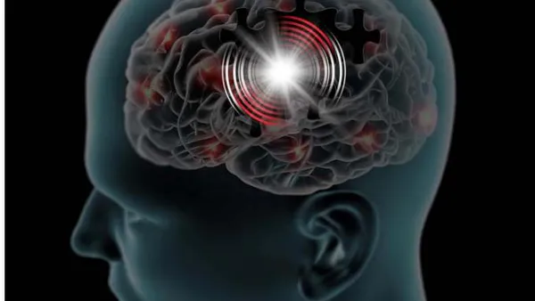
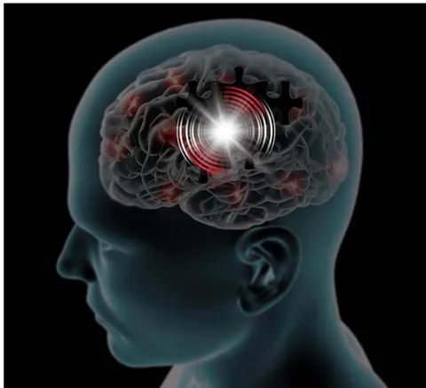
Like all forms of energy, mental energy would be expected to induce magnetic fields. Likewise, the neurons of the brain induce magnetic fields as they depolarize and repolarize. Hence, the mind and the brain are naturally poised to communicate in the same language—electromagnetic energy. Besides helping to explain both the emerging data on NDEs and the experimental observations of Cerf and his colleagues, a duality of mind and brain could, for the first time, explain the distinction between unconscious and conscious mental processing. Unconscious mental processing would occur independent of brain function, whereas conscious mental processing would occur when neurologically-induced magnetic fields synchronized with mentally-induced magnetic fields (Figure 1). This synchronization process hypothetically explains the familiar time-delay when the mind attempts to formulate a thought or draw a memory into consciousness. Consciousness, in this sense, could more aptly be called "corporeal consciousness" because it occurs in conjunction with neurological function. This is in contrast to "incorporeal consciousness," which would occur independent of neurological function [11]. Note also that unconscious mental processing, being electromagnetic but independent of neurological function, would proceed at a speed of approximately 300,000,000 meters/second (the speed of electromagnetic waves). This is in contrast to conscious mental processing, which, being dependent upon neurological function, would proceed at the relatively slow speed of about 150 meters/second (the speed of salutatory conduction) [39]. This difference, together with the uncoupling of the mind from bodily sensory systems during an NDE, could explain why NDEers experience such a dramatic expansion of consciousness when they separate from their physical bodies [31-34].
Further evidence that the mind is capable of functioning independent of the brain comes from the observation that children who are born without a cerebral cortex are conscious [40], and in their pioneering work, Wilder Penfield and others found that awareness of self and environment were fully preserved as they surgically removed relatively large areas of the cortex to treat refractory seizures [40, 41].
That leads to the question of where in the body the mind is located. Based on the observation that injury to anybody-part other than the head leaves corporeal consciousness intact, it is evident that the mind is located in the head. Also, with the exception of damage to the neurological system, damage to any part of the body can be perceived by the mind. That implies that the mind-body connection must be dependent upon intact neurological function. The only part of the neurological system that is in the head is the brain. Therefore, the mind-body connection must occur in the brain.
Although it would be difficult to pinpoint where in the brain the mind is located, the topography and functional anatomy of the brain provide important clues. It is well-recognized that virtually all sensory input is relayed directly to the thalamus. It is also known that the thalamus remains a part of the conversation as the input is being processed by the cerebral cortex and other parts of the brain [42]. Furthermore, even mild damage to the thalamus can result in a vegetative state [43]. Conversely, deep brain stimulation of the thalamus has been found to be of some benefit in rousing patients from a minimally conscious state [44, 45]. Hence, it appears that the thalamus, which has been called "the gateway to the mind," could be acting as a functional interface that allows the mind to monitor and control virtually every function of the brain and body [11]. That would place the mind, or at least its primary area of focus, at the core of the brain.
b) Practical Application of Mind-Brain Dynamics to the Diagnosis and Treatment of Mental Illness
The idea that the mind and the brain are two distinctly different entities that interact with each other could begin to explain how treatment with psychotherapy alone and medication alone can achieve similar results both psychologically and neurologically [46]. Therapies that are aimed directly at changing the way one thinks would have secondary affects on the brain because everything that is processed by the mind would simultaneously be processed by the brain. Conversely, therapies that are aimed directly at modulating brain function would have secondary effects on the mind because everything that is processed by the brain would simultaneously be processed by the mind. Thus, for example, cognitive-behavioral therapy, which changes the way one thinks and feels, would retrain circuits in the brain because changes in cognitive-emotional processing alter neuronal firing patterns. Conversely, pharmacological therapy, which modulates the activity of specific neuronal circuits, would retrain one's thoughts and emotions because changes in neuronal signaling cause changes in mental and emotional processing.
The big question when it comes to therapy, however, is which form would be most effective for which patient? To answer that question, one would first need to determine which of the two—the mind or the brain—was the primary driver of the symptoms. One would then need to determine which form of therapy, when used to treat the appropriate part of the cognitive-emotional system, would be best for which patient. However, the answer to both of these questions would depend upon an accurate understanding of what causes psychiatric symptoms to begin with.
Although the precise cause of psychiatric symptoms remains unclear, an emerging hypothesis contends that psychiatric symptoms are driven by pathological hyperactivity in symptom-related circuits in the brain. According to the multi-circuit neuronal hyperexcitability (MCNH) hypothesis of psychiatric disorders, pathological hyperactivity in anxiety circuits causes elevated and persistent feelings of anxiety; pathological hyperactivity in depressive circuits causes elevated and persistent feelings of depression; and pathological hyperactivity in cognitive circuits causes racing thoughts and obsessional thinking [47]. Yet, that would still fall short of explaining why the symptom-related circuits in the brain become pathologically hyperactive.
However, a possible answer to that question is supplied by the gene research. A number of large, multicenter gene association studies have found that persons who suffer from common psychiatric disorders, such as anxiety, depression, bipolar disorder, and schizophrenia, have gene variants whose protein products fail to adequately regulate the firing of neurons [48-61]. Now then, given that all of the most common psychiatric disorders are essentially different combinations of the same symptoms, it would not be unreasonable to think that all of these disorders could be rooted in a shared physiological abnormality; namely, neuronal hyperexcitability. Hyperexcitable neurons would just fire too easily and fail to shut off when they should. Indeed, this aligns with the neurophysiological abnormalities that have been observed on functional [62] and electroencephalographic [63] studies of depression.
Now imagine that an affected person were confronted with a stressful situation. The hyperexcitability of the neurons would cause all of the person's anxious thoughts to run through his or her mind more times than they should, and it would cause all of the person's uneasy emotions to be abnormally intense and persistent. In addition to being experienced as inappropriately excessive worry and anxiety, the added mental and emotional tension would cause the related circuits in the brain to be further stimulated, thus creating a vicious cycle of mutual overstimulation between the mind and the brain. Moreover, this vicious cycle would, over time, be further amplified by "primed burst potentiation," a natural kindling effect through which neurons that are repeatedly stimulated become increasingly responsive to further stimulation [64].
Another factor that would add fuel to the fire is the tendency for neuronal circuits to compete for dominance. From the study of epilepsy, it is known that pathologically hyperactive circuits tend to inhibit the activity of competing circuits [65]. This phenomenon would tend to prevent the mind from shifting attention to less anxious and more productive thoughts. In other words, it would leave the mind and the brain caught in the "default mode," a psychophysiological state of unproductive internal processing that has been observed on functional imaging of clinical depression and other neuropsychiatric disorders [66]. It could also lead to aberrant circuit induction. This process, which is analogous to a short-circuit in a wired electrical system, hypothetically involves the inappropriate stimulation of relatively hypoactive circuits by pathologically hyperactive circuits [67]. As the feeder circuits quiet down due to synaptic fatigue [68], the freshly activated receiver circuits cause the person's thoughts and emotions to shift accordingly, thus driving the "bipolar switch" [67]. With all of this abnormal electrical activity hijacking the cognitive-emotional system, it is not surprising that affected persons are so easily overwhelmed, so emotionally unstable, and so plagued with self-doubt.
This raises the question of what really drives patients to seek treatment. The natural assumption is that they are driven to seek treatment by the factors that they say drove them to seek treatment. However, as one can see from the forgoing discussion, those factors can be abnormally amplified and distorted by poorly restrained discharges from the brain. Yet in actual practice, neither patients nor their healthcare providers have any reliable way of knowing this. In the 1900s, mild cognitive-emotional distortions were referred to as "neuroses," and severe cognitive-emotional distortions are still referred to as "psychoses." According to the MCNH hypothesis, various forms of psychosis are created when, due to the amplifying effect of neuronal hyperexcitability, the intensity of mentally-generated thoughts and emotions becomes as high or higher than the intensity of thoughts and emotions that would normally be driven by input from the eyes, ears, and other sensory organs. Hypothetically, the margin between internally-driven thoughts and emotions, which are normally of lower intensity, and externally-driven thoughts and emotions, which are normally of higher intensity, is what allows a person to distinguish internal from external reality. Of course, the distorting effect of neuronal hyperexcitability can easily be recognized in severely psychotic patients; but the distorting effect can be more difficult to recognize in patients whose complaints are less out-of-line with reality. If the therapist then begins to work with this distorted content in such patients, he or she would unwittingly be attempting to treat a neurological problem with a psychological intervention. By analogy, it would be like trying to correct impaired vision by talking about it. The difference, however, is that talking about a visual impairment cannot do further damage to the eye; whereas, talking about neurologically-distorted thoughts and emotions can cause further damage by continuing to stir the pot, particularly in a person whose hyperexcitable brain is continuing to distort everything that he or she thinks and feels. Most experienced psychotherapists can readily attest to the risk of regression when intensive psychotherapy is attempted with more severely disturbed patients (presumably those with higher levels of neuronal hyperexcitability), and the renowned Austrian psychiatrist Sigmund Freud, due to the same concerns, was careful to avoid psychoanalyzing psychotic-range patients [69].
In contrast to persons with hyperexcitable brains, those with normoexcitable brains would be relatively resistant to cognitive-emotional stress, and they would be even more resistant to developing psychiatric symptoms. That raises the possibility that most, if not all, persons who present for psychotherapy have hyperexcitable brains. Additional support for this idea comes from the observation that the vast majority of persons who initially seek the care of a psychotherapist rarely need to continue psychotherapy once, upon being referred to a prescriber, their neurological function is normalized with anticonvulsant drugs. Another observation that suggests that most persons who seek psychotherapy have hyperexcitable brains is that such persons are rarely satisfied with their treatment until they either become willing to accept medical therapy or they establish natural brain-calming habits and routines, such as stress-reduction, establishment of an early sleep schedule, regular exercise, avoidance of psychostimulants, and minimization of refined sugar. Consistent with this observation, the Royal Australian and New Zealand College of Psychiatrists is now, for the first time, recommending attention to diet, regular exercise, and sleep hygiene as "non-negotiable first steps" in the treatment of major depressive disorder [70].
Another important factor to consider is that the majority of studies that compare the effectiveness of psychotherapy alone to pharmacotherapy alone involve the use of antidepressants, and antidepressants are not the appropriate treatment for neuronal hyperexcitability [67, 71, 72]. Still, such studies yield comparable results [73], an observation that calls psychotherapy into question as much as the use of antidepressants. That is not to say that psychotherapy, as a therapeutic tool, is unhelpful, but only to say that most persons who seek psychotherapy would be better served if they were to simultaneously be assessed for neuronal hyperexcitability. If this common condition could be identified and treated successfully early in the course of psychotherapy, the distorting element of the patient's distress would be minimized, and the therapy could focus more on matters that truly were rooted in psychology, such as attitude, values, and priorities. Some of the aforementioned psychotherapeutic techniques do just that, whereas others analyze the patient's distressing thoughts and emotions.
What all of the psychotherapeutic techniques have in common, however, is that they aim to reduce intrapsychic tension. Reducing intrapsychic tension has both direct and indirect benefits; it benefits the mind directly by bringing psychological relief, and it benefits the brain indirectly by reducing mental stimulation of the brain. However, as previously discussed, intrapsychic tension can be difficult to reduce when the pathologically hyperactive brain is keeping the mind bathed in stress. That underscores the importance of pharmacotherapy. If the brain could be quieted directly through anticonvulsant drugs (or any of the aforementioned medical therapies), the interference from the brain would be reduced, thus explaining why medical therapy tends to work faster than psychotherapy [46] but not as well as when combined with psychotherapy [74].
Notwithstanding the potential benefits of medical therapy, it should be noted that antidepressants, psychostimulants, and some of the other medical therapies that were referenced earlier stimulate some parts of the brain while calming others. For example, SSRIs increase neuronal firing in the cerebral cortex [75] but reduce neuronal firing in the amygdala [76], and rTMS can be used to either stimulate or inhibit the activity of specific neuronal circuits [77, 78]. Although increasing the activity of specific circuits can be therapeutic, it can also be counter-therapeutic, depending on how it affects the circuit-specific imbalances that are driving the patient's symptoms. This is the MCNH explanation for the paradoxical effects that neuroactivating medical therapies, particularly antidepressant and psychostimulant therapies, can have. With these two classes of drugs topping the list of the most commonly prescribed medications, and the prevalence of psychiatric and substance use disorders at epidemic proportions, the need to better understand how these drugs and other medical therapies are affecting the mind and brain is evident.
IV. ASSESSING THE RELATIVE IMPORTANCE OF THE NEURONAL HYPEREXCITABILITY TRAIT
But even if neuronal hyperexcitability were at the root of psychiatric symptoms, it would not discount the importance of numerous other factors, such as family upbringing, childhood trauma, ongoing stressors, and personal choices. However, an analysis of the family pedigrees of persons who exhibit signs of mental illness is quite revealing. Although family, twin, and adoption studies have historically failed to identify a classic Mendelian pattern of inheritance for any of the common psychiatric disorders, a reconceptualization of psychiatric symptoms as the symptomatic expression of the neuronal hyperexcitability trait does reveal a classic Mendelian distribution. That distribution is strikingly autosomal dominant! [47]. In other words, in those families that are affected, probands who develop either subsyndromal or more obvious signs and symptoms of mental illness, such as a diagnosable psychiatric, functional physical, or substance use disorder, almost always appear in a classic autosomal dominant distribution. Moreover, a predictable subset of children in these families are completely unaffected despite being raised in the same households by the same parents. These so-called "survivors," who typically appear in an autosomal recessive distribution, are presumably those who did not inherit one of the gene variants that have been linked to neuronal hyperexcitability. These observations combine to suggest that: 1) all of the most common psychiatric and functional physical disorders are rooted in the same biological abnormality; 2) all of these disorders may be driven by polymorphisms of a single gene locus; and 3) the hypothesized abnormality may be the most important predisposing factor in the development of these disorders. While recognizing their profound importance, these observations should be interpreted with caution because they are based on informally-obtained family pedigrees (approximately 300) rather than tightly controlled studies [67, 79].
V. THE CHALLENGE OF IDENTIFYING THE NEURONAL HYPEREXCITABILITY TRAIT
Although the phenomenon of neuronal hyperexcitability as a possible driver of psychiatric symptoms has been described previously [47, 80], its significance has been sorely overlooked. This is largely due to the elusive nature of the neuronal hyperexcitability trait. The reasons for the difficulty identifying the trait are complex and multi-faceted. Some, but not all, will be discussed here for the purpose of illustration.
The most fundamental reason that the neuronal hyperexcitability trait has been so difficult to identify is that the trait has heretofore been undetectable by any form of laboratory testing, neuroimaging, or electroencephalography. Hyperexcitable neurons, like a hive of irritable bees, cannot be distinguished from normoexcitable neurons until the metaphorical bees are disturbed. However, even then, the brain does not become hyperactive as a whole. Rather, the pathological hyperactivity occurs in the brain's microcircuitry[81], where it can easily be overlooked or considered to be normal on diagnostic studies. The same challenge is experienced clinically, as carriers of the trait can be completely asymptomatic until something or someone begins to stress them. However, even when symptoms begin to appear, they are commonly accepted as normal both because the neuronal hyperexcitability trait is harbored by such a large fraction of society and because the symptoms primarily involve the same cognitive-emotional states that every person may experience from time to time.
Another reason that the neuronal hyperexcitability trait has remained so difficult to identify is that stress-inducing circumstances are highly specific to the individual and, in most cases, only really known by the individual. This makes it difficult to assess the appropriateness of the symptoms to the circumstances that seem to precipitate them. Also, due to the variable time-course of kindling, symptom-onset can be delayed by days, weeks, or months [82], thus adding to the difficulty of assessing the appropriateness of the symptoms.
Yet another reason that the neuronal hyperexcitability trait has remained so elusive is that the diagnosis of psychiatric disorders has traditionally been symptom-based rather than pathology-based. Hence, the signs and symptoms of neuronal hyperexcitability, which can be highly diverse due to the high diversity of neuronal circuits and firing patterns, are generally viewed as different syndromes rather than as exacerbations of a shared neurophysiological abnormality [83, 84]. This, in turn, has treatment implications that can lead clinicians even further down the wrong path due to current prescribing habits. Since the development of the monoamine hypothesis of depression, prescribers have been strongly entrained to treat most psychiatric disorders with antidepressants. However, based on resting vital-sign measurements (the diagnostic value of which will be discussed later), the neuronal hyperexcitability trait is harbored by approximately 4 out of 10 persons [67, 85, 86]. This estimate is corroborated by the fact that anticonvulsants and other brain-calming drugs had, throughout most of recorded history, been the mainstay of medical treatment for a wide range of emotional and physical ailments [87]. Today, in the wake of the antidepressant revolution, the use of anticonvulsants has been relegated to bipolar spectrum disorder [67, 88]. The problem with this diagnostically-based change is that bipolar spectrum disorder is often misdiagnosed as unipolar depressive disorder [89-92]. This error is further complicated by the fact that antidepressants can have beneficial effects in bipolar spectrum patients despite the fact that they do not address the core physiological abnormality in the disorder [72]. All of these barriers to recognizing the neuronal hyperexcitability trait underscore the need to more easily identify the trait.
VI. TOWARD AN OBJECTIVE METHOD OF IDENTIFYING THE NEURONAL HYPEREXCITABILITY TRAIT
In recent years, an explosion of clinical studies has identified an association between resting vital-sign measurements and the later development of various psychiatric and general medical conditions. In a longitudinal study involving more than one million men in Sweden, Latvala et al. [93] found that subtle elevations in resting heart rate (RHR) were predictive of the later development of generalized anxiety disorder, obsessive-compulsive disorder, and schizophrenia. Similarly, Blom et al. [94] found that adolescent girls with emotional disorders had increased resting respiratory rates (RRR) in comparison to healthy controls. Persons with higher resting heart and respiratory rates have also been found to be at increased risk of developing a wide range of chronic physical illnesses, including diabetes [95-98], high blood pressure [99-101], cardiovascular disease [102-107], cerebrovascular disease [108-110], cancer [110-112], dementia [113], and all-cause mortality [110, 114]. The subtle vital-sign elevations with which these illnesses are associated are thought to be the consequence of a tonic elevation in basal neurological activity in those persons who inherit the genes for neuronal hyperexcitability [115]. This is the MCNH explanation for why the lifespan of persons with severe mental illness tends to be much shorter than the general population [115]. The reason that psychiatric and "functional" physical symptoms would tend to precede the development of diagnosable physical abnormalities is that the cognitive-emotional system is more expressive of neuronal excitation than other organs and systems of the body [116]. The physical consequences would tend to be delayed because they would express the gradual erosive effects of neuronal hyperexcitability, which can take years or even decades to occur [115]. Thus, there is mounting evidence that the neuronal hyperexcitability trait can be identified objectively [67, 115]. It has been estimated that, in the absence of any significant cardiorespiratory disease, confounding medications, or substances of abuse, an RHR above 75 beats/min or an RRR above 15 breaths/min is indicative of the neuronal hyperexcitability trait. Parenthetically, in the more than 100 consecutive outpatients that have been studied thus far, resting heart and respiratory rate measurements have proven to be more sensitive in detecting the neuronal hyperexcitability trait than formal clinical assessments.
VII. DISCUSSION
The goal of this review was to address the question that every behavioral health clinician faces—that of deciding whether a patient should be treated with psychotherapy, medical therapy, or a combination of the two. Short of an objective way for either the patient or the clinician to make this determination, self-referral is generally the decisive factor in determining which type of therapy a patient receives, at least initially. As discussed earlier, this is potentially faulty because most patients have limited insight into the psychophysiological underpinnings of their distress, and even experienced clinicians are often unable to tell how much of the patient's distress is rooted in psychological factors and how much is rooted in biological factors. However, the idea that the inherited trait of neuronal hyperexcitability can drive the same symptoms as purely psychological factors, taken together with the idea that the trait can be identified through resting vital-sign measurements, has the potential to objectivize, for the first time, which type of therapy—psychological or biological—a patient should receive. It also has the potential to determine what percentage of patients who present for behavioral health services are carriers of the neuronal hyperexcitability trait.
Under the current system of referral and treatment selection, many patients may be receiving the wrong type of therapy. Some may be receiving psychotherapy when they should be receiving medical therapy, and some may be receiving medical therapy when they should be receiving psychotherapy. There may also be some who are receiving one form of therapy or the other when in fact they should be receiving both forms of therapy simultaneously. Also, because the neuronal hyperexcitability trait continues to be so elusive, some patients may be receiving the wrong type of medication [71, 87, 117].
Fortuitously, all of this could be about to change with the growing recognition that resting vital-sign measurements offer an objective way to determine which form of treatment a patient should receive. Beyond that, recognizing neuronal hyperexcitability as the core abnormality in mental illness could bring with it a highly treatable biological target. This too would be a first in psychiatry because the current system of diagnosis and treatment is symptom-based rather than pathology-based. Guided by the MCNH hypothesis, any patient who was determined, based on resting vital-sign measurements, to have a hyperexcitable brain could first be educated about the natural ways to calm the brain, such as stress reduction, establishment of an early sleep schedule, regular exercise, and the other lifestyle habits that were discussed earlier. Patients with moderate-to-severe symptoms could also be offered anticonvulsant therapy, as the degree of improvement achieved through lifestyle changes alone is typically limited to about . Anticonvulsants, which, based on their putative mechanism of action, could more aptly be called "neuroregulators" [118], go right to the root of the problem. They reduce the excitability of the neurological system, thereby compensating for the gene abnormality that is believed to underlie the neuronal hyperexcitability trait. Moreover, unlike commonly prescribed medications, such as antidepressants, psychostimulants, and antipsychotics, all of which alter the activity of specific receptors and circuits in the brain, neuroregulators simply normalize brain function. This is a healthier approach because the brain, in most of the common psychiatric disorders, is not misfiring but rather over-firing. Hence, if a given neuroregulator were ineffective at reducing symptoms, it could appropriately be replaced with another neuroregulator rather than switching to a different class of drugs; and if one neuroregulator were only partially effective, a second one could be added, and so on. This approach, which could be called "focused neuroregulation" [119], would optimize the effectiveness of neuroregulators and minimize the need for medications that can have unpredictable, conflicting, and sometimes paradoxical effects [7, 72, 117]. As for those patients whose resting vital signs fell below the minimum cutoffs, psychotherapy alone could be recommended as first-line treatment. In such cases, the therapy would be addressing a problem that was fundamentally psychological rather than just helping a patient cope with a problem that was fundamentally neurological. Thus, the MCNH hypothesis in conjunction with resting vital-sign measurements has the potential to fast-track patients to the most efficient and effective treatment approach. Moreover, because resting vital signs can be measured in the comfort of one's own home, prospective patients would be able to perform the initial screening themselves. In an era of cellphones, smart watches, and a host of new health-tracking devices, this triage system could not be any easier or more practical.
VIII. DIRECTIONS FOR FUTURE RESEARCH
Urgently needed are clinical studies aimed at determining the effectiveness of focused neuroregulation in those patients who, irrespective of their DSM diagnosis, present with an RHR above 75 beats/min or an RRR above 15 breaths/min. This approach would allow researchers to circumvent the problem of overlapping and co-occurring diagnoses and focus on determining which psychiatric symptoms would be most responsive to focused neuroregulation. A high response rate would help validate the use of resting vital-signs as markers of neuronal hyperexcitability. Also, by calculating the fraction of patients who exceed the resting vital-sign cutoffs, insight could be gained into the epidemiology of neuronal hyperexcitability and the sensitivity of resting vital-sign measurements as biomarkers of the neuronal hyperexcitability trait. If promising, such pilot studies could be followed by head-to-head prospective studies comparing the short and long-term effectiveness of this objectively-based method of diagnosis and treatment to standard (symptom-based) treatment.
Additionally, because resting vital signs appear to be constitutionally elevated in carriers of the neuronal hyperexcitability trait [93], prevention studies could be done to determine the benefits of prophylactic neuroregulator therapy in those members of severely affected families who have upper-end-of-normal resting vital signs but have not yet manifested any clear evidence of mental illness. Adjustments of prophylactic medication in such persons could be guided by the response of resting vital signs to the medication and by reassessing for signs and symptoms that may become more clinically apparent only after they are reduced. Also in these studies, the relative importance of resting vital signs as predictive markers of mental illness and the specificity of these markers could be determined by tracking the progress of siblings whose vital signs fell below the hypothesized cutoffs as well as those whose resting vital signs fell above the hypothesized cutoffs but who decided against prophylactic therapy.
Finally, to validate the hypothesis that the vulnerability to developing any of a wide range of psychiatric and functional physical symptoms is rooted in polymorphisms of single gene loci, comprehensive family diagnostic studies could be performed to determine the inheritance pattern of these symptoms and their associated psychiatric disorders as a clinically heterogenous but genetically related group. A classic Mendelian distribution would provide further support for the MCNH hypothesis and potentially pave the way for future research using CRISPR-Cas9 technology [120], offering exciting possibilities for targeted gene therapies [121].
IX. CONCLUSION
By recognizing the cognitive-emotional system as a dynamic interplay between mind and brain and reconceptualizing psychiatric symptoms as pathological hyperactivity in symptom-related circuits in the brain, the MCNH hypothesis, in conjunction with resting vital-sign measurements, has the potential to revolutionize the treatment of mental illness. Rather than treating patients based on subjective assessments and personal skill sets, treatment selection could, for the first time, be based on quantitative biomarkers. Resting vital-sign measurements provide an objective, evidence-based, and easily accessible way to identify the neuronal hyperexcitability trait, an inherited neurophysiological abnormality that is hypothesized to be at the root of most psychiatric and functional physical symptoms. In addition to improving diagnostic accuracy and guiding treatment selection, targeting the neuronal hyperexcitability trait informs the use of focused neuroregulation, a safer, faster, and more effective treatment approach for those patients who are determined, based on resting vital-sign measurements, to have a biologically-based psychiatric disorder. By identifying the neuronal hyperexcitability trait, the challenge of overlapping and co-occurring psychiatric diagnoses is circumvented and the use of medications that have unpredictable, conflicting, and sometimes paradoxical effects can be minimized. Targeting the neuronal hyperexcitability trait also has the potential to ward off psychiatric symptoms before they even begin and reduce the risk of developing any of the wide range of chronic health conditions with which this highly prevalent trait has been associated. In short, the MCNH hypothesis, in conjunction with resting vital-sign measurements, has the potential to change the face of modern psychiatry by transforming the treatment of mental illness from a symptom-based practice to a biologically-based practice. By seizing this unprecedented opportunity, we can strive toward a future in which behavioral healthcare, like other fields of medicine, is aimed at specific pathological processes, thus streamlining care, speeding recovery, and overcoming the long-held stigma of mental illness.
Conflicts of Interest
The author declares that he has no competing interests.